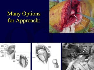
Hip Dislocations andFemoral
Head Fractures
Fernando Serna, MD
John T. Gorczyca, MD
University of Rochester Medical Center
Created March 2004; First Revision January 2006
Second Revision July 2009
2.
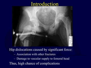
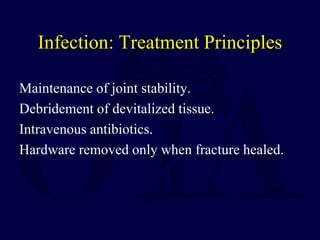
Introduction
Hip dislocations causedby significant force:
– Association with other fractures
– Damage to vascular supply to femoral head
Thus, high chance of complications
3.
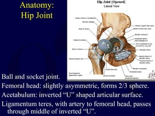
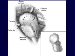
Anatomy:
Hip Joint
Ball andsocket joint.
Femoral head: slightly asymmetric, forms 2/3 sphere.
Acetabulum: inverted “U” shaped articular surface.
Ligamentum teres, with artery to femoral head, passes
through middle of inverted “U”.
4.
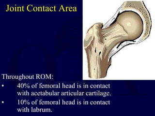
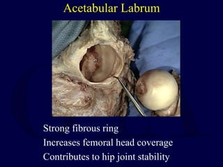
Joint Contact Area
ThroughoutROM:
• 40% of femoral head is in contact
with acetabular articular cartilage.
• 10% of femoral head is in contact
with labrum.
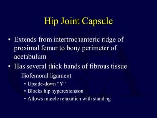
Hip Joint Capsule
•Extends from intertrochanteric ridge of
proximal femur to bony perimeter of
acetabulum
• Has several thick bands of fibrous tissue
Iliofemoral ligament
• Upside-down “Y”
• Blocks hip hyperextension
• Allows muscle relaxation with standing
7.
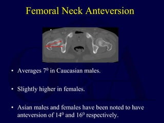
Femoral Neck Anteversion
•Averages 70 in Caucasian males.
• Slightly higher in females.
• Asian males and females have been noted to have
anteversion of 140 and 160 respectively.
8.
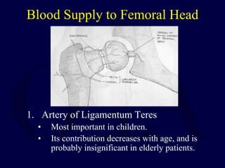
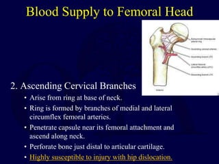
Blood Supply toFemoral Head
1. Artery of Ligamentum Teres
• Most important in children.
• Its contribution decreases with age, and is
probably insignificant in elderly patients.
9.
Blood Supply toFemoral Head
2. Ascending Cervical Branches
• Arise from ring at base of neck.
• Ring is formed by branches of medial and lateral
circumflex femoral arteries.
• Penetrate capsule near its femoral attachment and
ascend along neck.
• Perforate bone just distal to articular cartilage.
• Highly susceptible to injury with hip dislocation.
10.
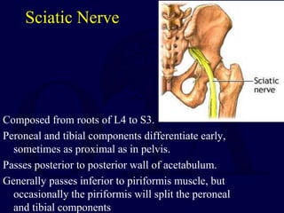
Sciatic Nerve
Composed fromroots of L4 to S3.
Peroneal and tibial components differentiate early,
sometimes as proximal as in pelvis.
Passes posterior to posterior wall of acetabulum.
Generally passes inferior to piriformis muscle, but
occasionally the piriformis will split the peroneal
and tibial components
11.
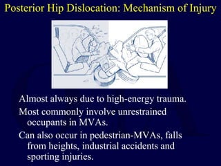
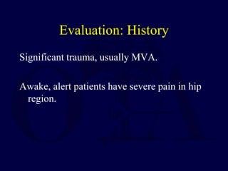
Posterior Hip Dislocation:Mechanism of Injury
Almost always due to high-energy trauma.
Most commonly involve unrestrained
occupants in MVAs.
Can also occur in pedestrian-MVAs, falls
from heights, industrial accidents and
sporting injuries.
12.
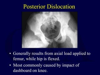
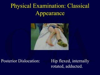
Posterior Dislocation
• Generallyresults from axial load applied to
femur, while hip is flexed.
• Most commonly caused by impact of
dashboard on knee.
13.
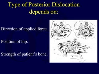
Type of PosteriorDislocation
depends on:
Direction of applied force.
Position of hip.
Strength of patient’s bone.
14.
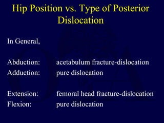
Hip Position vs.Type of Posterior
Dislocation
In General,
Abduction: acetabulum fracture-dislocation
Adduction: pure dislocation
Extension: femoral head fracture-dislocation
Flexion: pure dislocation
15.
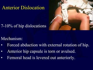
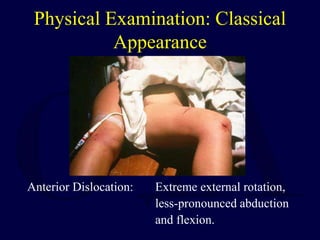
Anterior Dislocation
7-10% ofhip dislocations
Mechanism:
• Forced abduction with external rotation of hip.
• Anterior hip capsule is torn or avulsed.
• Femoral head is levered out anteriorly.
16.
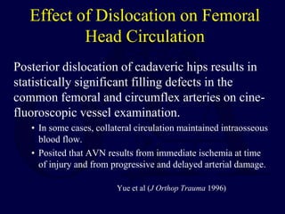
Effect of Dislocationon Femoral
Head Circulation
Posterior dislocation of cadaveric hips results in
statistically significant filling defects in the
common femoral and circumflex arteries on cine-
fluoroscopic vessel examination.
• In some cases, collateral circulation maintained intraosseous
blood flow.
• Posited that AVN results from immediate ischemia at time
of injury and from progressive and delayed arterial damage.
Yue et al (J Orthop Trauma 1996)
17.
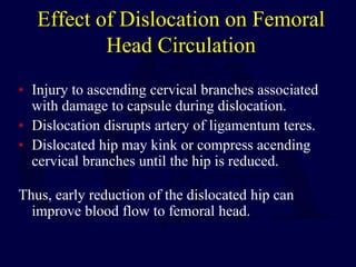
Effect of Dislocationon Femoral
Head Circulation
• Injury to ascending cervical branches associated
with damage to capsule during dislocation.
• Dislocation disrupts artery of ligamentum teres.
• Dislocated hip may kink or compress acending
cervical branches until the hip is reduced.
Thus, early reduction of the dislocated hip can
improve blood flow to femoral head.
18.
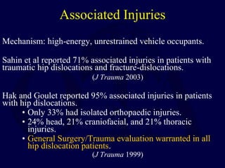
Associated Injuries
Mechanism: high-energy,unrestrained vehicle occupants.
Sahin et al reported 71% associated injuries in patients with
traumatic hip dislocations and fracture-dislocations.
(J Trauma 2003)
Hak and Goulet reported 95% associated injuries in patients
with hip dislocations.
• Only 33% had isolated orthopaedic injuries.
• 24% head, 21% craniofacial, and 21% thoracic
injuries.
• General Surgery/Trauma evaluation warranted in all
hip dislocation patients.
(J Trauma 1999)
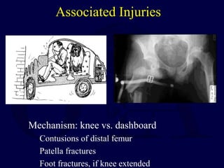
Associated Knee Injuries
25%(46 of 187) of hip injury patients had knee injury.
• 27 acetabulum fractures without dislocation, 10 pure hip
dislocation, and 9 acetabulum fx-dislocations.
• 85% had symptoms or clinical findings of knee injury.
• 13 fractures (7 patella, 5 supracondylar femur or tibial
plateau, 1 osteochondral), 9 ligamentous injury
(2 knee dislocations, 1 MCL, 1 LCL, 5 combined),
1 patellar tendon tear, and multiple wounds and contusions
• 75% had other injuries
•Underscores the need for vigilance in detecting these injuries.
Tabuenca and Truan (CORR 2000)
21.
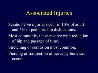
Associated Injuries
Sciatic nerveinjuries occur in 10% of adult
and 5% of pediatric hip dislocations.
Most commonly, these resolve with reduction
of hip and passage of time.
Stretching or contusion most common.
Piercing or transection of nerve by bone can
occur.
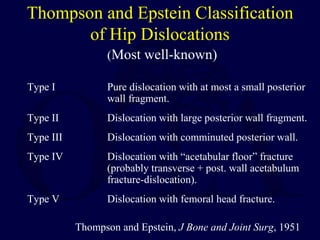
Thompson and EpsteinClassification
of Hip Dislocations
(Most well-known)
Type I Pure dislocation with at most a small posterior
wall fragment.
Type II Dislocation with large posterior wall fragment.
Type III Dislocation with comminuted posterior wall.
Type IV Dislocation with “acetabular floor” fracture
(probably transverse + post. wall acetabulum
fracture-dislocation).
Type V Dislocation with femoral head fracture.
Thompson and Epstein, J Bone and Joint Surg, 1951
24.
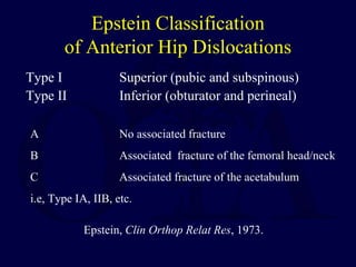
Epstein Classification
of AnteriorHip Dislocations
Type I Superior (pubic and subspinous)
Type II Inferior (obturator and perineal)
A No associated fracture
B Associated fracture of the femoral head/neck
C Associated fracture of the acetabulum
i.e, Type IA, IIB, etc.
Epstein, Clin Orthop Relat Res, 1973.
25.
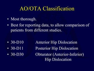
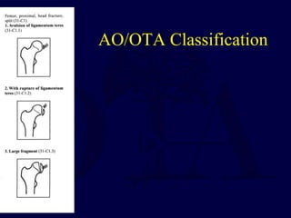
AO/OTA Classification
• Mostthorough.
• Best for reporting data, to allow comparison of
patients from different studies.
• 30-D10 Anterior Hip Dislocation
• 30-D11 Posterior Hip Dislocation
• 30-D30 Obturator (Anterior-Inferior)
Hip Dislocation
Physical Examination
• Painto palpation of hip.
• Pain with attempted motion of hip.
• Possible neurological impairment:
Thorough exam essential!
31.
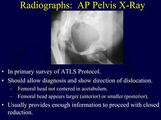
Radiographs: AP PelvisX-Ray
• In primary survey of ATLS Protocol.
• Should allow diagnosis and show direction of dislocation.
– Femoral head not centered in acetabulum.
– Femoral head appears larger (anterior) or smaller (posterior).
• Usually provides enough information to proceed with closed
reduction.
32.
Reasons to ObtainMore
X-Rays Before Hip Reduction
• View of femoral neck inadequate to rule out
fracture.
• Patient requires CT scan of abdomen/pelvis for
hemodynamic instability
– and additional time to obtain 2-3 mm cuts through
acetabulum + femoral head/neck would be minimal.
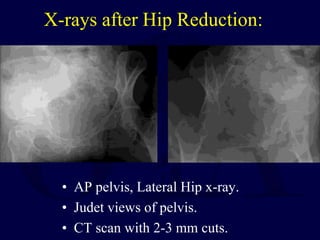
33.
X-rays after HipReduction:
• AP pelvis, Lateral Hip x-ray.
• Judet views of pelvis.
• CT scan with 2-3 mm cuts.
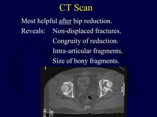
34.
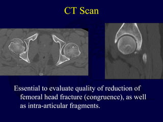
CT Scan
Most helpfulafter hip reduction.
Reveals: Non-displaced fractures.
Congruity of reduction.
Intra-articular fragments.
Size of bony fragments.
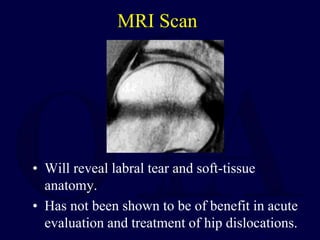
35.
MRI Scan
• Willreveal labral tear and soft-tissue
anatomy.
• Has not been shown to be of benefit in acute
evaluation and treatment of hip dislocations.
Emergent Reduction
• Allowsrestoration of flow through occluded or
compressed vessels.
• Requires proper anesthesia.
• Requires “team” (i.e. more than one person).
38.
Time to reduction
•Controversy in the literature regarding appropriate
timing to reduction
• Marchetti, Steinberg, and Coumas (J Orthop Trauma
1996) found no statistically significant difference in
outcomes in posterior fracture-dislocations when reduced
greater or less than 6 hrs from time of injury
• Mehlman et al (CORR 2000) demonstrated a 20X greater
risk of AVN in pediatric traumatic hip dislocations if
reduction delayed > 6 hrs
• Sahin et al (J Trauma 2003) demonstrated better prognosis
in hip dislocations and fracture-dislocations reduced within
12 hrs
• Universally agreed that the earlier the better
39.
Anesthesia
• General anesthesiawith muscle relaxation facilitates
reduction, but is not necessary.
• Conscious sedation is acceptable.
• Attempts at reduction with inadequate analgesia/
sedation will cause unnecessary pain, create muscle
spasm, and make subsequent attempts at reduction
more difficult.
40.
General Anesthesia if:
•Patient is to be intubated emergently in
Emergency Room.
• Patient is being transported to Operating
Room for emergent head, abdominal or
chest surgery.
• Take advantage of opportunity.
41.
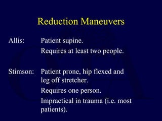
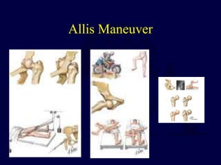
Reduction Maneuvers
Allis: Patientsupine.
Requires at least two people.
Stimson: Patient prone, hip flexed and
leg off stretcher.
Requires one person.
Impractical in trauma (i.e. most
patients).
42.
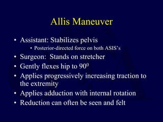
Allis Maneuver
• Assistant:Stabilizes pelvis
• Posterior-directed force on both ASIS’s
• Surgeon: Stands on stretcher
• Gently flexes hip to 900
• Applies progressively increasing traction to
the extremity
• Applies adduction with internal rotation
• Reduction can often be seen and felt
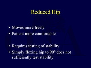
Reduced Hip
• Movesmore freely
• Patient more comfortable
• Requires testing of stability
• Simply flexing hip to 900 does not
sufficiently test stability
45.
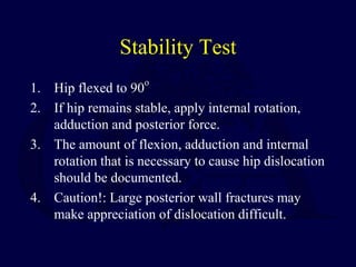
Stability Test
1. Hipflexed to 90
o
2. If hip remains stable, apply internal rotation,
adduction and posterior force.
3. The amount of flexion, adduction and internal
rotation that is necessary to cause hip dislocation
should be documented.
4. Caution!: Large posterior wall fractures may
make appreciation of dislocation difficult.
46.
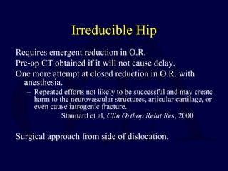
Irreducible Hip
Requires emergentreduction in O.R.
Pre-op CT obtained if it will not cause delay.
One more attempt at closed reduction in O.R. with
anesthesia.
– Repeated efforts not likely to be successful and may create
harm to the neurovascular structures, articular cartilage, or
even cause iatrogenic fracture.
Stannard et al, Clin Orthop Relat Res, 2000
Surgical approach from side of dislocation.
47.
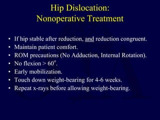
Hip Dislocation:
Nonoperative Treatment
•If hip stable after reduction, and reduction congruent.
• Maintain patient comfort.
• ROM precautions (No Adduction, Internal Rotation).
• No flexion > 60
o
.
• Early mobilization.
• Touch down weight-bearing for 4-6 weeks.
• Repeat x-rays before allowing weight-bearing.
48.
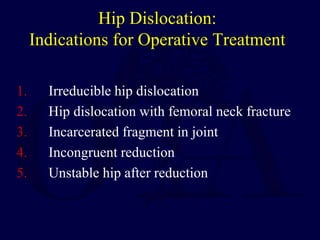
Hip Dislocation:
Indications forOperative Treatment
1. Irreducible hip dislocation
2. Hip dislocation with femoral neck fracture
3. Incarcerated fragment in joint
4. Incongruent reduction
5. Unstable hip after reduction
49.
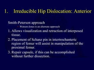
1. Irreducible HipDislocation: Anterior
Smith-Peterson approach
• Watson-Jones is an alternate approach
1. Allows visualization and retraction of interposed
tissue.
2. Placement of Schanz pin in intertrochanteric
region of femur will assist in manipulation of the
proximal femur.
3. Repair capsule, if this can be accomplished
without further dissection.
50.
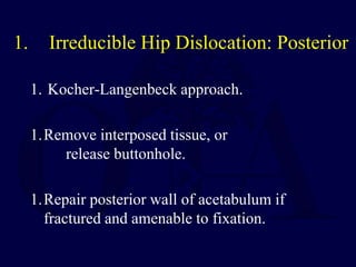
1. Kocher-Langenbeck approach.
1.Removeinterposed tissue, or
release buttonhole.
1.Repair posterior wall of acetabulum if
fractured and amenable to fixation.
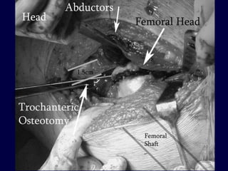
1. Irreducible Hip Dislocation: Posterior
51.
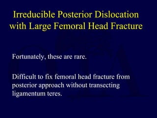
Irreducible Posterior Dislocation
withLarge Femoral Head Fracture
Fortunately, these are rare.
Difficult to fix femoral head fracture from
posterior approach without transecting
ligamentum teres.
52.
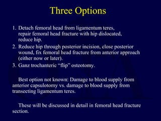
Three Options
1. Detachfemoral head from ligamentum teres,
repair femoral head fracture with hip dislocated,
reduce hip.
2. Reduce hip through posterior incision, close posterior
wound, fix femoral head fracture from anterior approach
(either now or later).
3. Ganz trochanteric “flip” osteotomy.
Best option not known: Damage to blood supply from
anterior capsulotomy vs. damage to blood supply from
transecting ligamentum teres.
These will be discussed in detail in femoral head fracture
section.
53.
2. Hip Dislocationwith Femoral
Neck Fracture
Attempts at closed reduction potentiate chance of fracture
displacement with consequent increased risk of AVN.
If femoral neck fracture is already displaced, then the
ability to reduce the head by closed means is markedly
compromised.
Thus, closed reduction should not be attempted.
54.
2. Hip Dislocationwith Femoral
Neck Fracture
Usually the dislocation is posterior.
Thus, Kocher-Langenbeck approach.
If fracture is non-displaced, stabilize fracture
with parallel lag screws first.
If fracture is displaced, open reduction of
femoral head into acetabulum, reduction of
femoral neck fracture, and stabilization of
femoral neck fracture.
55.
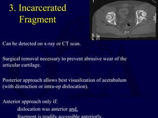
3. Incarcerated
Fragment
Can bedetected on x-ray or CT scan.
Surgical removal necessary to prevent abrasive wear of the
articular cartilage.
Posterior approach allows best visualization of acetabulum
(with distraction or intra-op dislocation).
Anterior approach only if:
dislocation was anterior and,
56.
4. Incongruent Reduction
CausedBy:
• Acetabulum Fracture (weight-bearing portion).
• Femoral Head Fracture (any portion).
• Interposed tissue.
Goal: achieve congruence by removing interposed tissue
and/or reducing and stabilizing fracture.
57.
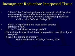
• 25% (9/35)of pediatric patients with traumatic hip dislocation
required surgery to remove interposed soft tissue and/or
osteochondral fragments to achieve congruent hip reduction.
Vialle et al, J Pediatric Orthop 2005
• 92% (33/36) of adults had loose bodies detected
arthroscopically.
• 21% (7/33) had normal x-rays and CT.
–Clinical significance of soft-tissue interposition is not clear if joint
congruent.
–? Benefit of routine arthroscopy.
Mullis and Dahner, J Orthop Trauma, 2006
Incongruent Reduction: Interposed Tissue
58.
5. Unstable Hipafter Reduction
• Due to posterior wall and/or femoral head fracture.
• Requires reduction and stabilization fracture.
• Labral detachment or tear
– Highly uncommon cause of instability.
– Its presence in the unstable hip would justify surgical repair.
– MRI may be helpful in establishing diagnosis.
59.
Results of Treatment
•Large range: from normal to severe pain and
degeneration.
• In general, dislocations with associated femoral head or
acetabulum fractures fare worse.
• Dislocations with fractures of both the femoral head and
the acetabulum have a strong association with poor
results.
• Irreducible hip dislocations have a strong association
with poor results.
– 13/23 (61%) poor and 3/23 (13%) fair results.
McKee, Garay, Schemitsch, Kreder, Stephen. Irreducible
fracture-dislocation of the hip: a severe injury with a poor
prognosis. J Orthop Trauma. 1998.
60.
Complications of HipDislocation
• Avascular Necrosis (AVN): 1-20%
– Several authors have shown a positive correlation
between duration of dislocation and rate of AVN.
– Results are best if hip reduced within six hours.
• Mehlman et al (CORR 2000) demonstrated a 20X greater
risk of AVN in pediatric traumatic hip dislocations if
reduction delayed > 6 hrs
• Also demonstrated that bone scan results can be misleading,
and thus routine screening is not recommended
61.
Post-traumatic Osteoarthritis
• Canoccur with or without AVN
• May be unavoidable in cases with severe cartilaginous
injury.
• Incidence increases with associated femoral head or
acetabulum fractures.
16% osteoarthritis in uncomplicated hip dislocations and up
to 88% in dislocations associated with severe acetabular
fractures
• Upudhyay et al, J Bone Joint Surg. (Br.), 1983.
• Efforts to minimize osteoarthritis are best directed at
achieving anatomic reduction of injury and preventing
abrasive wear between articular carrtilage and sharp bone
edges.
62.
Recurrent Dislocation
Rare, unlessan underlying bony instability has not
been surgically corrected (e.g. excision of large
posterior wall fragment instead of ORIF).
Some cases involve pure dislocation with inadequate
soft-tissue healing – may benefit from surgical
imbrication (rare).
Can occur from detached labrum, which would
benefit from repair (rare).
63.
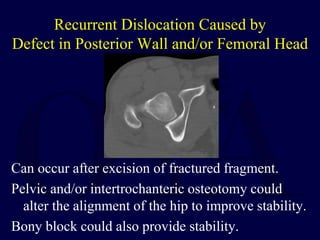
Recurrent Dislocation Causedby
Defect in Posterior Wall and/or Femoral Head
Can occur after excision of fractured fragment.
Pelvic and/or intertrochanteric osteotomy could
alter the alignment of the hip to improve stability.
Bony block could also provide stability.
64.
Delayed Diagnosis ofHip Dislocation
Increased incidence in multiple trauma patients.
More common if patient has altered sensorium.
Results in: more difficult closed reduction.
higher incidence of AVN.
In NO Case should a hip dislocation be treated
without reduction.
65.
Sciatic Nerve Injury
Occursin up to 20% of adult and 5% of pediatric
patients with hip dislocation.
Peroneal nerve affected more commonly than
tibial
Nerve stretched, compressed or transected.
With reduction: 40% complete resolution
25-35% partial resolution
66.
Sciatic Nerve Palsy:
IfNo Improvement after 3–4 Weeks
EMG and Nerve Conduction Studies for
baseline information and for prognosis.
Allows localization of injury in the event that
surgery is required.
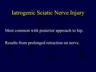
Iatrogenic Sciatic NerveInjury
Most common with posterior approach to hip.
Results from prolonged retraction on nerve.
71.
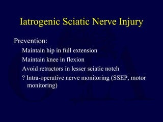
Iatrogenic Sciatic NerveInjury
Prevention:
Maintain hip in full extension
Maintain knee in flexion
Avoid retractors in lesser sciatic notch
? Intra-operative nerve monitoring (SSEP, motor
monitoring)
72.
Thromboembolism
Hip dislocation =high risk patient.
Prophylactic treatment with:
• low molecular weight heparin, or
• coumadin
Early postoperative mobilization.
Discontinue prophylaxis after 2-6 weeks (if
patient mobile).
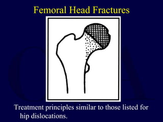
Femoral Head Fractures–
Mechanism
• Fracture occurs by shear as femoral head
dislocates.
• With less hip flexion, femoral head
fracture tends to be larger.
75.
History and PhysicalExamination
Similar to patient with hip dislocation.
Patient posture may be less extreme due to
femoral head fracture.
76.
Classification of FemoralHead Fractures
Thompson and Epstein Type V
• Posterior hip dislocation with femoral head fracture
Epstein Type IB
• Anterosuperior dislocation with femoral head and/or
neck fracture
Epstein Type IIB
• Anteroinferior dislocation with femoral head and/or
neck fracture
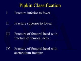
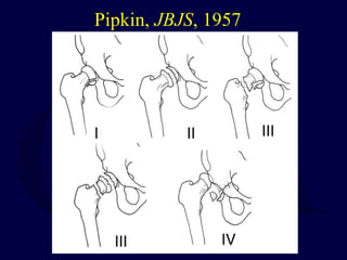
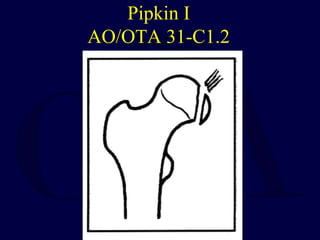
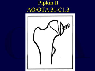
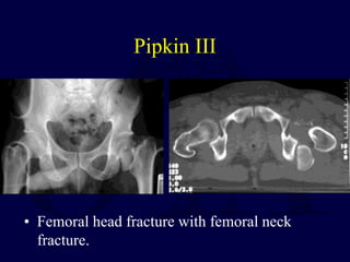
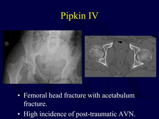
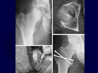
Pipkin Classification
I Fractureinferior to fovea
II Fracture superior to fovea
III Fracture of femoral head with
fracture of femoral neck
IV Fracture of femoral head with
acetabulum fracture
Pipkin IV
• Femoralhead fracture with acetabulum
fracture.
• High incidence of post-traumatic AVN.
84.
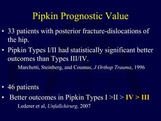
Pipkin Prognostic Value
•33 patients with posterior fracture-dislocations of
the hip.
• Pipkin Types I/II had statistically significant better
outcomes than Types III/IV.
Marchetti, Steinberg, and Coumas, J Orthop Trauma, 1996
• 46 patients
• Better outcomes in Pipkin Types I >II > IV > III
Lederer et al, Unfallchirurg, 2007
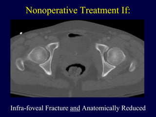
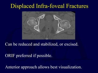
Displaced Infra-foveal Fractures
Canbe reduced and stabilized, or excised.
ORIF preferred if possible.
Anterior approach allows best visualization.
89.
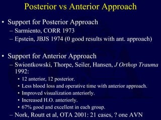
Posterior vs AnteriorApproach
• Support for Posterior Approach
– Sarmiento, CORR 1973
– Epstein, JBJS 1974 (0 good results with ant. approach)
• Support for Anterior Approach
– Swiontkowski, Thorpe, Seiler, Hansen, J Orthop Trauma
1992:
• 12 anterior, 12 posterior.
• Less blood loss and operative time with anterior approach.
• Improved visualization anteriorly.
• Increased H.O. anteriorly.
• 67% good and excellent in each group.
– Nork, Routt et al, OTA 2001: 21 cases, ? one AVN
90.
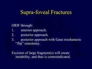
Supra-foveal Fractures
ORIF through:
1.anterior approach.
2. posterior approach.
3. posterior approach with Ganz trochanteric
“flip” osteotomy.
Excision of large fragment(s) will create
instability, and thus is contraindicated.
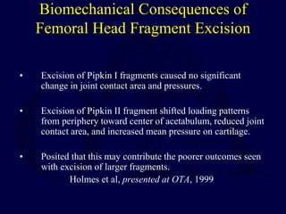
Biomechanical Consequences of
FemoralHead Fragment Excision
• Excision of Pipkin I fragments caused no significant
change in joint contact area and pressures.
• Excision of Pipkin II fragment shifted loading patterns
from periphery toward center of acetabulum, reduced joint
contact area, and increased mean pressure on cartilage.
• Posited that this may contribute the poorer outcomes seen
with excision of larger fragments.
Holmes et al, presented at OTA, 1999
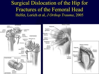
93.
Surgical Dislocation ofthe Hip for
Fractures of the Femoral Head
Helfet, Lorich et al, J Orthop Trauma, 2005
97.
Pipkin III Fractures
Highincidence of AVN and degeneration with displaced
fractures.
– Relative indication for hemiarthroplasty in older patient due
to this risk
– Attempt at ORIF warranted in active, younger patients
If femoral neck fracture is non-displaced, do not attempt
manipulative reduction of hip until femoral neck is
stabilized.
98.
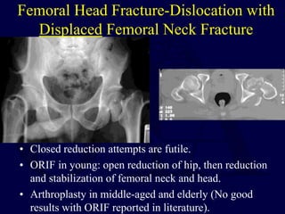
Femoral Head Fracture-Dislocationwith
Displaced Femoral Neck Fracture
• Closed reduction attempts are futile.
• ORIF in young: open reduction of hip, then reduction
and stabilization of femoral neck and head.
• Arthroplasty in middle-aged and elderly (No good
results with ORIF reported in literature).
99.
Femoral Head Fracture-Dislocationwith
Non-Displaced Femoral Neck Fracture
Must consider stabilizing femoral neck
fracture before performing reduction of hip.
100.
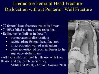
Irreducible Femoral HeadFracture-
Dislocation without Posterior Wall Fracture
• 72 femoral head fractures treated in 6 years
• 7 (10%) failed routine closed reduction.
• Radiographic findings in these 7:
• posterosuperior disclocations
• sagittal plane femoral head fractures
• intact posterior wall of acetabulum
• close apposition of proximal femur to the
supra-acetabular ilium.
• All had slight, but fixed hip flexion with knee
flexion and leg length discrepancy.
Mehta and Routt, J Orthop Trauma, 2008
101.
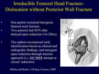
Irreducible Femoral HeadFracture-
Dislocation without Posterior Wall Fracture
• One patient sustained iatrogenic
femoral neck fracture.
• Two patients had AVN after
delayed open reduction (14-32hrs).
• The authors recommend early
identification based on clinical and
radiograhic findings, and emergent
open reduction through anterior
approach (i.e. DO NOT attempt at
closed reduction)
Mehta and Routt, J Orthop Trauma, 2008
102.
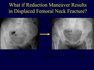
What if ReductionManeuver Results
in Displaced Femoral Neck Fracture?
103.
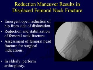
Reduction Maneuver Resultsin
Displaced Femoral Neck Fracture
• Emergent open reduction of
hip from side of dislocation.
• Reduction and stabilization
of femoral neck fracture.
• Assessment of femoral head
fracture for surgical
indications.
• In elderly, perform
arthroplasty.
104.
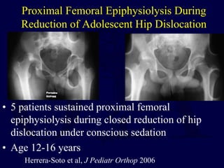
Proximal Femoral EpiphysiolysisDuring
Reduction of Adolescent Hip Dislocation
• 5 patients sustained proximal femoral
epiphysiolysis during closed reduction of hip
dislocation under conscious sedation
• Age 12-16 years
Herrera-Soto et al, J Pediatr Orthop 2006
105.
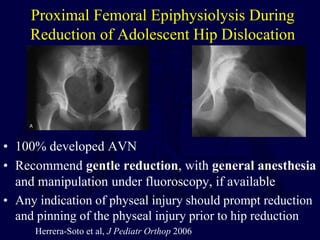
Proximal Femoral EpiphysiolysisDuring
Reduction of Adolescent Hip Dislocation
• 100% developed AVN
• Recommend gentle reduction, with general anesthesia
and manipulation under fluoroscopy, if available
• Any indication of physeal injury should prompt reduction
and pinning of the physeal injury prior to hip reduction
Herrera-Soto et al, J Pediatr Orthop 2006
106.
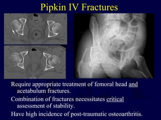
Pipkin IV Fractures
Requireappropriate treatment of femoral head and
acetabulum fractures.
Combination of fractures necessitates critical
assessment of stability.
Have high incidence of post-traumatic osteoarthritis.
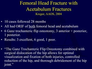
Femoral Head Fracturewith
Acetabulum Fractures
Kregor, AAOS, 2004
• 10 cases followed 28 months
• All had ORIF of both femoral head and acetabulum
• 6 Ganz trochanteric flip osteotomy, 3 anterior + posterior,
1 posterior.
• Results: 3 excellent, 6 good, 1 poor.
• “The Ganz Trochanteric Flip Osteotomy combined with
surgical dislocation of the hip allows for optimal
visualization and fixation of both injuries, controlled
reduction of the hip, and thorough debridement of the hip
joint.”
109.
Questions?
Return to
Lower Extremity
Index
Ifyou would like to volunteer as an author for the
Resident Slide Project or recommend updates to any of
the following slides, please send an e-mail to
ota@ota.org


![CTEV [ clubfoot] DR ARUN LAL ,DR MOHAMED ASHRAF travancore medical college k...](https://cdn.slidesharecdn.com/ss_thumbnails/ctevclubfootdrarunlaldrmohamedashraftravancoremedicalcollegekollamkeralaindia-260208063247-18fc466c-thumbnail.jpg?width=640&height=640&fit=bounds)

