This document discusses knee dislocations, including epidemiology, anatomy, pathomechanisms, classification, associated injuries, physical exam, and treatment principles. Some key points:
- Knee dislocations are rare injuries that often involve multiple ligament tears. Associated injuries like vascular injuries or peroneal nerve injuries are also common.
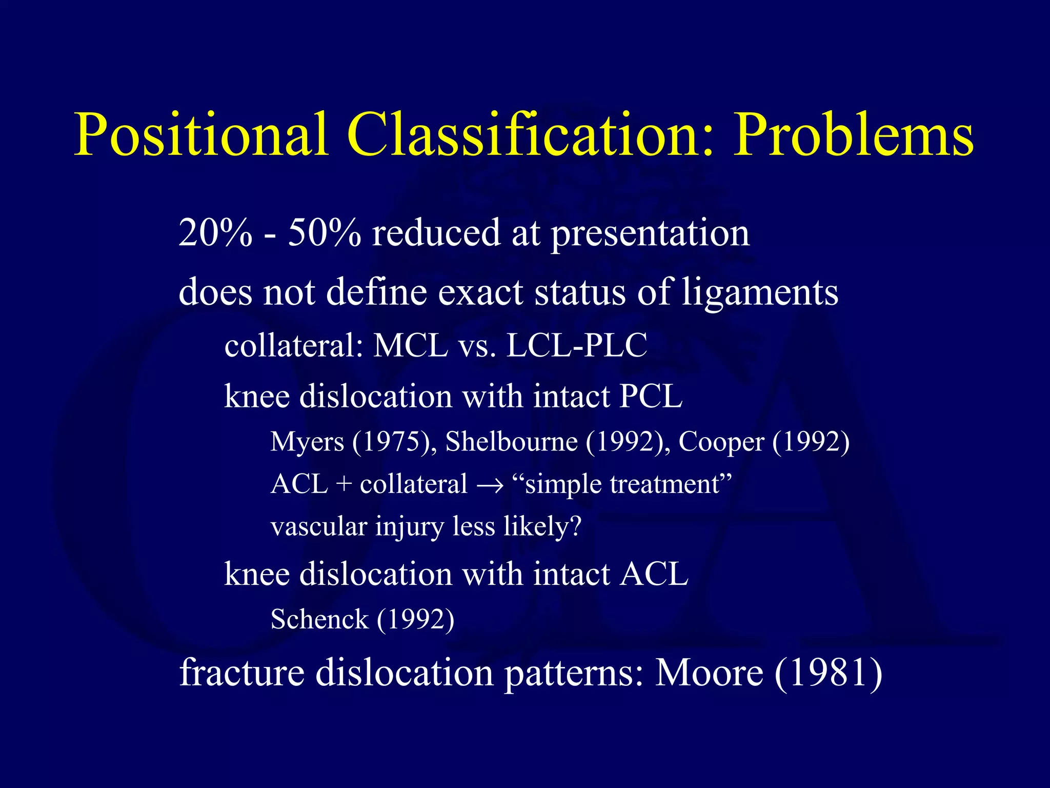
- An anatomic classification system focuses on the specific ligaments torn and has utility in guiding treatment and comparing outcomes. Type III injuries involving the PCL and MCL/LCL are most frequent.
- Vascular injuries occur in around 30% of cases. Revascularization is needed within 8 hours to avoid amputation. Arteriography is recommended if pulses are abnormal


![Vascular Injuries: Recommendations
[A] ischemic limb after reduction
immediate surgical exploration
injury and location predictable
arteriogram: only if additional associated proximal injury
[B] abnormal vascular status - viable limb
diminished pulses
decreased capillary refill
ABI < 0.9
“urgent” arteriogram](https://image.slidesharecdn.com/l06-kneedislocations-161225231936/75/L06-knee-dislocations-23-2048.jpg)
![Vascular Injuries: Recommendations
[C] normal vascular status and no ligament or
extremity surgery
normal PT/DP pulses and normal capillary refill
ABI > 0.90
careful observation with serial exams
vascular surgery and invasive radiology “available”
MRA/MRI
evaluate for non-occlusive (intimal) injury
sensitivity and specificity uncertain
arteriogram if abnormal](https://image.slidesharecdn.com/l06-kneedislocations-161225231936/75/L06-knee-dislocations-24-2048.jpg)
![Vascular Injuries: Recommendations
[D] normal vascular status - potential or
planned ligament or extremity surgery
normal PT/DP pulses and normal capillary refill
ABI > 0.90
careful observation with serial exams
vascular surgery and invasive radiology “available”
MRA/MRI as part of pre-operative evaluation
routine arteriogram within 24 - 48 hours
intimal injury
anticoagulation
no tourniquet
limited and delayed surgery (10-14 days)
no endoscopic PCL (tibial tunnel)](https://image.slidesharecdn.com/l06-kneedislocations-161225231936/75/L06-knee-dislocations-25-2048.jpg)


![Associated Injuries: Polytrauma
Mills WJ : Severe HO After High Energy Knee Dislocation: The
Predicitve Value of the Injury Severity Score; OTA 2001.
35 consecutive knee dislocations
Harborview Medical Center
associated injuries
23% popliteal artery
20% peroneal nerve
surgical treatment
29: open acute [< 4 weeks]
6: arthroscopic delayed [6 wk - 10 m]
CPM and early motion as wound permitted](https://image.slidesharecdn.com/l06-kneedislocations-161225231936/75/L06-knee-dislocations-33-2048.jpg)


![42 female
unrestrained front seat passenger MVA
multiple injuries
laparotomy
spleenectomy, hepatic packing
LC-1 pelvis
[R] knee dislocation
Case Example: KD-IIIL](https://image.slidesharecdn.com/l06-kneedislocations-161225231936/75/L06-knee-dislocations-70-2048.jpg)


