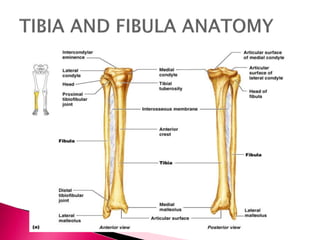
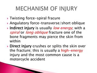
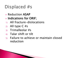
This document discusses tibia fractures, including:
1. Tibia fractures are more common than other long bone fractures and often result in open fractures.
2. Tibia fractures are caused by twisting, angulatory, or indirect/direct forces and are classified based on the soft tissue injury and fracture stability.
3. Treatment depends on the soft tissue condition, fracture severity, stability, and degree of contamination. Most are treated non-operatively but unstable or open fractures may require surgery.
This document discusses fractures of the tibia shaft. It begins by providing an overview of tibia anatomy, noting that it is the second largest bone and carries 80% of body weight from the femur to the foot. It then describes the anatomy of the tibia in more detail. Mechanisms of tibia shaft fractures are discussed as well as common fracture patterns, clinical presentation, diagnostic imaging, and treatment approaches including non-operative and operative options like intramedullary nailing and plating. Compartment syndrome is also covered as a potential complication.
1. Fractures of the clavicle and scapula are uncommon but can result from high-energy trauma.
2. Clavicle fractures most commonly occur in the middle third and are usually treated conservatively with sling immobilization.
3. Scapula fractures involve the body, neck, glenoid, coracoid, or acromion and are often associated with life-threatening injuries requiring assessment by ATLS protocols. Most are also treated initially with sling immobilization.
(9)external fixation indications and techniques(bonatus)Drpraveen Kumar
External fixation involves placing pins or wires connected to bars outside the skin to stabilize bone fragments. It provides relative stability and healing with callus formation. Advantages include minimal damage to soft tissues and blood supply. Disadvantages include restricted motion and pin tract infections. Indications include open fractures, fractures with soft tissue compromise, periarticular fractures, polytrauma, pelvic fractures, and children's fractures. Constructs can be uni-plane, bi-plane, multi-plane, or rings. Stability increases with larger pins, more pins closer to fractures, more bars, and smaller rings. Complications include neurovascular injury, pin loosening, pin tract infections, joint stiffness, malalignment, and malunion/non
Tibial fractures can occur in the tibial plateau or tibial shaft. Tibial plateau fractures comprise 1% of all fractures and are usually caused by high-energy injuries involving axial loading and valgus/varus forces on the knee. They are often associated with soft tissue injuries. Tibial shaft fractures are commonly caused by direct trauma from motor vehicle accidents and may be open or closed fractures. Treatment depends on the fracture type but commonly involves internal fixation methods like intramedullary nailing or plating. Complications can include malunion, delayed healing, and infections.
Isabelle, a 23-year-old woman, was in a high-speed motor vehicle accident where her car collided head-on with another vehicle going 90 km/hour. She experienced an airbag deployment and hit her head on the windshield and knees on the dashboard, resulting in a brief loss of consciousness. She was brought to the emergency department complaining of neck, knee, and hip pain. One injury that can occur in high-energy trauma like this is a hip dislocation, which will be the focus of the discussion. Hip dislocations are classified based on their location (posterior, anterior, central) and can have complications like avascular necrosis if not properly reduced. Closed reduction techniques like the Allis
This document discusses fractures of the neck of the femur. It begins with an introduction and anatomy section describing the structure of the femoral neck. It then covers the classification, etiology, clinical presentation, diagnosis, treatment and complications of femoral neck fractures. Key points include that these fractures most commonly occur in the elderly due to falls, and treatment depends on the fracture type and patient age/health but may involve internal fixation, hemiarthroplasty or total hip replacement. Complications can include nonunion, avascular necrosis and osteoarthritis.
This document outlines the principles of amputation, including definitions, indications, types, pre-operative evaluation, operative techniques, post-operative care, and complications. It notes that amputation is an ancient procedure that should be viewed not as a failure of treatment, but as the first step towards allowing a patient to return to a more comfortable life. The document emphasizes the multidisciplinary nature of amputation and importance of both surgical and post-operative care in achieving the best outcomes for patients.
This document discusses hip dislocations, including anatomy, classification, clinical features, imaging, treatment approaches, and complications. It describes the ball-and-socket anatomy of the hip joint and ligaments that provide stability. Hip dislocations are most commonly posterior or anterior, depending on the direction the femoral head is displaced from the acetabulum. Treatment involves closed or open reduction, sometimes along with fixation of any fractures. Complications can include myositis ossificans or avascular necrosis leading to osteoarthritis.
The document discusses fractures of the bones in the foot, including the metatarsals and phalanges. It describes the anatomy of the foot bones and their divisions. It discusses the causes, presentations, findings and treatments for different types of metatarsal fractures, including stress fractures, shaft fractures, neck fractures, and fractures of the fifth metatarsal. It also covers phalangeal fractures of the toes. Treatment options discussed include closed reduction, percutaneous pinning, open reduction and internal fixation with plates or screws.
This document discusses supracondylar fractures of the humerus, which occur most commonly in children ages 5-10 years old. It describes the anatomy of the elbow joint and mechanisms of injury for supracondylar fractures. The Gartland classification system grades the fractures from non-displaced to severely displaced. Treatment depends on the fracture type, with non-displaced fractures treated conservatively and displaced fractures requiring closed or open reduction with pin fixation. Complications can include vascular injury, nerve injury, compartment syndrome, malunion, and elbow stiffness.
Compartment syndrome occurs when increased pressure within a closed muscle compartment reduces blood flow, potentially causing tissue death. It is caused by factors that increase swelling such as fractures. Symptoms include pain disproportionate to the injury that worsens with stretching of muscles. Diagnosis involves measuring compartment pressure. Early fasciotomy, in which fascia is cut to release pressure, can prevent complications if performed within 6-8 hours of onset. Later surgery risks muscle death and contractures.
A 55-year-old patient presented with right leg pain, abdominal pain, and head injury following a motor vehicle accident. Compartment syndrome is defined as elevated interstitial pressure within a closed muscle compartment, resulting in microvascular compromise. It is considered an orthopedic emergency. Compartment syndrome most commonly affects the lower leg, forearm, and thigh. It requires prompt diagnosis and fasciotomy to prevent permanent muscle and nerve damage.
1) Fractures of the humeral shaft can result from a fall on an outstretched hand or direct blow to the arm.
2) Treatment involves either hanging casts or surgery depending on the severity of the fracture and any complications.
3) Complications can include injury to the radial nerve and non-union of the bone fragments, so careful assessment of nerve function is important both before and after treatment.
This document provides information on pelvic fractures, including:
- Pelvic fractures account for about 5% of skeletal injuries and most commonly occur in road traffic accidents.
- The pelvic ring is composed of the sacrum and two innominate bones joined by ligaments to provide stability.
- Pelvic fractures can result from lateral compression, anteroposterior compression, or vertical shear forces.
- Treatment may involve non-operative management for minor injuries or operative stabilization using external or internal fixation for more severe injuries.
Shoulder dislocations have been documented as far back as ancient Egypt. The most common type is anterior dislocation, which accounts for 60% of cases. Reduction techniques described include external rotation, scapular manipulation, Milch, Stimson, traction-countertraction, and Spaso. Post-reduction, most patients are immobilized for 3 weeks if under 30 or begin mobilization after 1 week if over 30. Recurrent dislocation is a major complication, seen in 50-90% of patients under 20.
This document discusses proximal humerus fractures, including:
- They are common in older patients and often result from low-energy falls.
- Classification systems include the AO/OTA system and Neer system, which categorizes fractures as one, two, three, or four-part based on displacement of fragments.
- Nondisplaced or minimally displaced one-part fractures are most common and are typically treated non-operatively with rest and sling immobilization.
supracondylar fracture humerus in childrenHardik Pawar
Supracondylar fractures of the humerus are the most common elbow injuries in children, making up approximately 60% of cases. They typically occur as a result of a fall onto an outstretched hand in children aged 5-7 years old. Radiographs are used to classify fractures as non-displaced (Type I), displaced with an intact posterior cortex (Type II), or completely displaced (Type III). Posteromedial displacement is more common than posterolateral. Physical examination focuses on evaluating neurovascular status and detecting any S-shaped deformity, with nerve injuries occurring in up to 16% of cases.
This document discusses various types of femoral fractures including: femoral head fractures, femoral neck fractures, intertrochanteric fractures, subtrochanteric fractures, and distal femur fractures. It provides details on mechanisms of injury, clinical presentation, imaging, classification systems, and treatment approaches for each type of femoral fracture. Nonoperative and operative treatment options are described depending on the fracture pattern and patient factors.
This document discusses the anatomy and treatment of femoral shaft fractures. It notes that the femur is the largest bone surrounded by large muscles. It describes the muscles that act on the femur and surrounding fascia. Types of femoral shaft fractures are described along with signs, symptoms, imaging and classifications. Treatment involves stabilization, possible surgery with intramedullary nailing or plating, and considerations like age and fracture pattern.
The Achilles tendon is the largest tendon in the body, originating from the gastrocnemius and soleus muscles and inserting on the calcaneal tuberosity. It lacks a true synovial sheath and is surrounded by a paratenon with visceral and parietal layers that allows 1.5cm of tendon glide. The tendon has a blood supply from the musculotendinous junction, osseous insertion, and multiple vessels on the anterior surface of the paratenon. Ruptures most commonly occur in the watershed area 4cm proximal to the insertion in those aged 30-40 years old during eccentric loading. Treatment involves diagnosis, primary care, and either operative
Ankle fractures are common injuries that require careful evaluation to identify bony and soft tissue damage. The ankle is a complex hinge joint supported by ligaments and the tibia, fibula, talus, and deltoid ligament. Classification systems like Lauge-Hansen and Weber are used to characterize fracture patterns and guide management, which may involve closed treatment for stable injuries or surgery to restore ankle anatomy and stability for unstable fractures. Radiographs are important for diagnosis but CT or MRI may be needed to fully evaluate injury extent.
Galeazzi fracture-dislocation is a fracture of the distal or middle third of the radius shaft combined with dislocation of the distal radioulnar joint. It most often occurs in males due to indirect trauma from a fall on an outstretched hand with rotation. Radiographs show the radial fracture and dislocation of the distal radioulnar joint. Treatment involves open reduction and internal fixation of the radial fracture with a plate while restoring length and stability of the distal radioulnar joint. The forearm is then immobilized in supination for 4-6 weeks to heal.
The document discusses fracture of the shaft of the femur. It begins by describing the anatomy of the femur bone and its role in weight bearing. It then discusses the clinical presentation, mechanisms, imaging, and management of femoral shaft fractures. Femoral shaft fractures are typically caused by high-energy trauma and present with thigh pain and swelling. Imaging includes x-rays to diagnose the fracture. Management involves resuscitation, splinting, and either non-operative treatment with traction or operative treatment with intramedullary nailing or plating depending on the fracture pattern and patient factors.
This document discusses nonunion fractures, including definitions, causes, classification, evaluation, and management. Some key points:
- Nonunion occurs when a fracture fails to heal in the expected time and is unlikely to heal without further intervention. Delayed union is when healing is delayed but still possible with treatment.
- Causes of nonunion include poor vascularity, instability, infection, and patient factors like smoking or diabetes. Types of nonunion include hypertrophic, atrophic, necrotic, and defect.
- Evaluation involves standard radiographs and stress views. Treatment includes non-operative options like bracing or stimulation, or operative options like plating, nailing, bone grafting, and correction
This document provides an overview of acetabular fractures including:
- Anatomy of the acetabulum and its components
- Mechanisms and classifications of acetabular fractures
- Evaluation through radiographs and CT scans
- Management considerations including operative vs non-operative treatment and various surgical approaches
- Specifics on fracture types, indications for surgery, timing of surgery, and surgical approaches for different fractures
The document contains detailed information on evaluating and treating acetabular fractures.
The document discusses various types of lower extremity trauma including fractures of the hip, femur, knee, tibia, and ankle. For each injury, the document describes the mechanism of injury, classification systems, treatment options, and important clinical considerations. Management involves restoring anatomy, protecting soft tissues, preventing complications, and allowing for early mobilization depending on the specific fracture pattern and patient factors.
This document summarizes common lower limb fractures, including fractures of the femur (hip), tibia, fibula, patella, ankle, calcaneus, and metatarsals. It describes the location and classification of these fractures, along with examples of X-ray images demonstrating various fracture patterns such as femoral neck, tibial plateau, lateral malleolus, and Jones fractures of the 5th metatarsal. Classification systems are outlined for femoral neck, ankle, and calcaneal fractures based on their location and degree of displacement.
This document discusses open fractures of the tibial diaphysis (shaft). It notes that open tibia fractures account for about 1/4 of all tibial fractures and occur more commonly in the tibia than any other long bone. Treatment involves thorough debridement and irrigation of the soft tissue wound, antibiotics, and stabilization of the bone fracture, which may be via internal or external fixation depending on the fracture pattern and soft tissue injury severity. Complications include infection, nonunion, malunion, and compartment syndrome. Outcomes depend highly on the severity of soft tissue and neurovascular damage based on the Gustilo-Anderson classification.
1. Fracture healing involves inflammation, callus formation, consolidation, and remodeling. The type and location of bone formed depends on factors like fracture type, gap condition, fixation rigidity, and loading.
2. Fracture healing is divided into cortical bone healing and cancellous bone healing. Complications include malunion, delayed union, and nonunion.
3. Nonunion is established when a fracture shows no progressive healing for 3 months after at least 9 months. Treatment depends on the type of nonunion and may involve electrical stimulation, external fixation, or surgical techniques like bone grafting and internal or external fixation.
This document provides information on hip dislocations and femoral head fractures. It begins with an introduction noting that hip dislocations caused by significant force are associated with other fractures and damage to the vascular supply of the femoral head, resulting in a high chance of complications. It then discusses anatomy, mechanisms of injury, evaluation, classification, clinical management including emergent treatment and reduction, and indications for operative versus nonoperative treatment. The key points are that hip dislocations
1) Closed tibial shaft fractures are a common injury, with over 492,000 occurring per year in the US. They can be treated nonoperatively with casting or surgically with intramedullary nailing, plating, or external fixation.
2) Intramedullary nailing is now the preferred surgical treatment, as studies have shown it results in higher union rates and fewer complications compared to other options. However, anterior knee pain remains a common complication after nailing.
3) External fixation is generally reserved for open or periarticular fractures, as it is associated with higher malunion rates compared to nailing. Plating risks higher infection and soft tissue complications.
Tibia plateau, shaft, and plafond fractures require careful surgical treatment to restore alignment and allow for early range of motion. For tibia plateau fractures, obtaining anatomic reduction of the articular surface and restoring axial alignment are key surgical goals. Dual incision and plating may be necessary for complex bicondylar or posteromedial fragment fractures. Tibia shaft fractures are typically treated with reamed intramedullary nailing, though plating can be considered. Staged treatment protocols involving temporary external fixation followed later by definitive fixation once soft tissues are ready are commonly used for tibia plafond fractures to minimize complications.
compound fracture tibia is common ortthopaedic problem so hereby providing a detailed management by consulting various orthopaedic books.
good luck..!!
This document discusses hip dislocations, including types, causes, signs, treatments, and complications. It describes three main types of hip dislocation - posterior, anterior, and central. Posterior dislocations are the most common and often result from dashboard injuries, causing the leg to appear short, adducted, internally rotated and flexed. Treatment involves closed or open reduction depending on the severity of the dislocation and any fractures. Complications can be early such as nerve palsies or late such as avascular necrosis and osteoarthritis.
This is a lecture presentation on applying external fixator on open fracture specially on tibia. This method is a classical method. Various new and dynamic fixators are there but the basics are the same.
This document provides an overview of lower extremity fractures, focusing on pelvic fractures, fractures of the neck of the femur, and intertrochanteric fractures of the femur. Key points include the relevant anatomy, mechanisms of injury, clinical evaluation, classification systems, treatment approaches, and potential complications for each type of fracture. Radiographic evaluation and the importance of assessing hemodynamic status and associated injuries are also discussed.
Fractures of the lower limb can result from high-energy trauma or osteoporosis in the elderly. Common fractures include the femur, patella, tibia, fibula, ankle, and bones of the foot. Treatment depends on the type and location of the fracture, ranging from closed reduction and casting to open reduction with internal fixation using plates, screws, or intramedullary nails. Pelvic fractures may also require surgical fixation depending on the forces involved and stability of the injury.
Pulmonary tuberculosis is an infectious disease caused by the bacteria Mycobacterium tuberculosis that mainly affects the lungs. It spreads through airborne droplets from the coughs or sneezes of infected individuals. Symptoms may include fatigue, fever, weight loss, and breathing difficulties. Diagnosis involves tests such as tuberculin skin tests, sputum smear and culture, chest x-rays and CT scans to look for signs of infection and damage in the lungs. Tuberculosis has affected humans for centuries and remains a global public health problem.
This document discusses fractures of the elbow and forearm. It describes the anatomy of the elbow joint and various types of fractures that can occur in the distal humerus, radial head, coronoid process, and olecranon. Treatment options for different fracture patterns include closed reduction, open reduction and internal fixation using plates, screws and tension band wiring. Complications like stiffness, non-union and nerve injuries are also discussed. Physiotherapy management aims to regain range of motion, muscle strength, and function.
1. The document discusses different types of fractures including closed and open fractures, complete and incomplete fractures, and classifications based on mechanism of injury.
2. It also summarizes fracture healing processes, complications of different treatment methods like traction, casting, plating and external fixation.
3. Key principles of treating open fractures are also provided, focusing on antibiotic prophylaxis, debridement, stabilization and wound cover.
Dr. Shubham Patel specializes in orthopaedics and treats fractures of the upper and lower limbs. Orthopaedics involves the musculoskeletal system. Common upper limb fractures include the clavicle, scapula, humerus, elbow, radius, and Colles' fracture of the wrist. Lower limb fractures include the femur neck and shaft, tibia, and hip dislocations. Treatment depends on the fracture type but may involve closed reduction, casting, surgery such as open reduction and plating, or hip dislocation reduction. Complications can include nonunion, malunion, stiffness, and avascular necrosis.
1. The clavicle is the only long bone that lies horizontally in the body and connects the thorax to the shoulder girdle.
2. Clavicle fractures are classified based on their location as medial, middle, or lateral thirds. Common causes are falls on an outstretched hand or lateral shoulder.
3. Treatment depends on the fracture type and patient factors, ranging from sling immobilization to surgical fixation with plates or screws.
This document discusses various injuries to the ankle and foot, including:
- The anatomy of the ankle joint and ligaments that support it.
- Common ankle injuries like sprains, fractures of the medial/lateral malleolus, and fractures of the calcaneum.
- Clinical features, radiological examinations, and treatment approaches for different types of ankle and foot injuries. Conservative treatment involves immobilization, while surgical treatment may be needed for displaced fractures or chronic injuries. Complications can include stiffness, arthritis, and long-term impairment.
This document outlines principles of fracture management including reduction, retention, and rehabilitation. It discusses various methods of reduction including closed and open reduction. Immobilization techniques like skin traction, skeletal traction, casting, and internal/external fixation are explained. Complications and indications for different techniques are also provided. The goal of fracture management is to reduce fractures, immobilize the bone during healing, and rehabilitate the injury through regaining function and range of motion.
Common lower limb injuries include fractures, dislocations, and subluxations of bones or joints. Posterior hip dislocations are the most common type of hip dislocation, often caused by an axial load on the flexed and adducted hip. They are diagnosed via x-ray and treated initially with closed reduction and immobilization. Complications can include avascular necrosis, stiffness, and late onset osteoarthritis. Femoral neck fractures are also common in the elderly and are classified using the Garden system to determine appropriate treatment.
Dislocation of the knee joint can be a serious injury, especially if there is damage to blood vessels which can lead to limb loss if missed. The knee can dislocate in various positions such as anteriorly, posteriorly, or medially/laterally. Over half of dislocations are anterior or posterior, which have a high risk of popliteal artery injury. Knee dislocations require reduction and splinting, followed by examination and imaging to check for injuries to ligaments, blood vessels, and nerves.
elbow and wrist and hand fracture with managementkajalgoel8
describing anatomy of the wrist and hand ..
what is fracture
mechanism of injury of all the fracture
classification of fracture
clinical features
radiologicals exminations
management of the fracture
paediatric injuries around the elbow
supracondylar elbow injuries
pulled elbow in paediatric age r
radiological signs around elbow in supracondylar fracture humerus
The document discusses various musculoskeletal injuries including sprains, strains, fractures, and dislocations. Sprains involve ligament tearing and are graded based on severity. Strains refer to muscle-tendon tearing. Fractures are described based on location, pattern, and displacement. Common musculoskeletal imaging modalities are also discussed.
Musculoskeletal injuries commonly occur from sports or daily activities and include sprains, strains, fractures and dislocations. Sprains involve ligament injuries and are graded based on the amount of ligament tearing. Strains refer to muscle-tendon injuries. Fractures and dislocations alter the normal relationship between bones. Imaging plays an important role in evaluating these injuries.
PT Management of Fractures of Condyles of FemurNavKalsi1
This document discusses the management of fractures of the femoral condyles. It begins by classifying distal femur fractures, which include fractures of the femoral condyles. It then describes the conservative and surgical treatment options for supracondylar fractures and intercondylar fractures of the femur. Conservative treatment involves traction and casting, while surgical options include external or internal fixation devices. Post-treatment physiotherapy aims to restore range of motion, strength, and function. Exercises and weight bearing status progress over 16 weeks as healing allows. Potential complications are also outlined.
A Colles' fracture is a fracture of the distal radius bone in the forearm, just above the wrist. It is caused by falling onto an outstretched hand and results in dorsal displacement of the wrist. Abraham Colles first described this type of fracture in 1814. Treatment depends on severity but may include casting, closed reduction, or open reduction and internal fixation. Complications can include malunion, complex regional pain syndrome, and arthritis.
This document provides information on supracondylar fractures of the humerus, which commonly occur in children between ages 5-8 from falls on an outstretched hand. It describes the anatomy of the elbow joint, types and classifications of supracondylar fractures, clinical features, treatment options including closed or open reduction and K-wire fixation, and complications such as nerve injuries, Volkmann's ischemia, malunion, myositis ossificans, and Volkmann's contracture. Supracondylar fractures can have serious early complications and require prompt diagnosis and treatment to prevent long-term issues.
Fractures, bone healing & principles of tx. of fracturesSimba Syed
This document discusses fractures and bone healing. It begins by providing statistics on common fractures, noting that fractures of the extremities are most common and the rates differ between age groups and sexes. It then describes different types of fractures based on the force and displacement. The document outlines the process of bone healing in four stages. It also discusses evaluating and treating fractures, including determining if reduction is needed, how to hold the reduction through various fixation methods, and indicators that a fracture has fully healed. Complications of fractures are noted. The goal is to restore optimal function while preventing issues and allowing early rehabilitation.
1) Bones provide structure and protection for the body while allowing movement at joints.
2) Fractures can be caused by direct or indirect forces and result in complete or incomplete breaks in the bone.
3) Clinical features of a fracture include pain, deformity, loss of function, and crepitus while diagnostic measures include x-rays.
An olecranon fracture is a break of the proximal end of the ulna bone where it forms part of the elbow joint. It most often occurs from a fall on an outstretched arm. Diagnosis is made through physical exam finding tenderness and a gap at the fracture site as well as x-rays. Treatment depends on the severity of the break, with minor fractures treated by casting and more severe displaced fractures requiring surgical fixation such as screws, plates or wires to stabilize the bone fragments. Complications can include stiffness, non-healing of the fracture and arthritis if not properly treated.
This document discusses fractures, including their definition, causes, types, clinical manifestations, diagnosis, management, and complications. It defines a fracture as a break in the continuity of bone structure. Fractures can be caused by trauma or pathology and are classified as open or closed, complete or incomplete. The clinical signs of a fracture include pain, swelling, deformity, and loss of function. Diagnosis involves history, physical exam, x-rays, and sometimes CT or MRI. Management focuses on realignment, immobilization, and rehabilitation through various methods like casting, traction, or surgery. Potential complications include delayed healing, nonunion, malunion, and infection.
fracture is the breakdown in the continutity of the bone alignment this has many types as the fracure this topic include its definition , etiology, pathophysiology, clinical menisfestation, diagnosis and its treatment which can be used by nursing students for taking care of the patient suffering from fracture and for learning for their examination and knowledge purpose
Similar to Tibia and fibula diaphysis, ankle and foot injuries (20)
This document defines terms related to septicemia in children such as bacteremia, sepsis, severe sepsis, and septic shock. It describes the risk factors, common pathogens, clinical manifestations, diagnostic workup, and management of sepsis in children. Sepsis is diagnosed clinically based on signs of infection meeting two or more SIRS criteria. The most common infections are pneumonia, bloodstream, skin, and urinary tract infections. Treatment involves ABCDE approach, managing shock, administering early antibiotics, and providing supportive care.
Pyogenic liver abscesses are rare in children, with 50% occurring in children under 6 years old. Risk factors include diabetes, underlying liver or pancreatic diseases, immunosuppression, and regular use of proton pump inhibitors. The most common causes are spread from intra-abdominal infections or bacteremia. Symptoms are non-specific but include fever, abdominal pain, and hepatomegaly. Ultrasound and CT scan can confirm the diagnosis. Treatment involves percutaneous or surgical drainage of pus combined with 4-6 weeks of antibiotics targeting likely pathogens. Prognosis has improved with early diagnosis and treatment but mortality can reach 12% if complications occur.
The document summarizes key information about inflammatory bowel disease (IBD) in children, including:
1) IBD comprises ulcerative colitis and Crohn's disease, which have distinct but overlapping characteristics. The peak incidence is in adolescents and young adults between 15-30 years of age.
2) Diagnosis involves clinical suspicion based on symptoms, exclusion of other illnesses, differentiation of UC vs Crohn's based on endoscopy and imaging, and identification of extraintestinal manifestations. Laboratory tests like fecal calprotectin can help distinguish IBD from non-inflammatory diarrhea.
3) Treatment depends on disease severity and location, ranging from 5-ASA for mild disease to immunosuppressants, bi
This document provides guidance on the treatment of diabetes mellitus in children. It discusses intensive insulin therapy for type 1 diabetes, which involves a basal dose of long-acting insulin plus additional rapid-acting insulin before meals to minimize blood glucose elevations. It also addresses conventional insulin regimens, dosing, monitoring, and managing other aspects of type 1 diabetes care. For type 2 diabetes, recommendations include lifestyle modifications, metformin as first-line treatment, and potentially insulin or liraglutide. The document also reviews diabetic ketoacidosis and hyperglycemic hyperosmolar state, their causes, symptoms, and treatment protocols.
Hyperparathyroidism in children is uncommon and usually results from a single benign adenoma. It involves excessive production of parathyroid hormone (PTH) which increases calcium levels and can cause symptoms like weakness, nausea, and bone pain. Diagnosis involves lab tests showing high calcium and PTH levels. Primary hyperparathyroidism is typically treated by surgically removing the adenoma, while secondary causes related to kidney disease are managed medically. Without treatment, effects on bones can cause fractures and deformities.
Prostaglandins are locally-acting lipid mediators derived from arachidonic acid through the cyclooxygenase pathway. They have diverse hormone-like effects and are produced throughout the body, acting on nearby cells through G-protein coupled receptors. Prostaglandins have many functions including regulating inflammation, sensitizing neurons to pain, inducing labor, and vasodilation. They are different from true hormones as they act locally rather than at a distance and are synthesized as needed rather than being stored.
Renal regulation of potassium balance is critical for maintaining normal potassium levels. The kidneys excrete most of the daily potassium intake and reabsorb potassium in the proximal tubule. In the distal tubule and collecting duct, aldosterone stimulates potassium secretion. Hyperkalemia occurs when potassium levels shift from cells to extracellular fluid or potassium excretion is decreased. Hypokalemia is usually caused by increased potassium excretion due to mineralocorticoid excess or drugs like diuretics. Both conditions can cause cardiac arrhythmias.
Hypertension in children and adolescents is increasing in prevalence. The actual prevalence of clinical hypertension is approximately 3.5%, while the prevalence of prehypertension is 2.2-3.5%. High blood pressure in childhood increases the risk of adult hypertension and metabolic syndrome. Both primary and secondary causes of hypertension can occur in children. Treatment involves lifestyle modifications like diet and exercise changes as well as pharmacological treatment with medications like ACE inhibitors, ARBs, or calcium channel blockers if lifestyle changes are not effective. The goals of treatment are to lower blood pressure below the 90th percentile or 130/80 mmHg to reduce future cardiovascular risks.
Hormones of pituitary gland and its disordersJoyce Mwatonoka
The document discusses hormones of the pituitary gland and disorders of the pituitary gland. It describes the hormones produced by the anterior and posterior pituitary gland, including growth hormone, thyroid stimulating hormone, prolactin, adrenocorticotropic hormone, gonadotrophins, oxytocin, and antidiuretic hormone. It then discusses specific pituitary disorders such as galactorrhea, gigantism, acromegaly, hypopituitarism, diabetes insipidus, and others.
This document discusses various types of anti-cancer drugs and their mechanisms of action. It describes six main categories: alkylating agents, antimetabolites, cytotoxic antibiotics, plant derivatives, hormones, and monoclonal antibodies. Alkylating agents form cross-links with DNA. Antimetabolites block metabolic pathways involved in DNA synthesis. Cytotoxic antibiotics directly damage DNA through intercalation or inhibiting topoisomerase enzymes. Plant derivatives like vinca alkaloids and taxanes inhibit microtubule formation. Hormones inhibit hormone-dependent tumor growth. Monoclonal antibodies target specific proteins on cancer cells to induce immune-mediated killing or inhibit growth factor receptors.
Reporting and interpretation of laboratory resultsJoyce Mwatonoka
This document discusses the reporting and interpretation of laboratory test results. It outlines key factors that affect the reliability of lab tests such as accuracy versus precision. Random and systematic errors can influence test results. Reference ranges may vary between laboratories and populations due to factors like age, sex, and location. Test results should be interpreted in the clinical context and considering biological variability. False positive and negative results are possible. Various conditions like exercise, medications, and nutrition can impact lab values in healthy individuals. The interpretation of culture results depends on the microorganism isolated.
Post-term or post-maturity pregnancy is defined as a pregnancy continuing beyond 42 completed weeks of gestation. The average incidence is about 10%. Post-term pregnancies carry increased risks for both mother and baby, including macrosomia, placental insufficiency, meconium aspiration syndrome, hypoglycemia, and stillbirth. Diagnosis involves confirming gestational age through menstrual history, clinical examination, and ultrasound. Management may involve induction of labor or continued monitoring, depending on fetal well-being as assessed by tests like biophysical profile and nonstress test. Preventing post-term pregnancy involves accurate dating using early ultrasound and monitoring pregnancies at risk of going past 42 weeks.
Pathogenic mechanisms of microbes of medical importanceJoyce Mwatonoka
The document summarizes the pathogenic mechanisms of microbes that are medically important. It discusses key terms and outlines various mechanisms including adherence, invasion, evasion of host defenses, and toxigenesis. Specifically, it describes how bacteria adhere to host cells using adhesins and receptors. It also explains how they invade tissues using invasins like hyaluronidase and collagenase. Bacteria can evade host defenses by inhibiting phagocytosis and surviving inside phagocytes. Some vary antigens to avoid immune responses. Toxins including exotoxins and endotoxins are also discussed.
Travel Clinic Cardiff: Health Advice for International TravelersNX Healthcare
Travel Clinic Cardiff offers comprehensive travel health services, including vaccinations, travel advice, and preventive care for international travelers. Our expert team ensures you are well-prepared and protected for your journey, providing personalized consultations tailored to your destination. Conveniently located in Cardiff, we help you travel with confidence and peace of mind. Visit us: www.nxhealthcare.co.uk
5-hydroxytryptamine or 5-HT or Serotonin is a neurotransmitter that serves a range of roles in the human body. It is sometimes referred to as the happy chemical since it promotes overall well-being and happiness.
It is mostly found in the brain, intestines, and blood platelets.
5-HT is utilised to transport messages between nerve cells, is known to be involved in smooth muscle contraction, and adds to overall well-being and pleasure, among other benefits. 5-HT regulates the body's sleep-wake cycles and internal clock by acting as a precursor to melatonin.
It is hypothesised to regulate hunger, emotions, motor, cognitive, and autonomic processes.
Computer in pharmaceutical research and development-Mpharm(Pharmaceutics)MuskanShingari
Statistics- Statistics is the science of collecting, organizing, presenting, analyzing and interpreting numerical data to assist in making more effective decisions.
A statistics is a measure which is used to estimate the population parameter
Parameters-It is used to describe the properties of an entire population.
Examples-Measures of central tendency Dispersion, Variance, Standard Deviation (SD), Absolute Error, Mean Absolute Error (MAE), Eigen Value
Summer is a time for fun in the sun, but the heat and humidity can also wreak havoc on your skin. From itchy rashes to unwanted pigmentation, several skin conditions become more prevalent during these warmer months.
Histololgy of Female Reproductive System.pptxAyeshaZaid1
Dive into an in-depth exploration of the histological structure of female reproductive system with this comprehensive lecture. Presented by Dr. Ayesha Irfan, Assistant Professor of Anatomy, this presentation covers the Gross anatomy and functional histology of the female reproductive organs. Ideal for students, educators, and anyone interested in medical science, this lecture provides clear explanations, detailed diagrams, and valuable insights into female reproductive system. Enhance your knowledge and understanding of this essential aspect of human biology.
Pictorial and detailed description of patellar instability with sign and symptoms and how to diagnose , what investigations you should go with and how to approach with treatment options . I have presented this slide in my 2nd year junior residency in orthopedics at LLRM medical college Meerut and got good reviews for it
After getting it read you will definitely understand the topic.
The skin is the largest organ and its health plays a vital role among the other sense organs. The skin concerns like acne breakout, psoriasis, or anything similar along the lines, finding a qualified and experienced dermatologist becomes paramount.
Giloy in Ayurveda - Classical Categorization and SynonymsPlanet Ayurveda
Giloy, also known as Guduchi or Amrita in classical Ayurvedic texts, is a revered herb renowned for its myriad health benefits. It is categorized as a Rasayana, meaning it has rejuvenating properties that enhance vitality and longevity. Giloy is celebrated for its ability to boost the immune system, detoxify the body, and promote overall wellness. Its anti-inflammatory, antipyretic, and antioxidant properties make it a staple in managing conditions like fever, diabetes, and stress. The versatility and efficacy of Giloy in supporting health naturally highlight its importance in Ayurveda. At Planet Ayurveda, we provide a comprehensive range of health services and 100% herbal supplements that harness the power of natural ingredients like Giloy. Our products are globally available and affordable, ensuring that everyone can benefit from the ancient wisdom of Ayurveda. If you or your loved ones are dealing with health issues, contact Planet Ayurveda at 01725214040 to book an online video consultation with our professional doctors. Let us help you achieve optimal health and wellness naturally.
These lecture slides, by Dr Sidra Arshad, offer a simplified look into the mechanisms involved in the regulation of respiration:
Learning objectives:
1. Describe the organisation of respiratory center
2. Describe the nervous control of inspiration and respiratory rhythm
3. Describe the functions of the dorsal and respiratory groups of neurons
4. Describe the influences of the Pneumotaxic and Apneustic centers
5. Explain the role of Hering-Breur inflation reflex in regulation of inspiration
6. Explain the role of central chemoreceptors in regulation of respiration
7. Explain the role of peripheral chemoreceptors in regulation of respiration
8. Explain the regulation of respiration during exercise
9. Integrate the respiratory regulatory mechanisms
10. Describe the Cheyne-Stokes breathing
Study Resources:
1. Chapter 42, Guyton and Hall Textbook of Medical Physiology, 14th edition
2. Chapter 36, Ganong’s Review of Medical Physiology, 26th edition
3. Chapter 13, Human Physiology by Lauralee Sherwood, 9th edition
4. Because of its subcutaneous position, the
tibia is more commonly fractured, and more
often sustains an open fracture, than any
other long bone
5. Twisting force-spiral fracure
Angulatory force-transverse/short oblique
Indirect injury is usually low energy; with a
spiral or long oblique fracture one of the
bone fragments may pierce the skin from
within
Direct injury crushes or splits the skin over
the fracture; this is usually a high-energy
injury and the most common cause is a
motorcycle accident
6. The behaviour of these injuries – and
therefore the choice of treatment –depends
on the following factors;
1. The state of the soft tissues; direct
proportion to risk of complications and #
healing. Closed fractures are best described
using Tscherne’s (Oestern and Tscherne,
1984) method; for open injuries, Gustilo’s
grading. Risk of infection 1% in Gustilo 1 -
30% in Gustilo 3c.
7. 2. The severity of the bone injury;
High-energy fractures are more damaging and
take longer to heal than low-energy fractures;
this is regardless of whether the fracture is
open or closed. Lowenergy breaks are
typically closed or Gustilo I or II, and spiral.
High-energy fractures are usually caused by
direct trauma and tend to be open (Gustilo III
A–C), transverse or comminuted
8. 3. Stability of the fracture
Consider whether it will displace if weight
bearing is allowed. Long oblique fractures
tend to shorten; those with a butterfly
fragment tend to angulate towards the
butterfly. Severely comminuted fractures are
the least stable of all, and the most likely to
need mechanical fixation
9. 4. Degree of contamination
In open fractures this is an important
additional variable
10. IC1 No skin lesion
IC2 No skin laceration but contusion
IC3 Circumscribed degloving
IC4 Extensive, closed degloving
IC5 Necrosis from contusion
11. The limb should be carefully examined for
signs of soft-tissue damage: bruising, severe
swelling, crushing or tenting of the skin, an
open wound, circulatory changes, weak or
absent pulses, diminution or loss of
sensation and inability to move the toes. Any
deformity should be noted before splinting
the limb.
Always be on the alert for signs of an
impending compartment syndrome
12. X-ray The entire length of the tibia and
fibula, as well as the knee and ankle joints,
must be seen. The type of fracture, its level
and the degree of angulation and
displacement are recorded
Spiral fractures without comminution are low
energy injuries
Transverse, short oblique and comminuted
fractures, especially if displaced or associated
with a fibula # at a similar level, are high
energy injuries
13. (1) To limit soft-tissue damage and preserve (or
restore, in the case of open fractures) skin
cover;
(2) To prevent – or at least recognize – a
compartment syndrome;
(3) To obtain and hold fracture alignment;
(4) To start early weightbearing (loading
promotes healing);
(5) To start joint movements as soon as
possible.
14. Most can be treated by non-operative
methods
Undisplaced or minimally displaced #; a full-
length cast from upper thigh to metatarsal
necks is applied with the knee slightly flexed
and the ankle at a right angle
Displacement of the fibular fracture, unless it
involves the ankle joint, is unimportant and
can be ignored.
15. If the fracture is displaced, it is reduced
under GA with x-ray control
Apposition need not be complete but
alignment must be near-perfect (no more
than 7 degrees of angulation) and rotation
absolutely perfect. A full-length cast is
applied as for undisplaced fractures (note,
however, that if placing the ankle at 0
degrees causes the fracture to displace, a few
degrees of equinus are acceptable)
16. The position is checked by x-ray; minor
degrees of angulation can still be corrected
by making a transverse cut in the plaster and
wedging it into a better position
Then observation for 48-72 hrs
If there is excessive swelling, the cast is split
Then discharged and allowed to bear minimal
weight with the aid of crutches
17. The immediate application of plaster may be
unwise if skin viability is doubtful, in which
case a few days on skeletal traction is useful
as a preliminary measure
A change from an above- to a below-the-
knee cast is possible around 4–6 weeks, when
the fracture becomes ‘sticky’. The cast is
retained (or renewed if it becomes loose) until
the fracture unites, which is around 8 weeks
in children but seldom under 12 weeks in
adults
18. If follow-up x-rays show unsatisfactory
fracture alignment
Unstable #s
Open #s
19. Closed intra medullary nailing
method of choice for internal fixation
a guide-wire, a nail, transverse locking
screws
For diaphyseal fractures, union can be
expected in over 95% of cases.
However, the method is less suitable for
fractures near the bone ends
20. Plate fixation
Best for metaphyseal fractures that are
unsuitable for nailing
Also sometimes used for unstable tibial shaft
fractures in children
Full weightbearing will need to be deferred
until some callus formation is evident on
xray, usually at 6–8 weeks.
21. External fixation
An alternative to closed nailing
Partial weightbearing is permitted from the
start and the external fixator can be replaced
by a functional brace once there are signs of
union
22. First check for surrounding soft tissue and
bone viability
Transverse fractures are usually stable after
reduction; they can be treated ‘closed’
Comminuted and segmental fractures, those
associated with bone loss, and indeed any
high-energy fracture that is inherently
unstable, require early surgical stabilization
23. For closed #s, external fixation and closed
nailing are equally suitable
For open #s, the use of internal fixation has
to be accompanied by debridement and
prompt cover of the exposed bone and
implant; alternatively, external fixation can be
used
24. Each of your feet has 26 bones, 33 joints, and
more than 100 tendons, muscles, and
ligaments
25.
26. Most common of all sports related injuries,
accounting for over 25% of cases
In more than 75% of cases it is the lateral
ligament complex that is injured, in particular
the anterior talofibular and calcaneofibular
ligaments
Medial ligament injuries are usually
associated with a fracture or joint injury.
27. They are 3 ligaments that resist inversion of the
ankle joint
They run from the lateral malleolus of
the fibula to the talus and calcaneus
Anterior talofibular ligament (ATFL)
◦ anterior component
◦ arises from distal fibula; inserts on the lateral talus
Calcaneofibular ligament
◦ middle component
◦ arises from distal fibula; inserts on the lateral calcaneus
Posterior talofibular ligament (PTFL)
◦ posterior component
◦ arises from distal fibular; inserts on the posterior talus
28.
29. Attaches the medial malleolus to multiple
tarsal bones
The ligament is composed of two layers;
The superficial layer has variable attachments
and crosses two joints while. Has 4 ligaments
The deep layer has talar attachments and
crosses one joint. It is intra-articular and has
2 ligaments
30. It occurs due to eversion and/or pronation
injury, or can be associated with lateral ankle
fractures
About 15% are ass/c ankle #
31. (1) Pain around the malleolus;
(2) Inability to take weight on the ankle
immediately after the injury;
(3) Inability to take four steps in the Emergency
Department;
(4) Bone tenderness at the posterior edge or tip
of the medial or lateral malleolus or the base
of the fifth metatarsal bone
Anteroposterior, lateral and ‘mortise’ (30-
degree oblique) views are done
32. P – protection; crutches, splint or brace
R – rest;
I – ice; for 20min every 2hours and after any
activity
C - compression
E – elevation
R – rehabilitation; supported return to function
Continued for 1–3 weeks depending on the
severity of the injury and the response to
treatment
Use of NSAIDs oral/topical for pain management
33. If no impovement after 2weeks, further review
and investigation are called for
Persistent problems at 12 weeks after injury,
despite physiotherapy, may signal the need for
operative treatment (ORIF)
Postoperatively the ankle is immobilized in
eversion for 2 weeks; a below-knee cast is then
applied for another 4 weeks, during which time
the patient can bear weight
Thereafter, a removable brace is worn (for 3
months) and exercises are encouraged. The brace
can be used from time to time for sports
activities
34. Fractures and fracture dislocations of the
ankle are common
Most are low-energy fractures of one or both
malleoli, usually caused by a twisting
mechanism
Less common are the more severe fractures
involving the tibial plafond, the pilon
fractures, which are high-energy injuries
often caused by a fall from a height
35. The patient stumbles and falls
Usually the foot is anchored to the ground
while the body lunges forward
The ankle is twisted and the talus tilts
and/or rotates forcibly in the mortise,
causing a low-energy fracture of one or both
malleoli, with or without associated injuries
of the ligaments
36.
37. The precise fracture pattern is determined by:
1) The position of the foot;
2) The direction of force at the moment of
injury
Classification
a) Lauge-Hansen (1950)
b) Danis and Weber (Müller et al., 1991), which
focuses on the fibular fracture
38.
39.
40.
41.
42. A transverse fracture of the fibula below the
tibiofibular syndesmosis
Perhaps associated with an oblique or vertical
fracture of the medial malleolus; this is
almost certainly an adduction (or adduction
and internal rotation) injury
43. An oblique fracture of the fibula in the
sagittal plane (better seen in the lateral xray)
at the level of the syndesmosis;
Often there is also an avulsion injury on the
medial side (a torn deltoid ligament or # of
the medial malleolus)
This is probably an external rotation injury
and it may be associated with a tear of the
anterior tibiofibular ligament
44. Above the level of the syndesmosis, the
tibiofibular ligament and part of the
interosseous membrane must have been torn
This is due to severe abduction or a
combination of abduction and external
rotation
Associated injuries are an avulsion # of the
medial malleolus (or rupture of the medial
collateral ligament), a posterior malleolar
fracture and diastasis of the tibiofibular joint
45. The patient usually presents with a history of
a twisting injury, usually with the ankle going
into inversion, followed by immediate pain,
swelling and difficulty weightbearing
Bruising often comes out soon after injury
The site of tenderness is important; if both
the medial and lateral sides are tender, a
double injury (bony or ligamentous) must be
suspected
46. At least 3 views are needed: AP, lateral and a
30-degree oblique ‘mortise’ view
The level of the fibula # is often best seen in
the lateral view
Diastasis may not be appreciated without the
mortise view
Further x-rays may be needed to exclude a
proximal fibular fracture
47. Swelling is usually rapid and severe
Ankle fractures are often unstable
Look for clues to the invisible ligament injury
a) Widening of the tibiofibular space
b) Asymmetry of the talotibial space
c) Widening of the medial joint space, or
d) Tilting of the talus
48. (1) The fibula must be restored to its full length
(2) The talus must sit squarely in the mortise,
with the talar and tibial articular surfaces
parallel;
(3) The medial joint space must be restored to
its normal width, i.e. the same width as the
tibio-talar space (about 4mm);
(4) Oblique x-rays must show that there is no
tibiofibular diastasis
49. An isolated, undisplaced Danis–Weber type A
fracture is stable and will need minimal
splintage, a firm bandage or stirrup brace is
applied
Undisplaced type B fractures; a below-knee
cast is applied with the ankle in the
anatomical position (+/- an overboot). Pretty
unstable, x-ray after 2 weeks to confirm that
the # remains undisplaced. The cast can
usually be discarded after 6–8 weeks, then
physio
50. Reduction ASAP
Indications for ORIF;
1) All fracture-dislocations
2) All type C #s
3) Trimalleolar #s
4) Talar shift or tilt
5) Failure to achieve or maintain closed
reduction
51. NB; A trimalleolar fracture is a # of the ankle
that involves the lateral malleolus, the medial
malleolus, and the distal posterior aspect of
the tibia, which can be termed the posterior
malleolus
52.
53. Internal fixation; plate and screws or tension-
band wiring can be used. After ORIF
movements should be regained before
applying a below-knee plaster cast
Postoperatively a ‘walking cast’ or removable
splintage boot is applied for 6 weeks
Prognosis depend upon anatomic reduction
High incidence of post-traumatic arthritis
54. EARLY
Vascular injury
Wound breakdown and infection
LATE
Incomplete reduction
Non-union
Joint stiffness (when the plaster is removed,
he or she must, until circulatory control is
regained, wear a crepe bandage and elevate
the leg whenever it is not being used actively)
56. Is a fracture of the distal part of the tibia,
involving its articular surface at the ankle
joint
Occurs when a large force drives the talus
upwards against the tibial plafond, like a
pestle (pilon) being struck into a mortar
Damage to the articular cartilage and the
subchondral bone may be broken into several
pieces; in severe cases, the comminution
extends some way up the shaft of the tibia
57.
58.
59. Control of soft tissue swelling is a priority;
this is best achieved either by elevation and
applying an external fixator across the ankle
joint
It may take 2– 3 weeks before the soft tissues
improve
Once the skin has recovered, an open
reduction and fixation with plates and screws
(usually with bone grafting) may be possible
60. The bones in the foot may be broken in many ways
including direct blows, crush injuries, falls and
overuse or stress
Initial treatment may include RICE (rest, ice,
compression, elevation). Rest may include the use
of crutches to limit weight bearing
X-rays often help make the dx but bone scan or CT
may be needed to help visualize the injury
Rx of foot #s depend upon which bone is broken
but many #s are treated with a compression
dressing, a stiff- soled shoe, and weight bearing as
tolerated
Some foot fractures require surgery to repair the
damage
Complications of foot fractures include non-union
at the fracture site, arthritis if a joint is involved,
and infection if the skin is broken