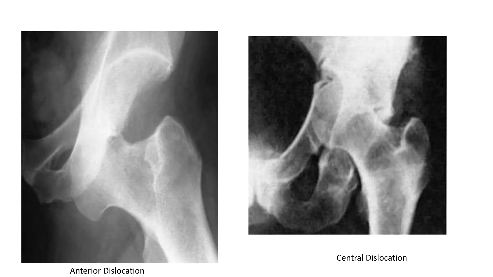
Dislocation of the Hip
By Chambega
Introduction
• Dislocation is a disarrangement of the normal relation
of the bones at a joint in which there complete loss of
contact between the two articular surfaces.
• The magnitude of force needed to dislocate the hip, is
so great that dislocation is often associated with
fractures (up to 50% of patients) either around the
joint or elsewhere in the same limb.
Epidemiology
• The majority of hip dislocations occur in 16- to 40-year-old
males involved in motor vehicle accidents.
• Almost all posterior hip dislocations result from motor
vehicle accidents.
• Anterior dislocations constitute 10% to 15% of traumatic
dislocations of the hip, with posterior dislocations
accounting for the remaining majority.
• Sciatic nerve injury is present in 10% to 20% of posterior
dislocations.
Anatomy
• The hip joint has a ball-and-socket configuration; synovial articulation
between the head of the femur and the acetabulum of the pelvis bone.
• Forty percent of the femoral head is covered by the bony acetabulum at
any position of hip motion. The effect of the labrum is to deepen the
acetabulum and increase the stability of the joint.
• The joint is supplemented by much stronger ligamentous condensations
(iliofemoral, pubofemoral, and ischiofemoral ligaments) that run in a
spiral fashion, preventing excessive hip extension.
• Hip joint is well confined by the virtue of its bony and soft tissue
anatomy.
Head of femur
Pubofemoral
ligament
Illiopubic
eminence
Ant. Inf. Iliac
spine
Main vascular supply is from
the lateral and medial
femoral circumflex arteries,
branches of the profunda
femoral artery.
An extracapsular vascular
ring is formed at the base of
the femoral neck with
ascending cervical branches
that pierce the hip joint at
the level of the capsular
insertion.
Classification of Hip dislocations
Hip dislocations are classified according to the direction of
the femoral head displacement:
• posterior (by far the commonest variety),
• anterior and
• central (a comminuted or displaced fracture of the acetabulum).
Posterior Dislocation
• Also known as “dashboard injury”
• They result from trauma to the flexed knee, with the hip in varying
degrees of flexion. The femur is thrust upwards and the femoral head is
forced out of its socket.
• Can be associated with fracture of the posterior wall of the acetabulum.
• The scenario is usually when someone seated in a truck or car, during a
road accident is thrown forward striking the knee against the dashboard.
• Seat-belt restraints can reduce the number of posterior hip dislocation.
Clinical features;
• Shortening of the limb
• Limb is adducted
• Internally rotated and
• Slightly flexed.
** if there is an associated
fracture of the femur, the injury
can be missed cause the limb
can adopt almost any position.
***insist x-rays should include the hip
and knee
Thompson and Epstein Classification of
Posterior Hip Dislocation.
Types Classifications
I Dislocation with no more than minor chip fractures.
II Dislocation with single large fragment of posterior
acetabular wall.
III Dislocation with comminuted fragments of posterior
acetabular wall.
IV Dislocation with fracture through acetabular floor.
V Dislocation with fracture through acetabular floor and
femoral head.
Management
• The dislocation must be reduced as soon as possible under general
anesthesia.
• In a majority of cases the reduction is performed closed, if not
achieved after two or three attempts an open reduction is required.
• Reduction is usually stable in type I injuries, but the hip has been
severely injured and needs to be rested, done by apply traction and
maintain it for a few days.
Cont…
• Type II fracture-dislocations are often treated by immediate open
reduction and anatomical fixation of the detached fragment, the
rationale being that many large posterior wall fragments either do not
reduce well or remain as a cause of instability even after reduction.
• Type III injuries are treated closed, but there may be retained
fragments and these should be removed by open operation.
• Types IV and V are treated initially by closed reduction. The
indications for surgery may depend on instability, retained fragments
or joint incongruity.
Allis Method
The patient is placed supine with
the surgeon standing above the
patient on the stretcher or table.
Initially, the surgeon applies in-line
traction while the assistant applies
counter traction by stabilizing the
patient’s pelvis. While increasing the
traction force, the surgeon should
slowly increase the degree of flexion
to approximately 70 degrees. Gentle
rotational motions of the hip as well
as slight adduction will often help
the femoral head to clear the lip of
the acetabulum. A lateral force to
the proximal thigh may assist in
reduction. An audible “clunk” is a
sign of a successful closed
reduction.
Complications
Early
• Sciatic nerve injury; nerve function tested and documented before reduction
attempted.
• Vascular injury; occasionally the superior gluteal artery
• Associated fracture shaft of femur
 Late
• Avascular necrosis
• Myositis ossificans; uncommon
• Unreduced dislocation; untreated dislocation
• Osteoarthritis; cartilage damage at time of dislocation, retained fragments in
the joint, ischemic necrosis femoral head
Anterior Dislocation
• Rare compared to posterior.
• Posteriorly directed force on an abducted and externally rotated hip
may cause the neck to impinge on the acetabular rim and lever the
femoral head out in front of its socket.
• The head will lie either superiorly (type I - pubic) or inferiorly (type II
–obturator).
• Clinically; the leg lies externally rotated, abducted and slightly flexed.
The head shows clinically as a prominent lump.
• X-ray; head is almost directly in front of its normal position, better
seen in a lateral film view.
Epstein Classification of Anterior Hip
Dislocations.
Types Classifications
I Superior dislocations, including pubic and subspinous.
IA No associated fractures.
IB Associated fracture or impaction of the femoral head.
IC Associated fracture of the acetabulum.
II Inferior dislocations, including obturator, and perineal.
IIA No associated fractures.
IIB Associated fracture or impaction of the femoral head.
IIC Associated fracture of the acetabulum.
Central Dislocation
• A fall on the side, or a blow over the greater
trochanter, may force the femoral head medially
through the floor of the acetabulum.
• Although this is called ‘central dislocation’, it is really a
fracture of the acetabulum.
Anterior Dislocation
Central Dislocation

Hip dislocation

  • 1.
    Dislocation of theHip By Chambega
  • 2.
    Introduction • Dislocation isa disarrangement of the normal relation of the bones at a joint in which there complete loss of contact between the two articular surfaces. • The magnitude of force needed to dislocate the hip, is so great that dislocation is often associated with fractures (up to 50% of patients) either around the joint or elsewhere in the same limb.
  • 3.
    Epidemiology • The majorityof hip dislocations occur in 16- to 40-year-old males involved in motor vehicle accidents. • Almost all posterior hip dislocations result from motor vehicle accidents. • Anterior dislocations constitute 10% to 15% of traumatic dislocations of the hip, with posterior dislocations accounting for the remaining majority. • Sciatic nerve injury is present in 10% to 20% of posterior dislocations.
  • 4.
    Anatomy • The hipjoint has a ball-and-socket configuration; synovial articulation between the head of the femur and the acetabulum of the pelvis bone. • Forty percent of the femoral head is covered by the bony acetabulum at any position of hip motion. The effect of the labrum is to deepen the acetabulum and increase the stability of the joint. • The joint is supplemented by much stronger ligamentous condensations (iliofemoral, pubofemoral, and ischiofemoral ligaments) that run in a spiral fashion, preventing excessive hip extension. • Hip joint is well confined by the virtue of its bony and soft tissue anatomy.
  • 5.
  • 6.
    Main vascular supplyis from the lateral and medial femoral circumflex arteries, branches of the profunda femoral artery. An extracapsular vascular ring is formed at the base of the femoral neck with ascending cervical branches that pierce the hip joint at the level of the capsular insertion.
  • 7.
    Classification of Hipdislocations Hip dislocations are classified according to the direction of the femoral head displacement: • posterior (by far the commonest variety), • anterior and • central (a comminuted or displaced fracture of the acetabulum).
  • 8.
    Posterior Dislocation • Alsoknown as “dashboard injury” • They result from trauma to the flexed knee, with the hip in varying degrees of flexion. The femur is thrust upwards and the femoral head is forced out of its socket. • Can be associated with fracture of the posterior wall of the acetabulum. • The scenario is usually when someone seated in a truck or car, during a road accident is thrown forward striking the knee against the dashboard. • Seat-belt restraints can reduce the number of posterior hip dislocation.
  • 9.
    Clinical features; • Shorteningof the limb • Limb is adducted • Internally rotated and • Slightly flexed. ** if there is an associated fracture of the femur, the injury can be missed cause the limb can adopt almost any position. ***insist x-rays should include the hip and knee
  • 11.
    Thompson and EpsteinClassification of Posterior Hip Dislocation. Types Classifications I Dislocation with no more than minor chip fractures. II Dislocation with single large fragment of posterior acetabular wall. III Dislocation with comminuted fragments of posterior acetabular wall. IV Dislocation with fracture through acetabular floor. V Dislocation with fracture through acetabular floor and femoral head.
  • 12.
    Management • The dislocationmust be reduced as soon as possible under general anesthesia. • In a majority of cases the reduction is performed closed, if not achieved after two or three attempts an open reduction is required. • Reduction is usually stable in type I injuries, but the hip has been severely injured and needs to be rested, done by apply traction and maintain it for a few days.
  • 13.
    Cont… • Type IIfracture-dislocations are often treated by immediate open reduction and anatomical fixation of the detached fragment, the rationale being that many large posterior wall fragments either do not reduce well or remain as a cause of instability even after reduction. • Type III injuries are treated closed, but there may be retained fragments and these should be removed by open operation. • Types IV and V are treated initially by closed reduction. The indications for surgery may depend on instability, retained fragments or joint incongruity.
  • 14.
    Allis Method The patientis placed supine with the surgeon standing above the patient on the stretcher or table. Initially, the surgeon applies in-line traction while the assistant applies counter traction by stabilizing the patient’s pelvis. While increasing the traction force, the surgeon should slowly increase the degree of flexion to approximately 70 degrees. Gentle rotational motions of the hip as well as slight adduction will often help the femoral head to clear the lip of the acetabulum. A lateral force to the proximal thigh may assist in reduction. An audible “clunk” is a sign of a successful closed reduction.
  • 15.
    Complications Early • Sciatic nerveinjury; nerve function tested and documented before reduction attempted. • Vascular injury; occasionally the superior gluteal artery • Associated fracture shaft of femur  Late • Avascular necrosis • Myositis ossificans; uncommon • Unreduced dislocation; untreated dislocation • Osteoarthritis; cartilage damage at time of dislocation, retained fragments in the joint, ischemic necrosis femoral head
  • 16.
    Anterior Dislocation • Rarecompared to posterior. • Posteriorly directed force on an abducted and externally rotated hip may cause the neck to impinge on the acetabular rim and lever the femoral head out in front of its socket. • The head will lie either superiorly (type I - pubic) or inferiorly (type II –obturator). • Clinically; the leg lies externally rotated, abducted and slightly flexed. The head shows clinically as a prominent lump. • X-ray; head is almost directly in front of its normal position, better seen in a lateral film view.
  • 17.
    Epstein Classification ofAnterior Hip Dislocations. Types Classifications I Superior dislocations, including pubic and subspinous. IA No associated fractures. IB Associated fracture or impaction of the femoral head. IC Associated fracture of the acetabulum. II Inferior dislocations, including obturator, and perineal. IIA No associated fractures. IIB Associated fracture or impaction of the femoral head. IIC Associated fracture of the acetabulum.
  • 18.
    Central Dislocation • Afall on the side, or a blow over the greater trochanter, may force the femoral head medially through the floor of the acetabulum. • Although this is called ‘central dislocation’, it is really a fracture of the acetabulum.
  • 19.