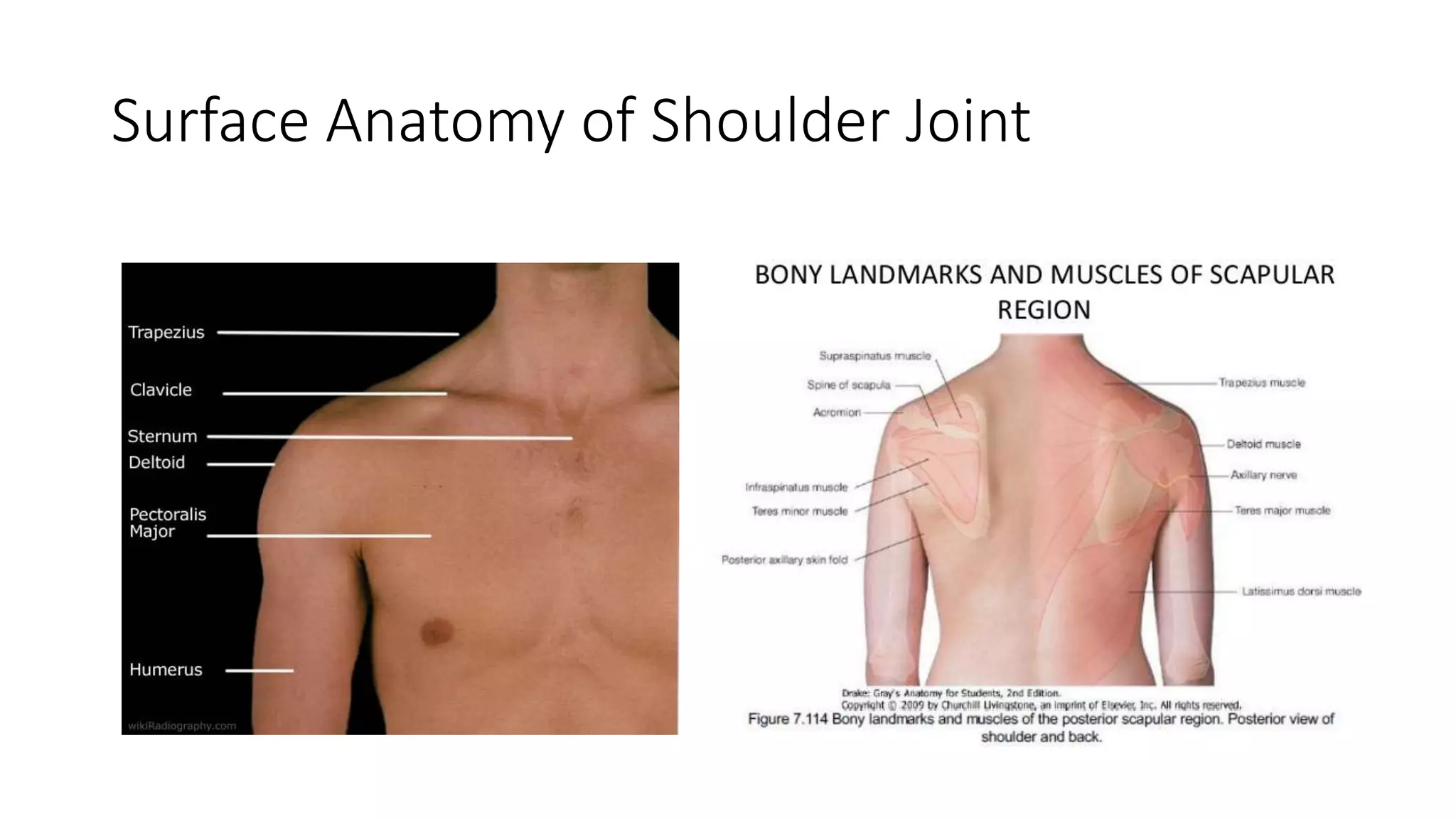
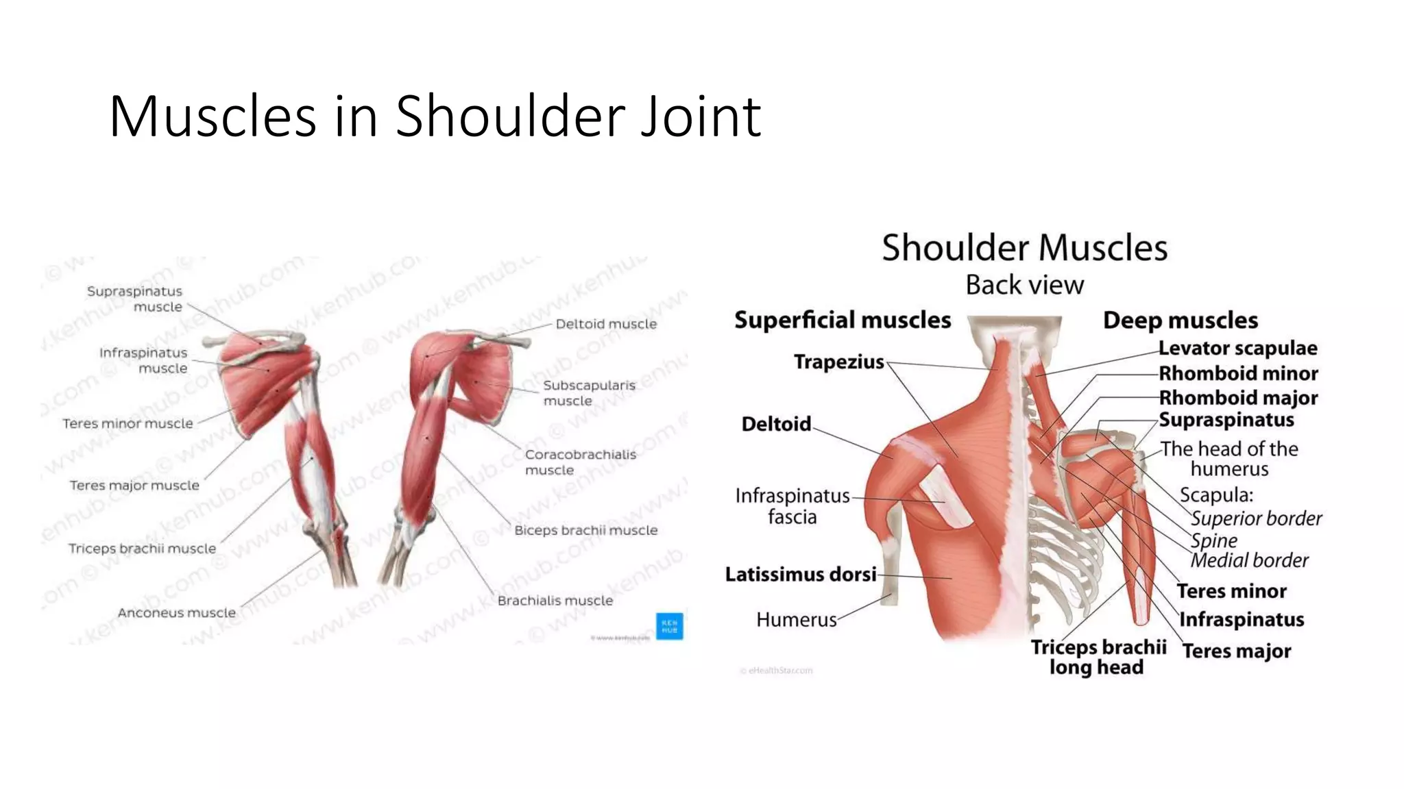
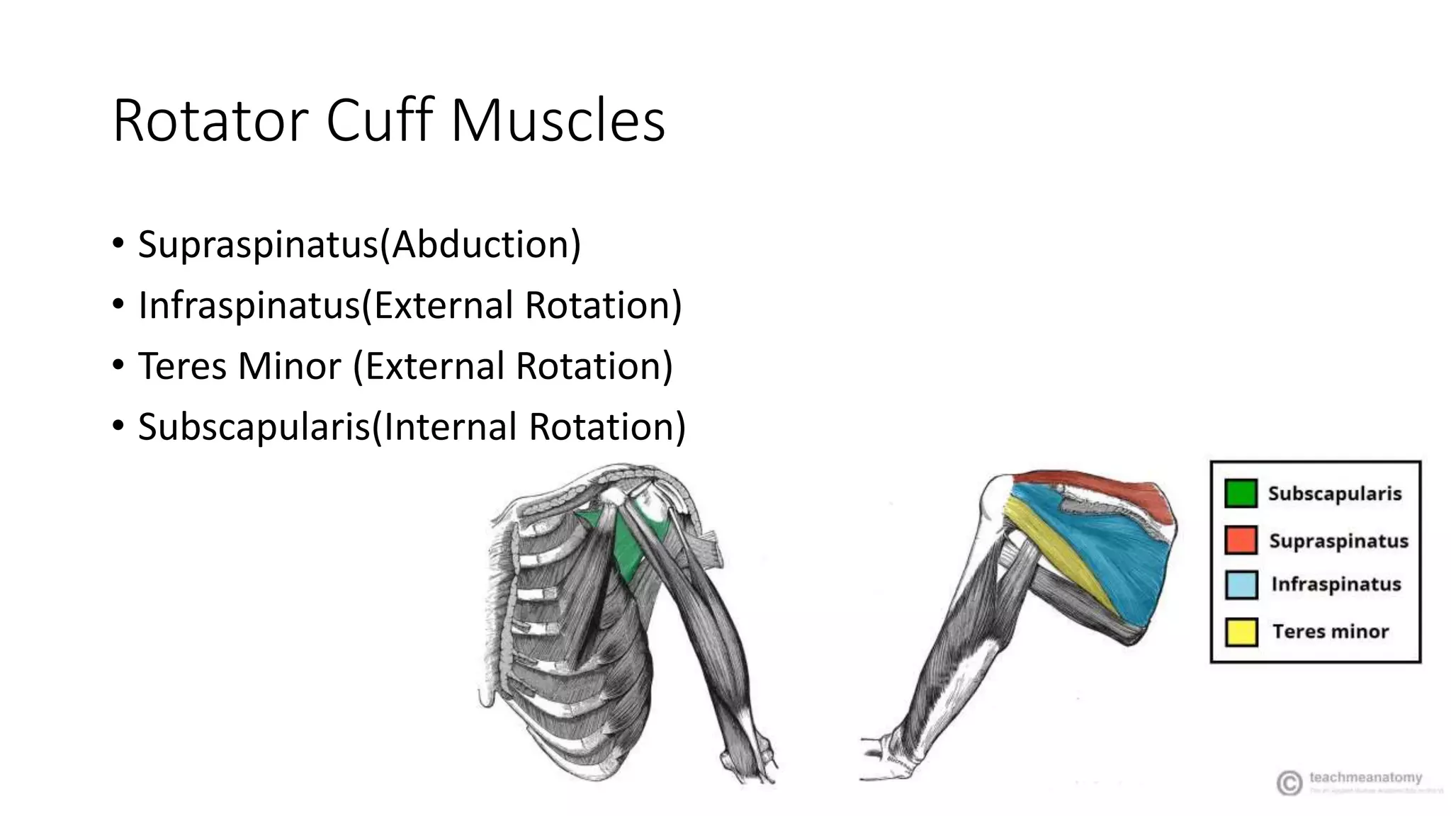
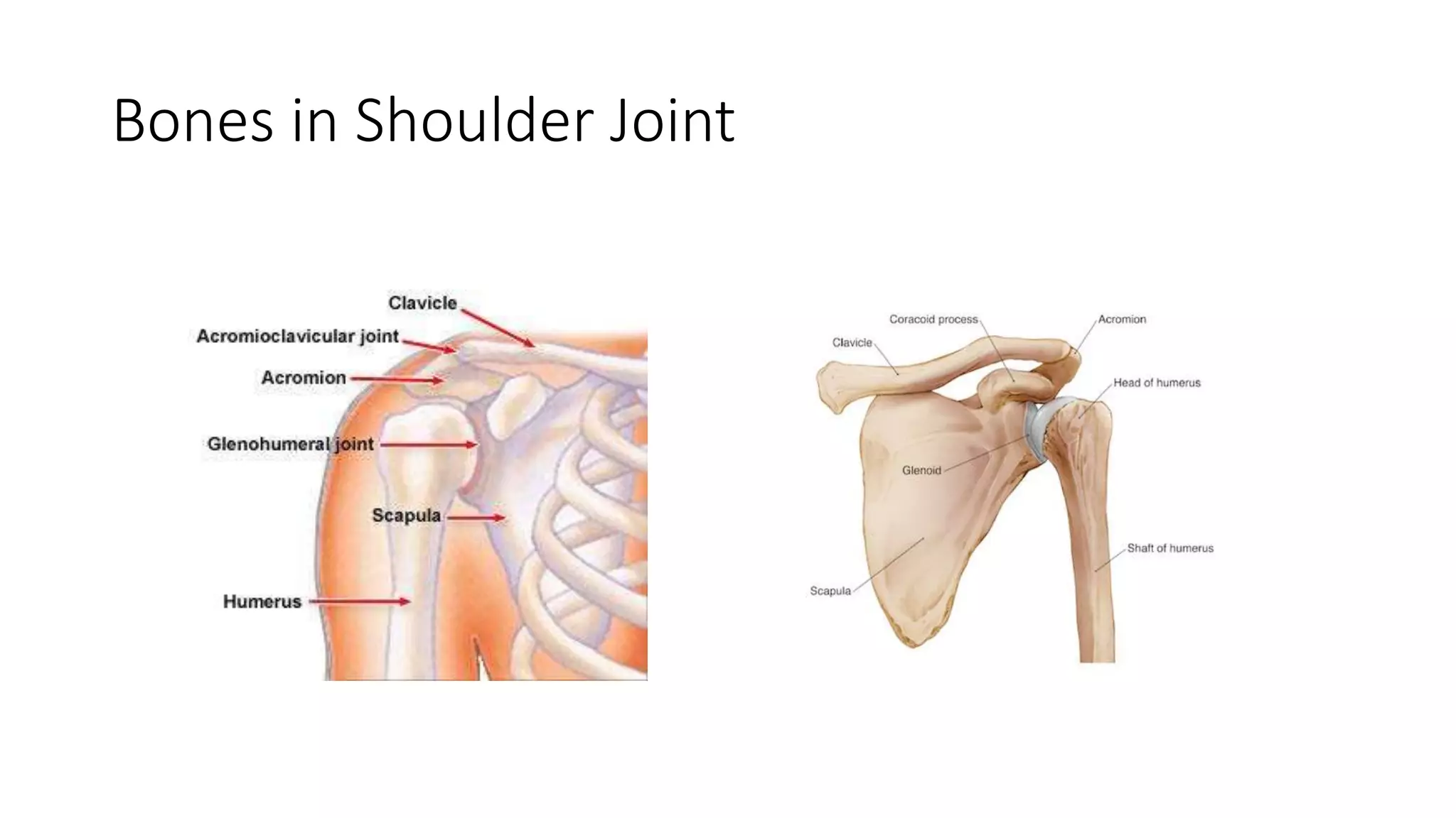
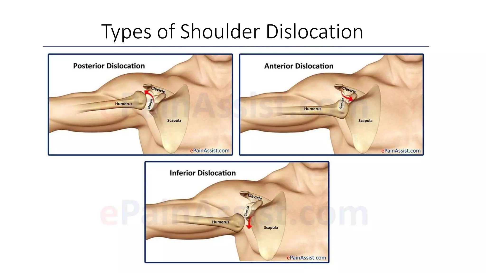
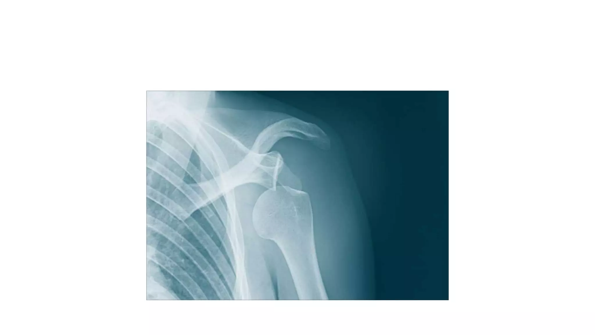
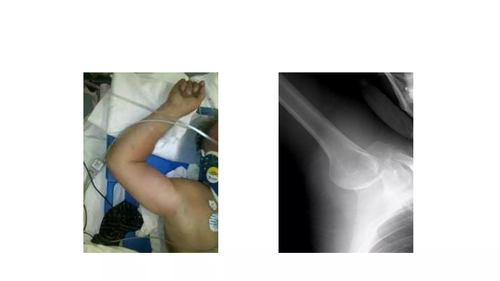
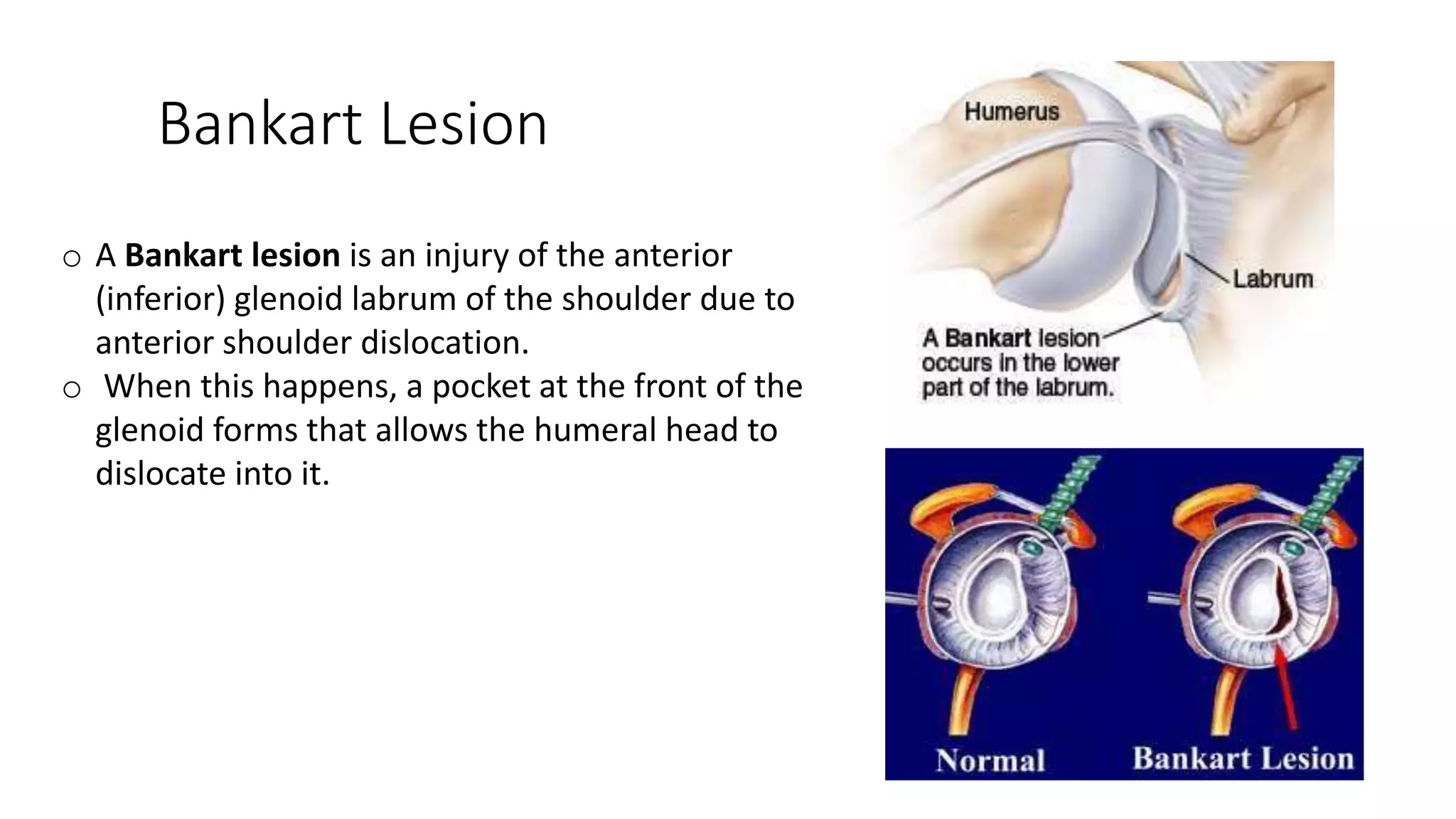
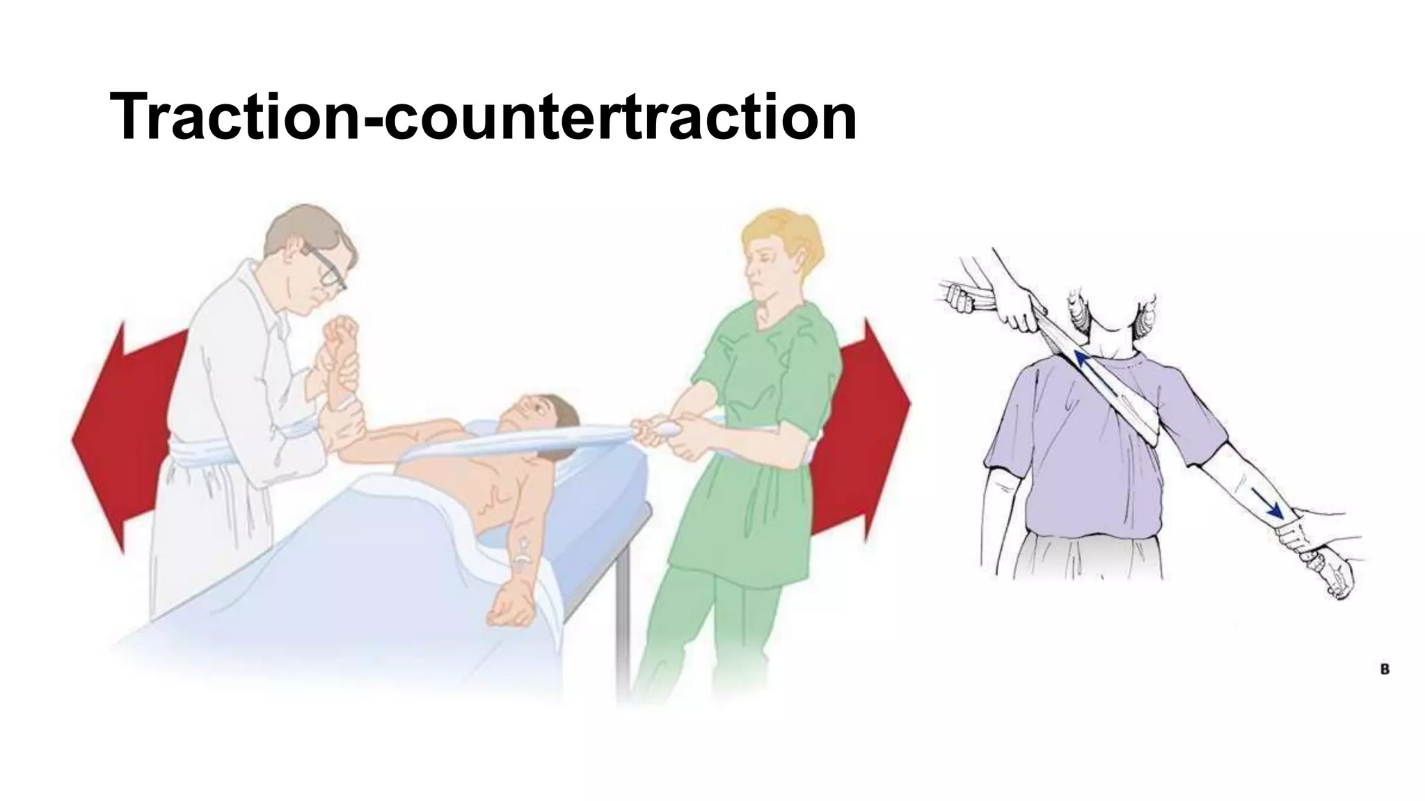
The document discusses shoulder dislocation, including the anatomy of the shoulder joint, causes of dislocation, signs and symptoms, types of dislocation, treatment options like closed reduction and surgery, rehabilitation, and complications. The most common type of dislocation is anterior dislocation, which can occur due to falls or impacts and results in the humeral head moving out of the glenoid socket in the front of the shoulder. Treatment depends on the severity of the dislocation and any associated injuries.
JOINT DISLOCATION of hip knee and shoulder PART-2.pptxrammmramm000
JOINT DISLOCATION of hip knee and shoulder
JOINT DISLOCATION of hip knee and shoulder JOINT DISLOCATION of hip knee and shoulder JOINT DISLOCATION of hip knee and shoulder JOINT DISLOCATION of hip knee and shoulder JOINT DISLOCATION of hip knee and shoulder JOINT DISLOCATION of hip knee and shoulder JOINT DISLOCATION of hip knee and shoulder JOINT DISLOCATION of hip knee and shoulder JOINT DISLOCATION of hip knee and shoulder JOINT DISLOCATION of hip knee and shoulder JOINT DISLOCATION of hip knee and shoulder JOINT DISLOCATION of hip knee and shoulder JOINT DISLOCATION of hip knee and shoulder
JOINT DISLOCATION of hip knee and shoulder JOINT DISLOCATION of hip knee and shoulder JOINT DISLOCATION of hip knee and shoulder JOINT DISLOCATION of hip kn
Overview of common dislocations and subluxations for undergraduate students. Includes clinical features, reduction methods and complications of commonly encountered injuries
Dislocations of the bone by dr amna hussainDureSameen19
A dislocation is a separation of two bones where they meet at a joint. This injury can be very painful and can temporarily deform and immobilize the joint. The most common locations for a dislocation are shoulders and fingers, but can also occur in elbows, knees and hips. The cause is often a fall or a blow, sometimes from playing a contact sport.
Earliest Galaxies in the JADES Origins Field: Luminosity Function and Cosmic ...Sérgio Sacani
We characterize the earliest galaxy population in the JADES Origins Field (JOF), the deepest
imaging field observed with JWST. We make use of the ancillary Hubble optical images (5 filters
spanning 0.4−0.9µm) and novel JWST images with 14 filters spanning 0.8−5µm, including 7 mediumband filters, and reaching total exposure times of up to 46 hours per filter. We combine all our data
at > 2.3µm to construct an ultradeep image, reaching as deep as ≈ 31.4 AB mag in the stack and
30.3-31.0 AB mag (5σ, r = 0.1” circular aperture) in individual filters. We measure photometric
redshifts and use robust selection criteria to identify a sample of eight galaxy candidates at redshifts
z = 11.5 − 15. These objects show compact half-light radii of R1/2 ∼ 50 − 200pc, stellar masses of
M⋆ ∼ 107−108M⊙, and star-formation rates of SFR ∼ 0.1−1 M⊙ yr−1
. Our search finds no candidates
at 15 < z < 20, placing upper limits at these redshifts. We develop a forward modeling approach to
infer the properties of the evolving luminosity function without binning in redshift or luminosity that
marginalizes over the photometric redshift uncertainty of our candidate galaxies and incorporates the
impact of non-detections. We find a z = 12 luminosity function in good agreement with prior results,
and that the luminosity function normalization and UV luminosity density decline by a factor of ∼ 2.5
from z = 12 to z = 14. We discuss the possible implications of our results in the context of theoretical
models for evolution of the dark matter halo mass function.
Cancer cell metabolism: special Reference to Lactate PathwayAADYARAJPANDEY1
Normal Cell Metabolism:
Cellular respiration describes the series of steps that cells use to break down sugar and other chemicals to get the energy we need to function.
Energy is stored in the bonds of glucose and when glucose is broken down, much of that energy is released.
Cell utilize energy in the form of ATP.
The first step of respiration is called glycolysis. In a series of steps, glycolysis breaks glucose into two smaller molecules - a chemical called pyruvate. A small amount of ATP is formed during this process.
Most healthy cells continue the breakdown in a second process, called the Kreb's cycle. The Kreb's cycle allows cells to “burn” the pyruvates made in glycolysis to get more ATP.
The last step in the breakdown of glucose is called oxidative phosphorylation (Ox-Phos).
It takes place in specialized cell structures called mitochondria. This process produces a large amount of ATP. Importantly, cells need oxygen to complete oxidative phosphorylation.
If a cell completes only glycolysis, only 2 molecules of ATP are made per glucose. However, if the cell completes the entire respiration process (glycolysis - Kreb's - oxidative phosphorylation), about 36 molecules of ATP are created, giving it much more energy to use.
IN CANCER CELL:
Unlike healthy cells that "burn" the entire molecule of sugar to capture a large amount of energy as ATP, cancer cells are wasteful.
Cancer cells only partially break down sugar molecules. They overuse the first step of respiration, glycolysis. They frequently do not complete the second step, oxidative phosphorylation.
This results in only 2 molecules of ATP per each glucose molecule instead of the 36 or so ATPs healthy cells gain. As a result, cancer cells need to use a lot more sugar molecules to get enough energy to survive.
introduction to WARBERG PHENOMENA:
WARBURG EFFECT Usually, cancer cells are highly glycolytic (glucose addiction) and take up more glucose than do normal cells from outside.
Otto Heinrich Warburg (; 8 October 1883 – 1 August 1970) In 1931 was awarded the Nobel Prize in Physiology for his "discovery of the nature and mode of action of the respiratory enzyme.
WARNBURG EFFECT : cancer cells under aerobic (well-oxygenated) conditions to metabolize glucose to lactate (aerobic glycolysis) is known as the Warburg effect. Warburg made the observation that tumor slices consume glucose and secrete lactate at a higher rate than normal tissues.
This presentation explores a brief idea about the structural and functional attributes of nucleotides, the structure and function of genetic materials along with the impact of UV rays and pH upon them.
Nutraceutical market, scope and growth: Herbal drug technologyLokesh Patil
As consumer awareness of health and wellness rises, the nutraceutical market—which includes goods like functional meals, drinks, and dietary supplements that provide health advantages beyond basic nutrition—is growing significantly. As healthcare expenses rise, the population ages, and people want natural and preventative health solutions more and more, this industry is increasing quickly. Further driving market expansion are product formulation innovations and the use of cutting-edge technology for customized nutrition. With its worldwide reach, the nutraceutical industry is expected to keep growing and provide significant chances for research and investment in a number of categories, including vitamins, minerals, probiotics, and herbal supplements.
Observation of Io’s Resurfacing via Plume Deposition Using Ground-based Adapt...Sérgio Sacani
Since volcanic activity was first discovered on Io from Voyager images in 1979, changes
on Io’s surface have been monitored from both spacecraft and ground-based telescopes.
Here, we present the highest spatial resolution images of Io ever obtained from a groundbased telescope. These images, acquired by the SHARK-VIS instrument on the Large
Binocular Telescope, show evidence of a major resurfacing event on Io’s trailing hemisphere. When compared to the most recent spacecraft images, the SHARK-VIS images
show that a plume deposit from a powerful eruption at Pillan Patera has covered part
of the long-lived Pele plume deposit. Although this type of resurfacing event may be common on Io, few have been detected due to the rarity of spacecraft visits and the previously low spatial resolution available from Earth-based telescopes. The SHARK-VIS instrument ushers in a new era of high resolution imaging of Io’s surface using adaptive
optics at visible wavelengths.
Slide 1: Title Slide
Extrachromosomal Inheritance
Slide 2: Introduction to Extrachromosomal Inheritance
Definition: Extrachromosomal inheritance refers to the transmission of genetic material that is not found within the nucleus.
Key Components: Involves genes located in mitochondria, chloroplasts, and plasmids.
Slide 3: Mitochondrial Inheritance
Mitochondria: Organelles responsible for energy production.
Mitochondrial DNA (mtDNA): Circular DNA molecule found in mitochondria.
Inheritance Pattern: Maternally inherited, meaning it is passed from mothers to all their offspring.
Diseases: Examples include Leber’s hereditary optic neuropathy (LHON) and mitochondrial myopathy.
Slide 4: Chloroplast Inheritance
Chloroplasts: Organelles responsible for photosynthesis in plants.
Chloroplast DNA (cpDNA): Circular DNA molecule found in chloroplasts.
Inheritance Pattern: Often maternally inherited in most plants, but can vary in some species.
Examples: Variegation in plants, where leaf color patterns are determined by chloroplast DNA.
Slide 5: Plasmid Inheritance
Plasmids: Small, circular DNA molecules found in bacteria and some eukaryotes.
Features: Can carry antibiotic resistance genes and can be transferred between cells through processes like conjugation.
Significance: Important in biotechnology for gene cloning and genetic engineering.
Slide 6: Mechanisms of Extrachromosomal Inheritance
Non-Mendelian Patterns: Do not follow Mendel’s laws of inheritance.
Cytoplasmic Segregation: During cell division, organelles like mitochondria and chloroplasts are randomly distributed to daughter cells.
Heteroplasmy: Presence of more than one type of organellar genome within a cell, leading to variation in expression.
Slide 7: Examples of Extrachromosomal Inheritance
Four O’clock Plant (Mirabilis jalapa): Shows variegated leaves due to different cpDNA in leaf cells.
Petite Mutants in Yeast: Result from mutations in mitochondrial DNA affecting respiration.
Slide 8: Importance of Extrachromosomal Inheritance
Evolution: Provides insight into the evolution of eukaryotic cells.
Medicine: Understanding mitochondrial inheritance helps in diagnosing and treating mitochondrial diseases.
Agriculture: Chloroplast inheritance can be used in plant breeding and genetic modification.
Slide 9: Recent Research and Advances
Gene Editing: Techniques like CRISPR-Cas9 are being used to edit mitochondrial and chloroplast DNA.
Therapies: Development of mitochondrial replacement therapy (MRT) for preventing mitochondrial diseases.
Slide 10: Conclusion
Summary: Extrachromosomal inheritance involves the transmission of genetic material outside the nucleus and plays a crucial role in genetics, medicine, and biotechnology.
Future Directions: Continued research and technological advancements hold promise for new treatments and applications.
Slide 11: Questions and Discussion
Invite Audience: Open the floor for any questions or further discussion on the topic.
Seminar of U.V. Spectroscopy by SAMIR PANDASAMIR PANDA
Spectroscopy is a branch of science dealing the study of interaction of electromagnetic radiation with matter.
Ultraviolet-visible spectroscopy refers to absorption spectroscopy or reflect spectroscopy in the UV-VIS spectral region.
Ultraviolet-visible spectroscopy is an analytical method that can measure the amount of light received by the analyte.
What is greenhouse gasses and how many gasses are there to affect the Earth.moosaasad1975
What are greenhouse gasses how they affect the earth and its environment what is the future of the environment and earth how the weather and the climate effects.
THE IMPORTANCE OF MARTIAN ATMOSPHERE SAMPLE RETURN.Sérgio Sacani
The return of a sample of near-surface atmosphere from Mars would facilitate answers to several first-order science questions surrounding the formation and evolution of the planet. One of the important aspects of terrestrial planet formation in general is the role that primary atmospheres played in influencing the chemistry and structure of the planets and their antecedents. Studies of the martian atmosphere can be used to investigate the role of a primary atmosphere in its history. Atmosphere samples would also inform our understanding of the near-surface chemistry of the planet, and ultimately the prospects for life. High-precision isotopic analyses of constituent gases are needed to address these questions, requiring that the analyses are made on returned samples rather than in situ.
Deep Behavioral Phenotyping in Systems Neuroscience for Functional Atlasing a...Ana Luísa Pinho
Functional Magnetic Resonance Imaging (fMRI) provides means to characterize brain activations in response to behavior. However, cognitive neuroscience has been limited to group-level effects referring to the performance of specific tasks. To obtain the functional profile of elementary cognitive mechanisms, the combination of brain responses to many tasks is required. Yet, to date, both structural atlases and parcellation-based activations do not fully account for cognitive function and still present several limitations. Further, they do not adapt overall to individual characteristics. In this talk, I will give an account of deep-behavioral phenotyping strategies, namely data-driven methods in large task-fMRI datasets, to optimize functional brain-data collection and improve inference of effects-of-interest related to mental processes. Key to this approach is the employment of fast multi-functional paradigms rich on features that can be well parametrized and, consequently, facilitate the creation of psycho-physiological constructs to be modelled with imaging data. Particular emphasis will be given to music stimuli when studying high-order cognitive mechanisms, due to their ecological nature and quality to enable complex behavior compounded by discrete entities. I will also discuss how deep-behavioral phenotyping and individualized models applied to neuroimaging data can better account for the subject-specific organization of domain-general cognitive systems in the human brain. Finally, the accumulation of functional brain signatures brings the possibility to clarify relationships among tasks and create a univocal link between brain systems and mental functions through: (1) the development of ontologies proposing an organization of cognitive processes; and (2) brain-network taxonomies describing functional specialization. To this end, tools to improve commensurability in cognitive science are necessary, such as public repositories, ontology-based platforms and automated meta-analysis tools. I will thus discuss some brain-atlasing resources currently under development, and their applicability in cognitive as well as clinical neuroscience.
9. Pathology
• The humeral head is stabilized within the
shoulder joint by the glenoid labrum, a fibro-
cartilaginous rim surrounding the glenoid cuff
that is attached to the tendon of the long
head of the biceps muscle.
• A tear or a violent stretch of these structures
causes the exit of the humeral head from the
glenoid, making the shoulder highly unstable.
10. • A shoulder dislocation can be complete or partial.
• In complete dislocation the head of the humerus moves entirely out
of the socket, requiring medical intervention
• whereas in partial dislocation, also named subluxation, the head of
the humerus slips out of the socket only temporarily to often
returning into place spontaneously.
• Both injuries can cause pain, arm weakness and swelling.
11. Reasons for Instability
• Shallow glenoid
• Extraordinary Rotation Of Muscle
• Vulnerability of upper limb to injury
• Underlying conditions eg. ligament laxity
12. Pathoanatomy of Dislocation
• Stretching/ tearing of capsule
• Avulsion of glenohumeral ligaments usually off the glenoid
• Labral injury
◦ Bankart lesion
• Impression fracture
◦ Hill-Sach lesion
• Rotator cuff tear
13. Clinical Picture
• Pain
• Holds injured limb with other hand close to trunk
• The shoulder is abducted and the elbow is kept
flexed
• Loss of the normal contour of the shoulder
• Anterior bulge of head of humerus may be visible
or palpable
• Empty glenoid socket
14. What causes a dislocated shoulder?
• Sports injuries.
• Accidents, including traffic accidents.
• Falling on your shoulder or outstretched
arm.
• Seizures and electric shocks, which can
cause muscle contractions that pull the
arm out of place.
15. Dislocated shoulder signs and symptoms may
include:
• Severe shoulder pain
• Swelling and bruising of your
shoulder or upper arm
• Numbness and/or weakness in
your arm, neck, hand, or fingers
• Trouble moving your arm
• Your arm seems to be out of place
• Muscle spasms in your shoulder
16. Risk Factors
• A previous shoulder dislocation or subluxation
predisposes to a second episode of the
pathology particularly in young men
(incidence of 80-90%).
• high risk is found in athletes involved in sports
such as football, rugby, hockey and skiing due
to the frequent contact impacts, throwing
activities and falls.
• Congenital conditions causing loosening of the
joints, such as Ehlers-Danlos Syndrome confer
an intrinsic poor stability of the shoulder joint
facilitating the exit of the humeral head.
• Weakness of the muscles around the shoulder
and core muscles due to lack of training can
predispose to a dislocation.
• Incorrect posture and inadequate sporting
technique are all contributing factors to a
shoulder dislocation.
17. Diagnosis
• A shoulder dislocation is diagnosed clinically when
significant pain, alterations in the appearance of
the shoulder anatomy and impaired movement of
the shoulder are present.
• The history of the mechanisms of injury and pre-
existing conditions are discussed with the doctor
and recorded
• Standard X-ray of the shoulder forms the first
diagnostic approach to confirm the type of
humeral head displacement and potentially
associated injuries to the surrounding bones.
• Additional damage to ligaments, vessels and
nerves is diagnosed by clinical examination,
computer tomogram (CT) scans, magnetic
resonance imaging (MRI), ultrasound and nerve
conduction studies.
19. Nonoperative treatment
• closed reduction is performed usually
under anaesthesia in the Emergency
Department.
• It consists of manual reposition the
humeral head in the glenoid using
different methods.
• This is followed by the immobilisation of
the shoulder for approximately four
weeks, aided by local treatment with ice
and/or heat and non-steroidal
antiinflammatory drugs (NSAIDs).
• At a later stage physiotherapy is
recommended.
20. Surgical treatment
• Surgery is performed if a closed reduction is
not successful or when a traumatic
dislocation is associated with injuries to the
labrum (e.g. Superior Labral Tear from
Anterior to Posterior also named SLAP tear) or
glenoid (Bankart lesion), damage of the
humeral head (Hill-Sachs lesion) or the
ligaments of the rotator cuff.
• These secondary pathologies produce
significant shoulder instability and require
surgical repair to prevent further dislocations.
• Various approaches are available including
arthroscopic surgery and open surgery.
• After surgery the shoulder is immobilised for
3-4 weeks prior to commence physical
therapy
21. Rehabilitation
• Physiotherapy is a key form of treatment following a shoulder
dislocation whether or not surgery has occurred. Strengthening the
muscles around the shoulder is essential for supporting the joint
stability provided by the shoulder ligaments. Therapy also aims at
restoring the range of motion of the shoulder following initial
immobilisation. Physiotherapy consists of a number of approaches:
• Use of a sling
• Massage
• Joint mobilisation
• Ice/heat treatment
• Physical exercise (pendular movements)
• Education in sport and daily activities
• Ergonometric postural correction
• Return to sport plan
• In case of persistent pain and/or movement restrictions the patient
can be treated with antiinflammatory treatment (NSAIDs) and local
steroid injection
22. Prevention
• Take care to avoid falls.
• Wear protective gear when you play
contact sports.
• Exercise regularly to maintain
strength and flexibility in your joints
and muscles.
24. Types of Dislocation of the Shoulder
• Mostly Anterior > 95 % of dislocations
• Posterior dislocation occurs < 5 %
• True Inferior dislocation (luxatio erecta) occurs <1%
• Superior dislocation occurs <1%
• Habitual - Non traumatic dislocation may present as Multi
directional dislocation due to generalized ligamentous laxity
and is Painless
25. Anterior Dislocation
◦ 97% of recurrent dislocations
◦ It is the most common dislocation
◦ cause inability to abduct the arm.
◦ loss in the round contour of the
shoulder.
◦ The most severe cases of anterior
dislocation of the shoulder are
associated with injury to the axillary
artery and the axillary nerve.
27. Posterior Dislocation
o 3% of shoulder dislocations
o caused by an external blow to the
front of the shoulder.
o This type of shoulder dislocation
can be the consequence of a high-
energy trauma and a fall due to
seizures.
o dislocations may also have
concurrent labral or rotator cuff
pathology
31. Inferior dislocation
• Inferior dislocation is rarely
seen.
• It is also called Luxatio Erecta
• It occurs when the humerus is
displaced below the joint.
• It is caused by a traumatic
impact pushing the shoulder
downwards.
32.
33.
34. Superior dislocation
• Superior dislocation is the least
frequent type of dislocation (1%).
• It occurs when the humeral head is
driven upward through the rotator
cuff.
• It can be associated with fracture of the
humerus, clavicle and acromion.
37. Complications of Shoulder Dislocation : Early
• Damage to
Axillary Nerve
Axillary Artery
And Ligaments
• Bone - Associated fracture
◦ Neck of humerus
◦ Greater or lesser tuberosity
• Hill Sach lesion
• Bankart lesion
38. Hill-Sachs lesion
o Hill–Sachs lesion, or Hill–Sachs fracture, is
a cortical depression in the posterolateral
head of the humerus.
o It results from forceful impaction of the
humeral head against the anteroinferior
glenoid rim when the shoulder is
dislocated anteriorly.
39. Bankart Lesion
o A Bankart lesion is an injury of the anterior
(inferior) glenoid labrum of the shoulder due to
anterior shoulder dislocation.
o When this happens, a pocket at the front of the
glenoid forms that allows the humeral head to
dislocate into it.
40. Complications of Shoulder Dislocation : Late
• Avascular necrosis of the head of the
Humerus(high risk with delayed
reduction)
• Heterotopic calcification (used to be
called Myositis Ossificans)
• Recurrent dislocation
45. Reference
• Gray_s Anatomy for Students 3rd Ed
• Moore - Clinically Oriented Anatomy 7th Ed by allmedicalstuff.com
• https://medlineplus.gov/dislocatedshoulder.html
• https://www.physio-pedia.com/Shoulder_Dislocation
• http://pathologies.lexmedicus.com.au/pathologies/shoulder-
dislocation-and-luxation