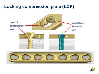
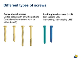
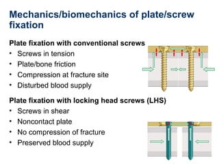
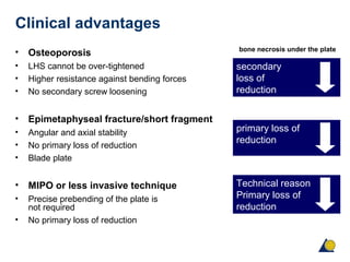
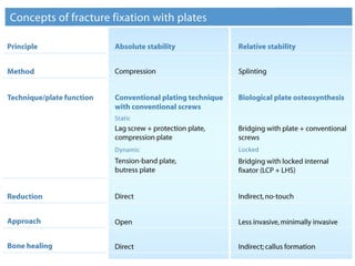
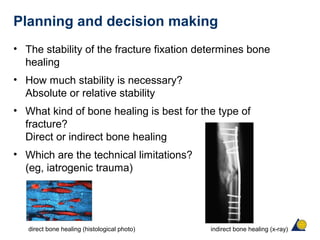
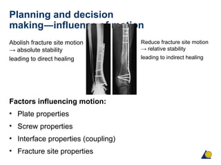
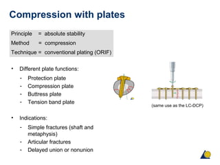
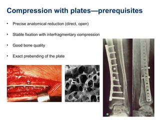
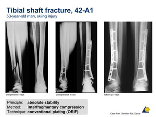
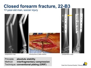
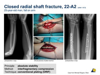
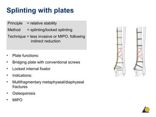
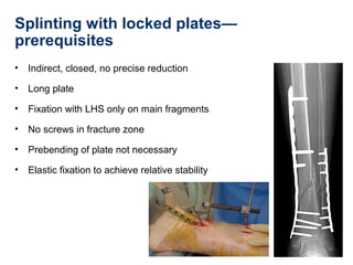
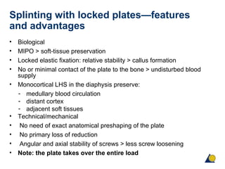
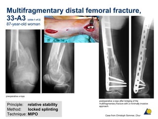
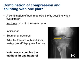
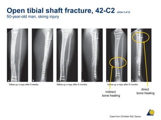
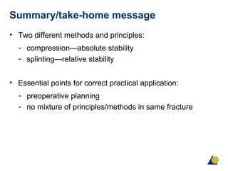
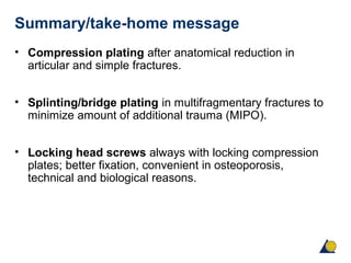
AOTrauma Principles Course
Fracture fixation using the
locking compression plate (LCP)
Michael Wagner, AT
Learning outcomes
• Understand the different functions of conventional and locking head
screws.
• Understand the different concepts of fracture fixation using locking
compression plates.
• Apply the principles to clinical usage—“know when which principle
needs to be applied in a clinical example”.
• Understand the advantages of locked plates in special clinical
circumstances (eg, osteoporosis).
Locking compression plate (LCP)
dynamic
compression
unit
conical and
threaded
unit
Different types of screws
Conventional screws
Cortex screw (with or without shaft)
Cancellous bone screw (with or
without shaft)
Locking head screws (LHS)
Self-tapping LHS
Self-drilling, self-tapping LHS
Mechanics/biomechanics of plate/screw
fixation
Plate fixation with conventional screws
• Screws in tension
• Plate/bone friction
• Compression at fracture site
• Disturbed blood supply
Plate fixation with locking head screws (LHS)
• Screws in shear
• Noncontact plate
• No compression of fracture
• Preserved blood supply
Functions of conventional screws (slide 1 of 2)
• Plate screw
- Preload and friction is applied to create force
- between plate and bone
• Lag screw
- Interfragmentary compression screw; always
- inserted before fixation of the plate with LHS
• Compression screw (eccentric screw)
- Positioned in the dynamic compression unit
(DCU)
• Position screw
- Keeps two fragments in position without
- compression
Functions of conventional screws (slide 2 of 2)
Reduction screw
Screw inserted through a
plate hole to pull fracture
fragments toward the plate
Reduction of a butterfly
fragment
(no interfragmentary
compression)
Drawbacks of fixation with conventional
screws
• Compression of the periosteum and resulting damage of
the blood supply to the bone
• Primary loss of reduction
• Secondary loss of reduction
primary loss of reduction secondary loss of reduction
Functions of locking head screws (LHS)
• Always in combination with a plate
• Never as lag screw
• Never cross an unreduced fracture with a
LHS
• Fixation screw
- Fix the plate to the bone
• Position screw
- Keeps two fragments in correct
relative anatomical position
Features and advantages of LHS
• Axial and angular stability
• Not preloaded
• Cannot be over-tightened
• Higher resistance against bending loads
• Monocortical insertion is possible (shaft and good bone quality
needed)
• No primary loss of reduction
• No or less screw loosening, no or less secondary loss of reduction
Features and advantages of locked
plates
• Screw-plate system with angular and axial stability
• Locked internal fixator/noncontact plate
• Individual blade plate
Biological advantages
• Reduced compression of the periosteum
• Protects blood supply to the bone
• Callus formation/bone healing under the plate
bone after plating with a DCP bone after plating with a LCP
Clinical advantages
• Osteoporosis
• LHS cannot be over-tightened
• Higher resistance against bending forces
• No secondary screw loosening
• Epimetaphyseal fracture/short fragment
• Angular and axial stability
• No primary loss of reduction
• Blade plate
• MIPO or less invasive technique
• Precise prebending of the plate is
not required
• No primary loss of reduction
secondary
loss of
reduction
primary loss of
reduction
Technical reason
Primary loss of
reduction
bone necrosis under the plate
Drawback of fixation with LHS
• Screw insertion is only possible in a 90° angle
• No bicortical insertion of self-drilling, self-tapping screws
(use self-tapping screws only for bicortical insertion
• Possible loss of the feel for the quality of the bone during
screw insertion and tightening
• Screw jamming and difficult implant removal
Planning and decision making
• The stability of the fracture fixation determines bone
healing
• How much stability is necessary?
Absolute or relative stability
• What kind of bone healing is best for the type of
fracture?
Direct or indirect bone healing
• Which are the technical limitations?
(eg, iatrogenic trauma)
direct bone healing (histological photo) indirect bone healing (x-ray)
Planning and decision
making―influence of motion
Abolish fracture site motion
→ absolute stability
leading to direct healing
Factors influencing motion:
• Plate properties
• Screw properties
• Interface properties (coupling)
• Fracture site properties
Reduce fracture site motion
→ relative stability
leading to indirect healing
Preoperative planning
• Factors determining the method and principle
• (grade of stability) of the fracture fixation:
• Length of plate and/or shape of plate
• Type and function of screw
• Position and number of screws
Characteristics of the LCP system:
different sizes and adjusted to anatomy
Standard plates
Special and anatomically preshaped plates
Compression with plates
Principle = absolute stability
Method = compression
Technique = conventional plating (ORIF)
• Different plate functions:
- Protection plate
- Compression plate
- Buttress plate
- Tension band plate
• Indications:
- Simple fractures (shaft and
metaphysis)
- Articular fractures
- Delayed union or nonunion
(same use as the LC-DCP)
Compression with plates—prerequisites
• Precise anatomical reduction (direct, open)
• Stable fixation with interfragmentary compression
• Good bone quality
• Exact prebending of the plate
Tibial shaft fracture, 42-A1
53-year-old man, skiing injury
Principle: absolute stability
Method: interfragmentary compression
Technique: conventional plating (ORIF)
first step
preoperative x-rays postoperative x-rays follow-up x-rays
Case from Christian Ryf, Davos
Closed forearm fracture, 22-B3
17-year-old man, soccer injury
1.
1.
3.
2.
Principle: absolute stability
Method: interfragmentary compression
Technique: conventional plating (ORIF)
preoperative x-rays postoperative x-rays
Case from Emanuel Gautier, Fribourg
clincal picture of plate insertion
Closed radial shaft fracture, 22-A2 (slide 1 of 2)
25-year-old man, fall on arm
Principle: absolute stability
Method: interfragmentary compression
Technique: conventional plating (ORIF)
preoperative x-rays postoperative x-rays
Case from Michael Wagner, Wien
clincal pictures of plate insertion
Closed radial shaft fracture, 22-A2 (slide 2 of 2)
25-year-old man, fall on arm
Case from Michael Wagner, Wien
follow-up x-rays and clinical pictures 6 weeks postoperatively
Articular multifragmentary tibial head
fracture, 41-C3 (slide 1 of 2)
19-year-old woman, fall from horse
Principle: absolute stability
Method: interfragmentary compression
Technique: conventional plating (ORIF) Case from Michael Wagner, Wien
preoperative x-rays, AP and lateral view CT scan in sagittal plane
CT scan on frontal plane
Articular multifragmentary tibial head
fracture, 41-C3 (slide 2 of 2)
19-year-old woman, fall from horse
Case from Michael Wagner, Wien
postoperative x-rays follow-up x-rays after 1 year
Splinting with plates
Principle = relative stability
Method = splinting/locked splinting
Technique = less invasive or MIPO, following
indirect reduction
• Plate functions:
• Bridging plate with conventional screws
• Locked internal fixator
• Indications:
• Multifragmentary metaphyseal/diaphyseal
fractures
• Osteoporosis
• MIPO
Splinting with locked plates—
prerequisites
• Indirect, closed, no precise reduction
• Long plate
• Fixation with LHS only on main fragments
• No screws in fracture zone
• Prebending of plate not necessary
• Elastic fixation to achieve relative stability
Splinting with locked plates—features
and advantages
• Biological
• MIPO > soft-tissue preservation
• Locked elastic fixation: relative stability > callus formation
• No or minimal contact of the plate to the bone > undisturbed blood
supply
• Monocortical LHS in the diaphysis preserve:
- medullary blood circulation
- distant cortex
- adjacent soft tissues
• Technical/mechanical
• No need of exact anatomical preshaping of the plate
• No primary loss of reduction
• Angular and axial stability of screws > less screw loosening
• Note: the plate takes over the entire load
Splinting with locked plates—
shortcomings and disadvantages
• Stability of the fixation depends on the rigidity of the
construct
• Plate takes over the entire load
• MIPO is a demanding/difficult technique (closed indirect
reduction)
Multifragmentary distal femoral fracture,
33-A3 (slide 1 of 2)
87-year-old woman
Principle: relative stability
Method: locked splinting
Technique: MIPO
preoperative x-rays
postoperative x-rays after bridging of the
multifragmentary fracture with a minimally invasive
approach.
follow-up x-rays
Case from Christoph Sommer, Chur
Multifragmentary distal femoral fracture,
33-A3 (slide 2 of 2)
87-year-old woman
postoperative x-rays after 3 months clinical pictures after 3 months follow-up x-rays after 5 months
Case from Christoph Sommer, Chur
Combination of compression and
splinting with one plate
• A combination of both methods is only possible when
two different
• fractures occur in the same bone.
• Indications
• Segmental fractures
• Articular fracture with additional
metaphyseal/diaphyseal fracture
• Note: never combine the
methods in one fracture!
Multifragmentary proximal tibial fracture,
41-C2
83-year-old woman (with osteoporosis), hit by car as pedestrian
Articular fracture
Principle: absolute stability
Method: interfragmentary compression
Technique: conventional plating (ORIF)
preoperative x-ray follow-up x-rays after 6 months follow-up x-rays after 1 year
Case from Michael Wagner, Wien
postoperative x-ray
Multifragmentary metaphyseal fracture
Principle: relative stability
Method: splinting
Technique: MIPO
Open tibial shaft fracture, 42-C2 (slide 1 of 2)
50-year-old man, skiing injury
Simple metaphyseal fracture
Principle: absolute stability
Method: compression
Technique: ORIF
preoperative x-ray reduction and fixation postoperative x-rays
Case from Christian Ryf, Davos
Multifragmentary shaft fracture
Principle: relative stability
Method: locked splinting
Technique: less invasive
Open tibial shaft fracture, 42-C2 (slide 2 of 2)
50-year-old man, skiing injury
follow-up x-rays after 6 weeks follow-up x-rays after 6 months
Case from Christian Ryf, Davos
follow-up x-rays after 8 months
indirect
bone healing
direct
bone healing
Summary/take-home message
• Two different methods and principles:
- compression—absolute stability
- splinting—relative stability
• Essential points for correct practical application:
- preoperative planning
- no mixture of principles/methods in same fracture
Summary/take-home message
• Compression plating after anatomical reduction in
articular and simple fractures.
• Splinting/bridge plating in multifragmentary fractures to
minimize amount of additional trauma (MIPO).
• Locking head screws always with locking compression
plates; better fixation, convenient in osteoporosis,
technical and biological reasons.

Lcp

  • 1.
    AOTrauma Principles Course Fracturefixation using the locking compression plate (LCP) Michael Wagner, AT
  • 2.
    Learning outcomes • Understandthe different functions of conventional and locking head screws. • Understand the different concepts of fracture fixation using locking compression plates. • Apply the principles to clinical usage—“know when which principle needs to be applied in a clinical example”. • Understand the advantages of locked plates in special clinical circumstances (eg, osteoporosis).
  • 3.
    Locking compression plate(LCP) dynamic compression unit conical and threaded unit
  • 4.
    Different types ofscrews Conventional screws Cortex screw (with or without shaft) Cancellous bone screw (with or without shaft) Locking head screws (LHS) Self-tapping LHS Self-drilling, self-tapping LHS
  • 5.
    Mechanics/biomechanics of plate/screw fixation Platefixation with conventional screws • Screws in tension • Plate/bone friction • Compression at fracture site • Disturbed blood supply Plate fixation with locking head screws (LHS) • Screws in shear • Noncontact plate • No compression of fracture • Preserved blood supply
  • 6.
    Functions of conventionalscrews (slide 1 of 2) • Plate screw - Preload and friction is applied to create force - between plate and bone • Lag screw - Interfragmentary compression screw; always - inserted before fixation of the plate with LHS • Compression screw (eccentric screw) - Positioned in the dynamic compression unit (DCU) • Position screw - Keeps two fragments in position without - compression
  • 7.
    Functions of conventionalscrews (slide 2 of 2) Reduction screw Screw inserted through a plate hole to pull fracture fragments toward the plate Reduction of a butterfly fragment (no interfragmentary compression)
  • 8.
    Drawbacks of fixationwith conventional screws • Compression of the periosteum and resulting damage of the blood supply to the bone • Primary loss of reduction • Secondary loss of reduction primary loss of reduction secondary loss of reduction
  • 9.
    Functions of lockinghead screws (LHS) • Always in combination with a plate • Never as lag screw • Never cross an unreduced fracture with a LHS • Fixation screw - Fix the plate to the bone • Position screw - Keeps two fragments in correct relative anatomical position
  • 10.
    Features and advantagesof LHS • Axial and angular stability • Not preloaded • Cannot be over-tightened • Higher resistance against bending loads • Monocortical insertion is possible (shaft and good bone quality needed) • No primary loss of reduction • No or less screw loosening, no or less secondary loss of reduction
  • 11.
    Features and advantagesof locked plates • Screw-plate system with angular and axial stability • Locked internal fixator/noncontact plate • Individual blade plate
  • 12.
    Biological advantages • Reducedcompression of the periosteum • Protects blood supply to the bone • Callus formation/bone healing under the plate bone after plating with a DCP bone after plating with a LCP
  • 13.
    Clinical advantages • Osteoporosis •LHS cannot be over-tightened • Higher resistance against bending forces • No secondary screw loosening • Epimetaphyseal fracture/short fragment • Angular and axial stability • No primary loss of reduction • Blade plate • MIPO or less invasive technique • Precise prebending of the plate is not required • No primary loss of reduction secondary loss of reduction primary loss of reduction Technical reason Primary loss of reduction bone necrosis under the plate
  • 14.
    Drawback of fixationwith LHS • Screw insertion is only possible in a 90° angle • No bicortical insertion of self-drilling, self-tapping screws (use self-tapping screws only for bicortical insertion • Possible loss of the feel for the quality of the bone during screw insertion and tightening • Screw jamming and difficult implant removal
  • 16.
    Planning and decisionmaking • The stability of the fracture fixation determines bone healing • How much stability is necessary? Absolute or relative stability • What kind of bone healing is best for the type of fracture? Direct or indirect bone healing • Which are the technical limitations? (eg, iatrogenic trauma) direct bone healing (histological photo) indirect bone healing (x-ray)
  • 17.
    Planning and decision making―influenceof motion Abolish fracture site motion → absolute stability leading to direct healing Factors influencing motion: • Plate properties • Screw properties • Interface properties (coupling) • Fracture site properties Reduce fracture site motion → relative stability leading to indirect healing
  • 18.
    Preoperative planning • Factorsdetermining the method and principle • (grade of stability) of the fracture fixation: • Length of plate and/or shape of plate • Type and function of screw • Position and number of screws
  • 19.
    Characteristics of theLCP system: different sizes and adjusted to anatomy Standard plates Special and anatomically preshaped plates
  • 20.
    Compression with plates Principle= absolute stability Method = compression Technique = conventional plating (ORIF) • Different plate functions: - Protection plate - Compression plate - Buttress plate - Tension band plate • Indications: - Simple fractures (shaft and metaphysis) - Articular fractures - Delayed union or nonunion (same use as the LC-DCP)
  • 21.
    Compression with plates—prerequisites •Precise anatomical reduction (direct, open) • Stable fixation with interfragmentary compression • Good bone quality • Exact prebending of the plate
  • 22.
    Tibial shaft fracture,42-A1 53-year-old man, skiing injury Principle: absolute stability Method: interfragmentary compression Technique: conventional plating (ORIF) first step preoperative x-rays postoperative x-rays follow-up x-rays Case from Christian Ryf, Davos
  • 23.
    Closed forearm fracture,22-B3 17-year-old man, soccer injury 1. 1. 3. 2. Principle: absolute stability Method: interfragmentary compression Technique: conventional plating (ORIF) preoperative x-rays postoperative x-rays Case from Emanuel Gautier, Fribourg clincal picture of plate insertion
  • 24.
    Closed radial shaftfracture, 22-A2 (slide 1 of 2) 25-year-old man, fall on arm Principle: absolute stability Method: interfragmentary compression Technique: conventional plating (ORIF) preoperative x-rays postoperative x-rays Case from Michael Wagner, Wien clincal pictures of plate insertion
  • 25.
    Closed radial shaftfracture, 22-A2 (slide 2 of 2) 25-year-old man, fall on arm Case from Michael Wagner, Wien follow-up x-rays and clinical pictures 6 weeks postoperatively
  • 26.
    Articular multifragmentary tibialhead fracture, 41-C3 (slide 1 of 2) 19-year-old woman, fall from horse Principle: absolute stability Method: interfragmentary compression Technique: conventional plating (ORIF) Case from Michael Wagner, Wien preoperative x-rays, AP and lateral view CT scan in sagittal plane CT scan on frontal plane
  • 27.
    Articular multifragmentary tibialhead fracture, 41-C3 (slide 2 of 2) 19-year-old woman, fall from horse Case from Michael Wagner, Wien postoperative x-rays follow-up x-rays after 1 year
  • 28.
    Splinting with plates Principle= relative stability Method = splinting/locked splinting Technique = less invasive or MIPO, following indirect reduction • Plate functions: • Bridging plate with conventional screws • Locked internal fixator • Indications: • Multifragmentary metaphyseal/diaphyseal fractures • Osteoporosis • MIPO
  • 29.
    Splinting with lockedplates— prerequisites • Indirect, closed, no precise reduction • Long plate • Fixation with LHS only on main fragments • No screws in fracture zone • Prebending of plate not necessary • Elastic fixation to achieve relative stability
  • 30.
    Splinting with lockedplates—features and advantages • Biological • MIPO > soft-tissue preservation • Locked elastic fixation: relative stability > callus formation • No or minimal contact of the plate to the bone > undisturbed blood supply • Monocortical LHS in the diaphysis preserve: - medullary blood circulation - distant cortex - adjacent soft tissues • Technical/mechanical • No need of exact anatomical preshaping of the plate • No primary loss of reduction • Angular and axial stability of screws > less screw loosening • Note: the plate takes over the entire load
  • 31.
    Splinting with lockedplates— shortcomings and disadvantages • Stability of the fixation depends on the rigidity of the construct • Plate takes over the entire load • MIPO is a demanding/difficult technique (closed indirect reduction)
  • 32.
    Multifragmentary distal femoralfracture, 33-A3 (slide 1 of 2) 87-year-old woman Principle: relative stability Method: locked splinting Technique: MIPO preoperative x-rays postoperative x-rays after bridging of the multifragmentary fracture with a minimally invasive approach. follow-up x-rays Case from Christoph Sommer, Chur
  • 33.
    Multifragmentary distal femoralfracture, 33-A3 (slide 2 of 2) 87-year-old woman postoperative x-rays after 3 months clinical pictures after 3 months follow-up x-rays after 5 months Case from Christoph Sommer, Chur
  • 34.
    Combination of compressionand splinting with one plate • A combination of both methods is only possible when two different • fractures occur in the same bone. • Indications • Segmental fractures • Articular fracture with additional metaphyseal/diaphyseal fracture • Note: never combine the methods in one fracture!
  • 35.
    Multifragmentary proximal tibialfracture, 41-C2 83-year-old woman (with osteoporosis), hit by car as pedestrian Articular fracture Principle: absolute stability Method: interfragmentary compression Technique: conventional plating (ORIF) preoperative x-ray follow-up x-rays after 6 months follow-up x-rays after 1 year Case from Michael Wagner, Wien postoperative x-ray Multifragmentary metaphyseal fracture Principle: relative stability Method: splinting Technique: MIPO
  • 36.
    Open tibial shaftfracture, 42-C2 (slide 1 of 2) 50-year-old man, skiing injury Simple metaphyseal fracture Principle: absolute stability Method: compression Technique: ORIF preoperative x-ray reduction and fixation postoperative x-rays Case from Christian Ryf, Davos Multifragmentary shaft fracture Principle: relative stability Method: locked splinting Technique: less invasive
  • 37.
    Open tibial shaftfracture, 42-C2 (slide 2 of 2) 50-year-old man, skiing injury follow-up x-rays after 6 weeks follow-up x-rays after 6 months Case from Christian Ryf, Davos follow-up x-rays after 8 months indirect bone healing direct bone healing
  • 38.
    Summary/take-home message • Twodifferent methods and principles: - compression—absolute stability - splinting—relative stability • Essential points for correct practical application: - preoperative planning - no mixture of principles/methods in same fracture
  • 39.
    Summary/take-home message • Compressionplating after anatomical reduction in articular and simple fractures. • Splinting/bridge plating in multifragmentary fractures to minimize amount of additional trauma (MIPO). • Locking head screws always with locking compression plates; better fixation, convenient in osteoporosis, technical and biological reasons.