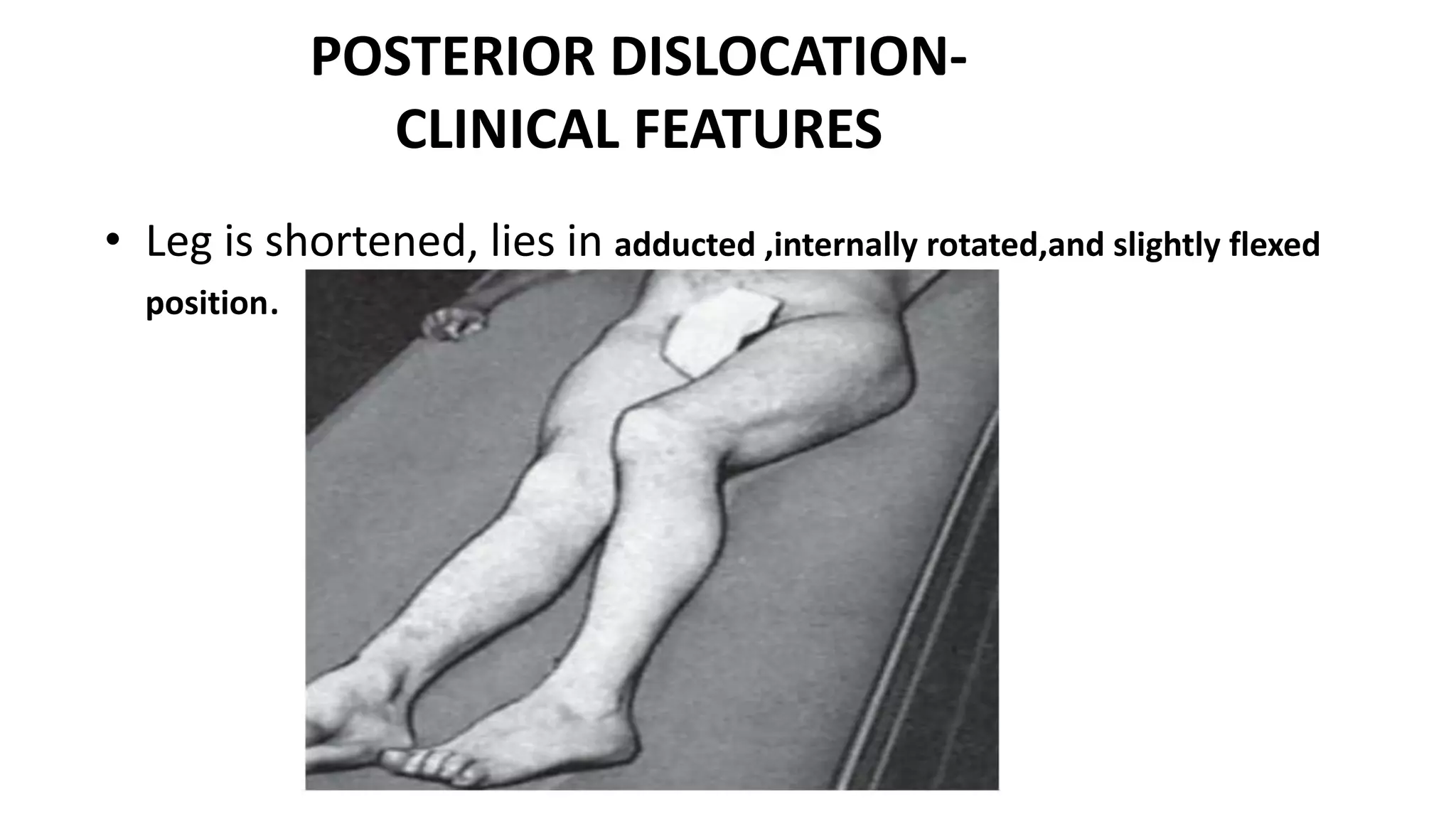
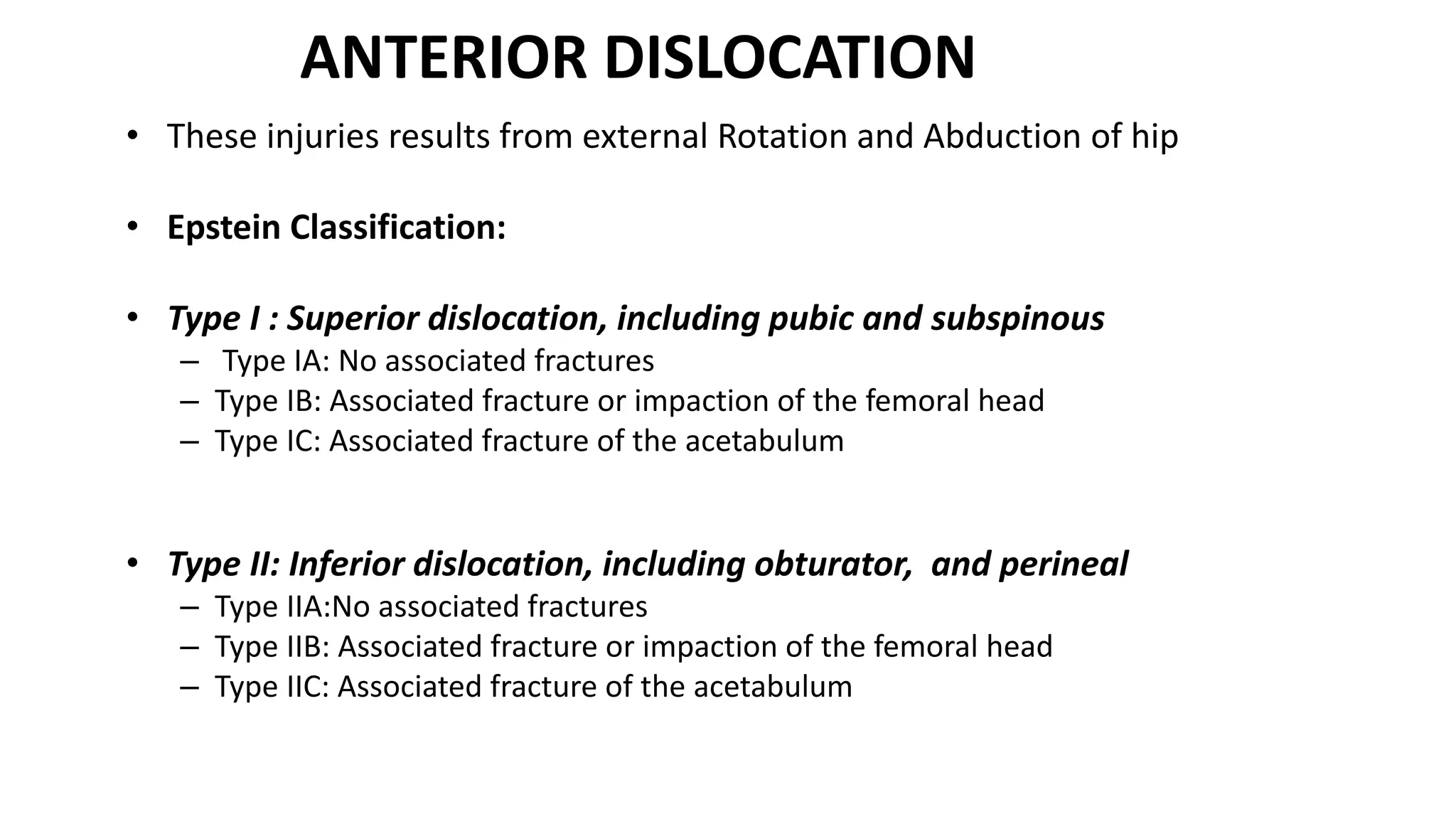
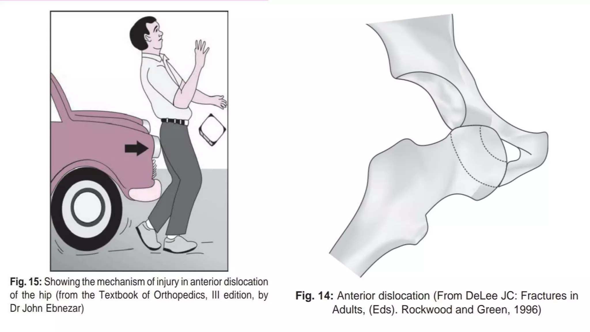
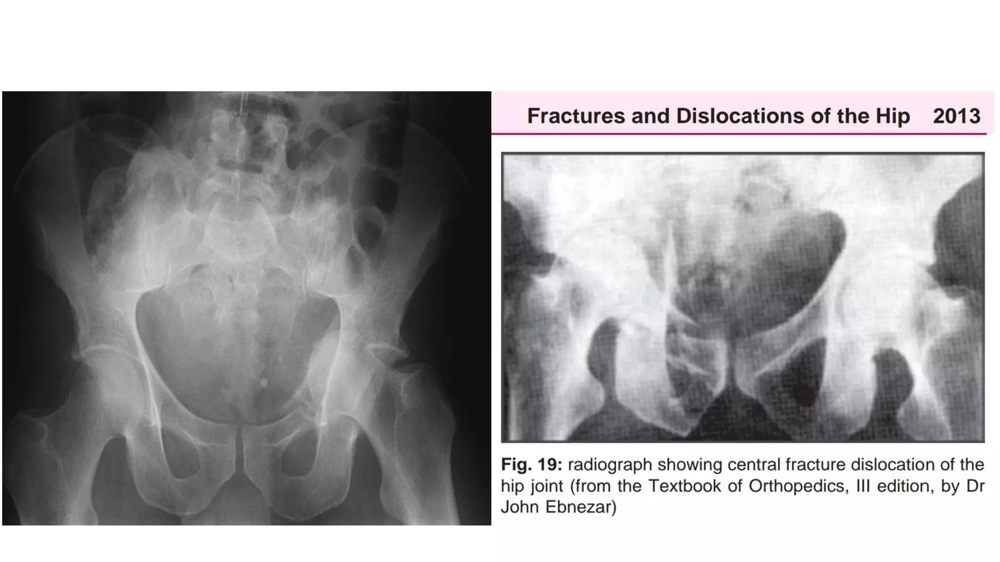
HIPDISLOCATION
Presented by- Dr.BIPUL BORTHAKUR
PROFESSOR, Dept of
Orthopaedics,SMCH,SILCHAR
ANATOMY
• Hip joint is a Ball and Socket type joint- Stability:-
-Anatomical configeration
-Capsule
-Ligaments
• 40 % of the femoral head in any position.
• But in Flexion,Adduction, Internal rotation is a vulnerable position
• The hip joint capsule is anteriorly attatched to intertrochanteric line and posteriorly it’s
1/2inch short.
LIGAMENTS AROUND HIP JOINT
• Inverted Y or Bigelow ligament- Strong ligament- Anteriorly
• Ischiofemoral ligament- posteriorly
• Iliofemoral ligament posteriorly
LIGAMENTS AROUND HIP JOINT-Anterior view
LIGAMENTS AROUND HIP JOINT-Posterior view
ANATOMY
• The main vascular supply to the femoral head is from Ascending branches of the medial
and lateral femoral circumflex arteries, branches of profunda femoral artery , which
arises from Femoral artery.
• An extra-capsular vascular ring is formed at the base of the femoral neck, it contributes
retinacular vessels which pierces the neck to supply blood to femoral head.
• The artery of the ligamentum teres, a brunch of the obturator artery, also contribute
blood supply from medial side.
MECHANISM OF INJURY
• Hip dislocation almost always results from high energy trauma, such as
– motor vehicle accidents,
– fall from height, or
– an industrial accident
– Posterior hip dislocations are much more common - 85-90%
THE FORCES COULD ARISE FROM;
1.The anterior part of flexed knee striking against an object
2.From the sole of feet with ipsilateral knee extended
3.From the greater trochanter
4.Rarely from posterior pelvis
CLASSIFICATION
• Posterior
- Epstein classification, Pipkin’s classification
• Anterior
- Obturator type , Ileal type
• Central- Nowadays discussed with acetabular fracture
POSTERIOR DISLOCATION
• These comprise 85 90 % of traumatic dislocation of the hip
• They results from trauma to the flexed knee, commonly in a seated car(eg:
dashboard injury) , the femur is thrust proximally and the femoral head is
forced posteriorly, often a piece of bone from acetabulum is sheared off.
• Thompson and Epstein Classification :
• Type I : Dislocation with no more than minor chip fractures
• Type II: Dislocation with single large fragment of posterior acetabular wall
• Type III: Dislocation with comminuted fragments of posterior acetabular wall
• Type IV: Dislocation with fracture through acetabular floor
• Type V: Disloction with fracture through acetabular floor and femoral head
POSTERIOR DISLOCATION-
CLINICAL FEATURES
• Leg is shortened, lies in adducted ,internally rotated,and slightly flexed
position.
ANTERIOR DISLOCATION
• These injuries results from external Rotation and Abduction of hip
• Epstein Classification:
• Type I : Superior dislocation, including pubic and subspinous
– Type IA: No associated fractures
– Type IB: Associated fracture or impaction of the femoral head
– Type IC: Associated fracture of the acetabulum
• Type II: Inferior dislocation, including obturator, and perineal
– Type IIA:No associated fractures
– Type IIB: Associated fracture or impaction of the femoral head
– Type IIC: Associated fracture of the acetabulum
CENTRAL FRACTURE – DISLOCATION
• Femoral head is driven through the medial wall of acetabulum toward the pelvic
cavity .
• The displacement of the head varied from the minimal to as much as the whole head
lying inside the pelvis.
• For this, skeletal traction is applied distally and laterally.
CLINICAL EVALUATION
• A full trauma survey is essential because of the high energy nature of these injuries.
• The classic appearance of an individual with a posterior hip dislocation is a patient in
severe pain with the hip in position of Flexion, internal rotation, and adduction.
• Patients with an anterior dislocation hold the hip in marked external rotation with mild
flexion and Abduction
• A careful neuro vascular examination is essential because injury to the sciatic nerve or
femoral neurovascular structures may occur at the time of dislocation
RADIOGRAPHIC EVALUATION
• An Anteroposterior (AP) radiograph of the pelvis is essential, as well as a cross table
lateral view of affected hip
• On AP view :
• In posteior dislocation, the affected femoral head will appear smaller than the normal
femoral head
• In anterior dislocation, the femoral head will appear slightly larger
• The shenton line should be smooth and continuous
• The realtive appearance of the greater and lesser trochanters may indicate pathological
internal or external rotation of the hip
RADIOGRAPHIC EVALUATION
• Use of 45 degree oblique views of the hip may be helpful to ascertain the presence of
osteochondral fragments, the integrity of the acetabulum, and the congruence of the
joint spaces
• CT scan should be obtained to detect the presence of intra articular fragments and to
rule out associated femoral head and acetabular fractures
TREATMENT
• Dislocation should be reduced on an urgent basis, to minimize the risk
of osteonecrosis of the femoral head
• Long term prognosis worsens if reduction is delayed more than 12
hours.
• Associated acetabular or femoral head fracture can be treated in the
subacute phase.
• Close reduction : Under sedation/Anaesthesia
• Techniques are-Allis, Stimpson and bigelow’s etc
• Supine, Prone, Lateral
TREATMENT
• 1.Allis Method:
• Patient is place supine with the surgeon standing above the patient on table.
• Initially, the surgeon applied in line traction while assistant applies countertraction
by stabilizing the patient’s pelvis.
• While increasing the traction force, the surgeon should slowly increase the degree
of Flexion to approximately 70 degree.
• Gentle rotational motion of the hip as well as slight adduction will often help the
femoral head to clear the hip of the acetabulum.
• A lateral force to the proximal thigh may assist in reduction.
• An audible “clunk” is a sign of a successful closed reduction
TREATMENT
• 2. Stimson Gravity Technique:
• The patient is placed prone on table with affected leg hanging off
the side of the table.
• In this position, the assistant immobilizes the pelvis, and the
surgeon applies an anterior directed, force on the proximal calf.
• Gentle rotation of the limb may assist in reduction
TREATMENT
• 3. Bigelow and Reverse Bigelow Maneuvers :
• The patient is in supine, and the surgeon applies longitudinal traction on the
limb,
• The adduction and internally rotated thigh is then flexed at least 90 degrees
• The femoral head is then levered into the acetabulum by abduction, external
rotation, and extension of the hip.
• In Reverse Bigelow Maneuvers, used for anterior dislocation, traction is again
applied in the line of deformity. The hip is then adducted, sharply internally
rotated and extended.
IMMOBILIZATION AFTER REDUCTION
• Post reduction CT scan is more important than pre-reduction to
confirm congruous reduction.
• Immobilization depends upon CT picture showing Acetabular or
Femoral injury
• Depends upon degree of injury
• MRI is important for Knee joint ligamental injuries in comparison to
Hip joint.
TREATMENT
• IF CLOSED REDUCTION SUCCESSFUL, DO STABILITY TEST(TELESCOPIC
TEST) AND THEN MAINTAIN REDUCTION WITH SPLINT.
• IF REDUCTION NOT POSSIBLE;
Causes;
- Infolding of Labrum- Button holdindg by capsule
and short rotators
-Sciatic nerve
-Bony fragments
TREATMENT –IF CLOSED REDUCTION FAILS
• Open reduction :
• Has to be done immediately
• Reconstuction of acetabulum is delayed
RECONSTRUCTION
• Reconstruction of Hip
• Reconstruction of acetabulum
Usually done in 2nd week following trauma
TREATMENT :Open reduction
• Approaches:
• Kocher-Langenbeck approach
– Posterior approach
– Done for the exploration of sciatic nerve, treatment of major posterior labral disruptionor
instability, and of Posterior acetabular fractures
• Smith- Peterson Approach
– Anterior approach
– Done for isolated femoral head fracture
• Watson-Jones approach
– Anterolateral approach
– Useful for most anterior dislocation and combined fracture of both femoral head and neck
PROGNOSIS
• 70-80 % good or excellent outcome in posterior dislocation.
• Anterior dislocation of hip are noted to have a higher incidence
of associated femoral head injuries .
COMPLICATIONS• Osteonecrosis (AVN)
– Seen in 5-40 % of injuries
– Increased risk associated with increased time untill reduction (>6-24 hours)
– Osteonecrosis may become clinically apparent several years after injury
• Post traumatic Osteoarthritis
– Most frequent long term complication.
– Incidence is more when associated with acetabular fractures or transchondral
fracture of femoral head
• Recurrent dislocation
• Neurovascular injury
– Sciatic nerve injury occurs in 10-20 % of hip dislocation
• Femoral head fracture
• Heterotopic ossification
In case of any suggestions/Questions
kindly message/contact on
Mob: +919435031719
Email: drbipulborthakur@gmail.com
drbborthakur@rediffmail.com
THANK YOU..

Hip dislocation

  • 1.
    HIPDISLOCATION Presented by- Dr.BIPULBORTHAKUR PROFESSOR, Dept of Orthopaedics,SMCH,SILCHAR
  • 2.
    ANATOMY • Hip jointis a Ball and Socket type joint- Stability:- -Anatomical configeration -Capsule -Ligaments • 40 % of the femoral head in any position. • But in Flexion,Adduction, Internal rotation is a vulnerable position • The hip joint capsule is anteriorly attatched to intertrochanteric line and posteriorly it’s 1/2inch short.
  • 3.
    LIGAMENTS AROUND HIPJOINT • Inverted Y or Bigelow ligament- Strong ligament- Anteriorly • Ischiofemoral ligament- posteriorly • Iliofemoral ligament posteriorly
  • 4.
    LIGAMENTS AROUND HIPJOINT-Anterior view
  • 5.
    LIGAMENTS AROUND HIPJOINT-Posterior view
  • 6.
    ANATOMY • The mainvascular supply to the femoral head is from Ascending branches of the medial and lateral femoral circumflex arteries, branches of profunda femoral artery , which arises from Femoral artery. • An extra-capsular vascular ring is formed at the base of the femoral neck, it contributes retinacular vessels which pierces the neck to supply blood to femoral head. • The artery of the ligamentum teres, a brunch of the obturator artery, also contribute blood supply from medial side.
  • 8.
    MECHANISM OF INJURY •Hip dislocation almost always results from high energy trauma, such as – motor vehicle accidents, – fall from height, or – an industrial accident – Posterior hip dislocations are much more common - 85-90% THE FORCES COULD ARISE FROM; 1.The anterior part of flexed knee striking against an object 2.From the sole of feet with ipsilateral knee extended 3.From the greater trochanter 4.Rarely from posterior pelvis
  • 9.
    CLASSIFICATION • Posterior - Epsteinclassification, Pipkin’s classification • Anterior - Obturator type , Ileal type • Central- Nowadays discussed with acetabular fracture
  • 10.
    POSTERIOR DISLOCATION • Thesecomprise 85 90 % of traumatic dislocation of the hip • They results from trauma to the flexed knee, commonly in a seated car(eg: dashboard injury) , the femur is thrust proximally and the femoral head is forced posteriorly, often a piece of bone from acetabulum is sheared off. • Thompson and Epstein Classification : • Type I : Dislocation with no more than minor chip fractures • Type II: Dislocation with single large fragment of posterior acetabular wall • Type III: Dislocation with comminuted fragments of posterior acetabular wall • Type IV: Dislocation with fracture through acetabular floor • Type V: Disloction with fracture through acetabular floor and femoral head
  • 12.
    POSTERIOR DISLOCATION- CLINICAL FEATURES •Leg is shortened, lies in adducted ,internally rotated,and slightly flexed position.
  • 15.
    ANTERIOR DISLOCATION • Theseinjuries results from external Rotation and Abduction of hip • Epstein Classification: • Type I : Superior dislocation, including pubic and subspinous – Type IA: No associated fractures – Type IB: Associated fracture or impaction of the femoral head – Type IC: Associated fracture of the acetabulum • Type II: Inferior dislocation, including obturator, and perineal – Type IIA:No associated fractures – Type IIB: Associated fracture or impaction of the femoral head – Type IIC: Associated fracture of the acetabulum
  • 18.
    CENTRAL FRACTURE –DISLOCATION • Femoral head is driven through the medial wall of acetabulum toward the pelvic cavity . • The displacement of the head varied from the minimal to as much as the whole head lying inside the pelvis. • For this, skeletal traction is applied distally and laterally.
  • 21.
    CLINICAL EVALUATION • Afull trauma survey is essential because of the high energy nature of these injuries. • The classic appearance of an individual with a posterior hip dislocation is a patient in severe pain with the hip in position of Flexion, internal rotation, and adduction. • Patients with an anterior dislocation hold the hip in marked external rotation with mild flexion and Abduction • A careful neuro vascular examination is essential because injury to the sciatic nerve or femoral neurovascular structures may occur at the time of dislocation
  • 22.
    RADIOGRAPHIC EVALUATION • AnAnteroposterior (AP) radiograph of the pelvis is essential, as well as a cross table lateral view of affected hip • On AP view : • In posteior dislocation, the affected femoral head will appear smaller than the normal femoral head • In anterior dislocation, the femoral head will appear slightly larger • The shenton line should be smooth and continuous • The realtive appearance of the greater and lesser trochanters may indicate pathological internal or external rotation of the hip
  • 23.
    RADIOGRAPHIC EVALUATION • Useof 45 degree oblique views of the hip may be helpful to ascertain the presence of osteochondral fragments, the integrity of the acetabulum, and the congruence of the joint spaces • CT scan should be obtained to detect the presence of intra articular fragments and to rule out associated femoral head and acetabular fractures
  • 24.
    TREATMENT • Dislocation shouldbe reduced on an urgent basis, to minimize the risk of osteonecrosis of the femoral head • Long term prognosis worsens if reduction is delayed more than 12 hours. • Associated acetabular or femoral head fracture can be treated in the subacute phase. • Close reduction : Under sedation/Anaesthesia • Techniques are-Allis, Stimpson and bigelow’s etc • Supine, Prone, Lateral
  • 25.
    TREATMENT • 1.Allis Method: •Patient is place supine with the surgeon standing above the patient on table. • Initially, the surgeon applied in line traction while assistant applies countertraction by stabilizing the patient’s pelvis. • While increasing the traction force, the surgeon should slowly increase the degree of Flexion to approximately 70 degree. • Gentle rotational motion of the hip as well as slight adduction will often help the femoral head to clear the hip of the acetabulum. • A lateral force to the proximal thigh may assist in reduction. • An audible “clunk” is a sign of a successful closed reduction
  • 27.
    TREATMENT • 2. StimsonGravity Technique: • The patient is placed prone on table with affected leg hanging off the side of the table. • In this position, the assistant immobilizes the pelvis, and the surgeon applies an anterior directed, force on the proximal calf. • Gentle rotation of the limb may assist in reduction
  • 29.
    TREATMENT • 3. Bigelowand Reverse Bigelow Maneuvers : • The patient is in supine, and the surgeon applies longitudinal traction on the limb, • The adduction and internally rotated thigh is then flexed at least 90 degrees • The femoral head is then levered into the acetabulum by abduction, external rotation, and extension of the hip. • In Reverse Bigelow Maneuvers, used for anterior dislocation, traction is again applied in the line of deformity. The hip is then adducted, sharply internally rotated and extended.
  • 31.
    IMMOBILIZATION AFTER REDUCTION •Post reduction CT scan is more important than pre-reduction to confirm congruous reduction. • Immobilization depends upon CT picture showing Acetabular or Femoral injury • Depends upon degree of injury • MRI is important for Knee joint ligamental injuries in comparison to Hip joint.
  • 32.
    TREATMENT • IF CLOSEDREDUCTION SUCCESSFUL, DO STABILITY TEST(TELESCOPIC TEST) AND THEN MAINTAIN REDUCTION WITH SPLINT. • IF REDUCTION NOT POSSIBLE; Causes; - Infolding of Labrum- Button holdindg by capsule and short rotators -Sciatic nerve -Bony fragments
  • 33.
    TREATMENT –IF CLOSEDREDUCTION FAILS • Open reduction : • Has to be done immediately • Reconstuction of acetabulum is delayed RECONSTRUCTION • Reconstruction of Hip • Reconstruction of acetabulum Usually done in 2nd week following trauma
  • 34.
    TREATMENT :Open reduction •Approaches: • Kocher-Langenbeck approach – Posterior approach – Done for the exploration of sciatic nerve, treatment of major posterior labral disruptionor instability, and of Posterior acetabular fractures • Smith- Peterson Approach – Anterior approach – Done for isolated femoral head fracture • Watson-Jones approach – Anterolateral approach – Useful for most anterior dislocation and combined fracture of both femoral head and neck
  • 35.
    PROGNOSIS • 70-80 %good or excellent outcome in posterior dislocation. • Anterior dislocation of hip are noted to have a higher incidence of associated femoral head injuries .
  • 36.
    COMPLICATIONS• Osteonecrosis (AVN) –Seen in 5-40 % of injuries – Increased risk associated with increased time untill reduction (>6-24 hours) – Osteonecrosis may become clinically apparent several years after injury • Post traumatic Osteoarthritis – Most frequent long term complication. – Incidence is more when associated with acetabular fractures or transchondral fracture of femoral head • Recurrent dislocation • Neurovascular injury – Sciatic nerve injury occurs in 10-20 % of hip dislocation • Femoral head fracture • Heterotopic ossification
  • 37.
    In case ofany suggestions/Questions kindly message/contact on Mob: +919435031719 Email: drbipulborthakur@gmail.com drbborthakur@rediffmail.com THANK YOU..