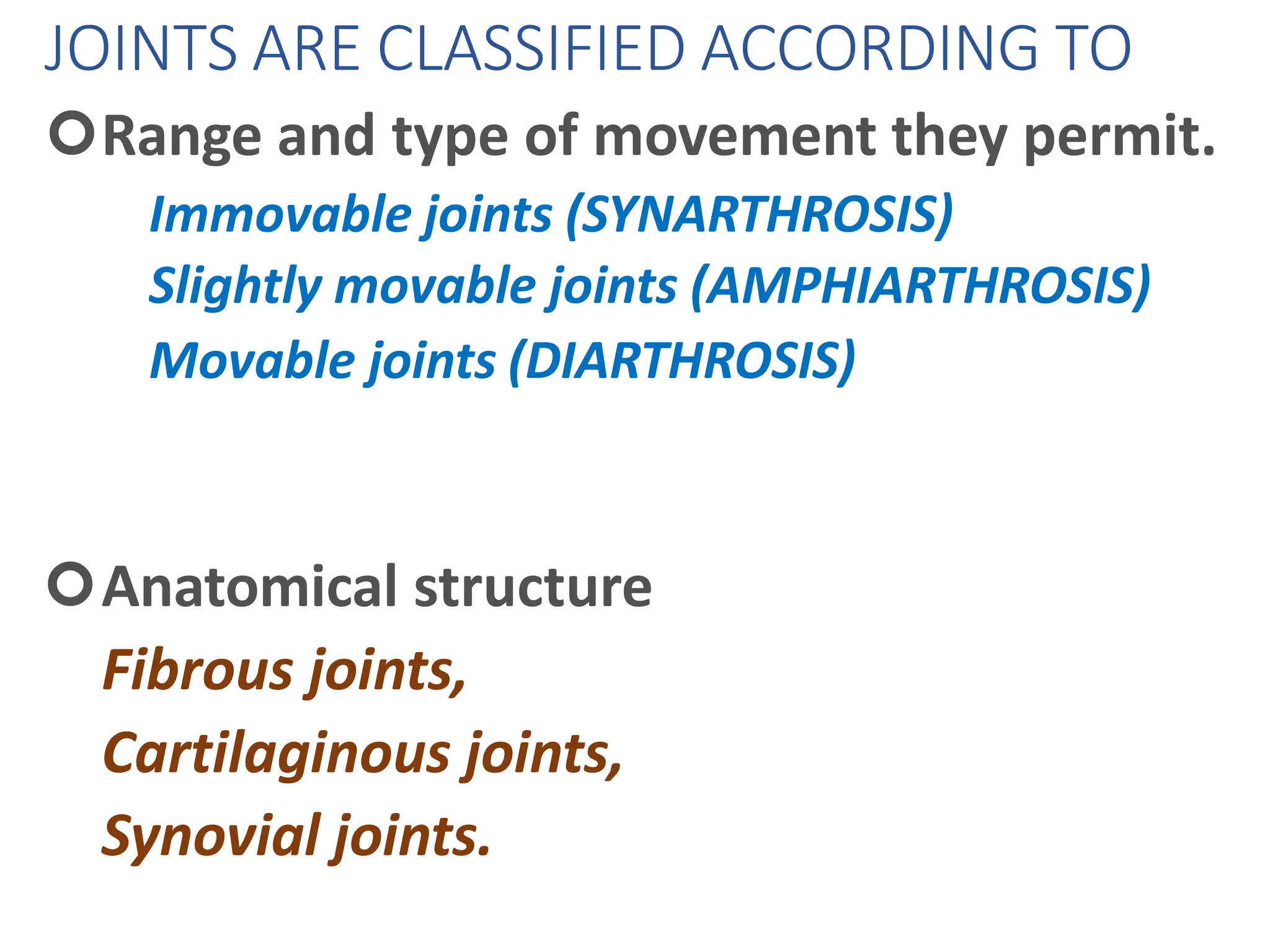
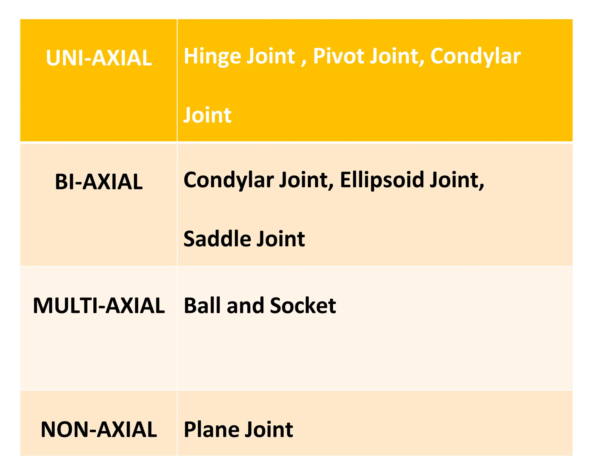
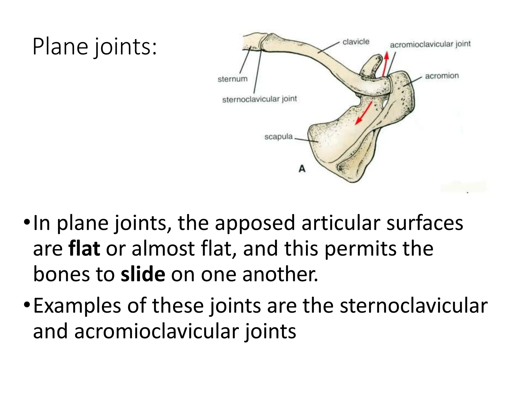
2. JOINTS ARE CLASSIFIED ACCORDING TO
Range and type of movement they permit.
Immovable joints (SYNARTHROSIS)
Slightly movable joints (AMPHIARTHROSIS)
Movable joints (DIARTHROSIS)
Anatomical structure
Fibrous joints,
Cartilaginous joints,
Synovial joints.
3. Functions:
• Permit movement
• Growth
• Molding during childbirth
• Concerned with differential growth
• Transmission of forces
4. Structural Classification
a) Fibrous e.g. i) Sutures,
ii) Syndesmosis
iii) Gomphosis
b) Cartilaginous e.g. i) Primary Cartilaginous
ii) Secondary cartilaginous
c) Synovial e.g. i) Uniaxial: Hinge, Pivot
ii) Biaxial: Condylar, Ellipsoid
iii) Multiaxial: Ball & Socket, Saddle
10. -Present only between bones of skull.
-Fibrous tissue is
continuous with
periosteum
-Sutures ossify and
fuse in middle age:
Called “synostoses”
SUTURES
bone—collagenous sutural ligament—bone
11. TYPES OF SUTURES
PLANE SUTURE
•Simple apposition of contiguous surfaces,
• usually rough and
reciprocally irregular,
Examples
• sutures between the
palatine bones,
• between the maxillae and
at the palatomaxillary sutures.
12. Denticulate Suture
Has small tooth like projections,
often widening towards their
ends to provide effective
interlocking.
When united by sutural
ligament and periosteum, such
sutures are almost completely
immobile
The lambdoid suture is a
denticulate suture.
14. Squamous suture
Where bones overlap,
as at the temporo-
parietal suture, a
squamous suture is
formed; the adjacent
bone surfaces are
reciprocally bevelled.
18. Cartilagenous joints
Articulating bones united by cartilage
Lack a joint cavity
Not highly movable
Two types
Synchondroses (singular:
synchondrosis)
Symphyses (singular: symphysis)
19. Synchondroses
Primary Cartilaginous Joints
Literally: “junction of cartilage”
A plate or bar of Hyaline cartilage unites the
bones
Immovable (synarthroses)
Examples:
Epiphyseal plates between epiphysis and
diaphysis of growing bone.
Joint between first rib’s costal cartilage and
manubrium of the sternum
23. The lambdoid suture is an example of a(n)
A. Diarthrosis
B. Amphiarthrosis
C. Synarthrosis
D. Synarthrosis and fibrous
24. The lambdoid suture is an example of a(n)
A. Diarthrosis
B. Amphiarthrosis
C. Synarthrosis
D. Synarthrosis and fibrous
25. Which of the following is an example of a
synchondrosis?
A. First sternocostal articulation
B. Humeroscapular articulation
C. Symphysis pubis
D. Tibiofibular articulation
26. Which of the following is an example of a
synchondrosis?
A. First sternocostal articulation
B. Humeroscapular articulation
C. Symphysis pubis
D. Tibiofibular articulation
27. The procedure arthroscopy refers to:
A. Examining interior of joint
B. Reducing inflammation of a bursa
C. Re-establishing blood supply of muscles
D. Repair of bone fractures
28. The procedure arthroscopy refers to:
A. Examining interior of joint
B. Reducing inflammation of a bursa
C. Re-establishing blood supply of muscles
D. Repair of bone fractures
29. Which of the following joints are held
together by cartilage?
A. Diarthrosis
B. Gomphosis
C. Symphysis
D. Synovial
30. Which of the following joints are held
together by cartilage?
A. Diarthrosis
B. Gomphosis
C. Symphysis
D. Synovial
31. The radioulnar joint is an example of a
A. Hinge
B. Pivot
C. Planar
D. Synarthroses
32. The radioulnar joint is an example of a
A. Hinge
B. Pivot
C. Planar
D. Synarthroses
33.
34. Synovial joints
DIARTHROSES
•Structure: bone—articular cartilage—
synovial fluid in cavity – articular cartilage—
bone
•Bond : surrounding sleeve of collagenous
fibrous capsule lined by synovial membrane;
extrinsic and intrinsic ligaments. Occasional
intracapsular ligaments, tendons, fat pads,
fibrocartilaginous discs or menisci.
37. General Structure of Synovial Joints
b
1. Articular cartilage
Hyaline
Spongy cushions absor
compression
Protects ends of bones
from being crushed
2. Joint (synovial) cavity
Potential space
Small amount of
synovial fluid
38. 3. Articular (joint) capsule
Two layered
Outer*: fibrous capsule of
dense irregular connective
tissue continuous with
periosteum.
Inner*: synovial membrane
of loose connective tissue
(makes synovial fluid)
Lines all internal joint surfaces
except the cartilages*
39. 4. Synovial fluid
Filtrate of blood
Contains special
glycoproteins
Nourishes cartilage and
functions as slippery
lubricant
5. Reinforcing ligaments
Capsular (most) –
thickened parts of capsule
Extracapsular
Intracapsular
40. 6. Nerves
Detect pain
Monitor stretch (one of the
ways of sensing posture
and body movements)
7. Blood vessels
Rich blood supply
Extensive capillary beds in
synovial membrane
(produce the blood filtrate)
41. Articular disc or
meniscus
(literally “crescent”)
Only some joints
Those with bone
ends of different
shapes or fitting poorly
Some to allow two kinds of movement (e.g. jaw)
Fibrocartilage
Examples: knee
TMJ (Temporomandibular joint)
sternoclavicular joint
42. •Fatty pads are found in
some synovial joints lying
between the synovial
membrane and the
fibrous capsule or bone.
•Examples are found in
the hip and knee joints.
43. Bursae and tendon sheaths
Contain synovial fluid
Not joints but often associated with them
Act like ball bearings
Bursa means “purse” in Latin
Flattened sac lined by synovial membrane
Where ligaments, muscles, tendons, or bones overlie each other and rub
together
Tendon sheath
Only on tendons subjected to friction
46. Plane joints:
•In plane joints, the apposed articular surfaces
are flat or almost flat, and this permits the
bones to slide on one another.
•Examples of these joints are the sternoclavicular
and acromioclavicular joints
47. Hinge joints:
•Hinge joints resemble the hinge on a door,
so that flexion and extension movements are
possible.
Examples of these joints are the elbow, knee, and
ankle joints
48. Pivot joints:
•In pivot joints, a central
bony pivot is
surrounded by a bony-
ligamentous ring and
rotation is the only
movement possible.
• E.g atlantoaxial and
superior radioulnar
joints
49. Condylar joints
•Condyloid joints have
two distinct convex
surfaces that
articulate with two concave surfaces.
•The movements of flexion, extension, with a
small amount of rotation.
•E.g. knee joint , temporo-mandibular joint
50. Ellipsoid joints
•In ellipsoid joints, an
elliptical convex
articular surface fits
into an elliptical
concave articular
surface. The movements of flexion, extension,
abduction, adduction and circumduction can
take place, but rotation is impossible.
•E.g. wrist joint, metacarpophalangeal joint
51. Saddle joints:
•The articular surfaces are
reciprocally concavoconvex
and resemble a saddle on a
horse's back.
•These joints permit flexion, extension,
abduction, adduction, and rotation.
•E.g. carpometacarpal joint of the thumb,
sternoclavicular joint
52. Ball and socket joints:
•In ball-and-socket joints,
a ball-shaped head of
one bone fits into a socket
like concavity of another.
•This arrangement permits free movements,
including flexion, extension, abduction,
adduction, medial rotation, lateral rotation, and
circumduction.
• E.g. shoulder and hip joints
53. Stability of Joints
The stability of a joint depends on three main
factors:
•the shape,
•size,
•arrangement of the articular surfaces;
•the ligaments;
•and the tone of the muscles around the joint.
54. MECHANISM OF LUBRICATION OF SYNOVIAL
JOINT
•Synovial fluid
•Hyaline cartilage
•Harvesian fatty pads
•Bursa
55. Nerve Supply of Joints
•The capsule and ligaments receive an
abundant sensory nerve supply.
•A sensory nerve supplying a joint also
supplies the muscles moving the joint and
the skin overlying the insertions of these
muscles, a fact that has been codified as
Hilton's law.
56. Classification of Synovial Joints
(According to the number of bones)
•SIMPLE JOINT Interphalangeal joint
•COMPOUND JOINT elbow ,wrist
•COMPLEX JOINT temporomandibular
joint
57.
58.
59. Cartilage Tendons Ligaments
Tough Attaches bone to
muscle
Attaches bone to
bone
Flexible Sturdy Elastic
At end of
bone
Non elastic Stabilise
Cushions Size changes
depending on
muscle
Made of many
fibres
Anchor Strong
60. Types of Joint Movement
• There are six different kinds of movement the
joints can allow
• These are:
– Extension
– Flexion
– Abduction
– Adduction
– Circumduction
– Rotation
61. Extension:
straightening or
extending a limb.
(Opening a Joint)
Flexion: Bending
or flexing a limb.
(Closing a joint)
Abduction: Moving
a limb away from the
centre line of the
body.
62. Rotation: This is a
turning or rotational
movement of a limb
or body part.
Circumduction:
The ability of a
limb to be moved
in circles.
Adduction:
moving a limb
towards the centre
line of the body
63. Plantarflexion (or plantar flexion)
Is the movement which increases
the approximate 90 degree angle
between the front part of the foot
and the shin, as when pressing
down on a car pedal
Dorsiflexion is the movement which
decreases the angle between the dorsum
(superior surface) of the foot and the leg, so
that the toes are brought closer to the shin.
The movement moving in opposite directions
is called plantarflexion. Put more simply: it
applies to the upward movement of the foot
at the ankle joint
64. Pronation – A rotation of the forearm
that moves the palm from an
anterior-facing position to a
posterior-facing position, or palm
facing down. This is not medial
rotation as this must be performed
when the arm is half flexed
Supination – The opposite of pronation,
the rotation of the forearm so that the palm
faces anteriorly, or palm facing up. The
hand is supine (facing anteriorly) in the
anatomical position