SlideShare a Scribd company logo
Joints In Human Body Dr. Usha (PT)
Assistant Professor
Introduction
Related Terms
1. Arthron (G. a joint). Compare with the terms arthrology,
synarthrosis, diarthrosis, arthritis, arthrodesis, etc.
2. Articulatio (L a joint); articulation (NA).
3. Junctura (L a joint).
4. Syndesmology (G. syndesmosis = ligament) is the study of
ligaments and related joints.
Definition
Joint is a junction between two or more bones or cartilages. It
is a device to permit movements.
However, immovable joints are primarily meant for growth,
and may permit moulding during childbirth.
There are more joints in a child than in an adult because as
growth proceeds some of the bones fuse together, e.g. the
ilium, ischium and pubis to form the pelvic bone; the two
halves of the infant frontal bone, and of the infant mandible;
the five sacral vertebrae and the four coccygeal vertebrae.
A. Structural Classification
1. Fibrous Joints
Sutures
Syndesmosis
Gomphosis
2. Cartilaginous
Joints
Primary
cartilaginous joints
or synchondrosis
Secondary
cartilaginous joints
or symphysis
3. Synovial Joints
Ball-and-socket or
spheroidal joints
Sellar or saddle joints
Condylar or bicondylar
joints
Ellipsoid joints
Hinge joints
Pivot or trochoid joints
Plane joints
B. Functional Classification (According
to the Degree of Mobility)
1.
• Synarthrosis (immovable), like fibrous joints
2.
• Amphiarthrosis (slightly movable), like
cartilaginous joints
3.
• Diarthrosis (freely movable), like synovial joints
Synarthroses
Fixed joints at which there is no
movement.
The articular surfaces are joined by
tough fibrous tissue.
Often the edges of the bones are
dovetailed into one another as in the
sutures of the skull.
Amphiarthroses
Joints at which slight movement is
possible.
A pad of cartilage lies between the bone
surfaces, and there are fibrous
ligaments to hold the bones and
cartilage in place.
The cartilages of such joints also act as
shock absorbers, e.g. the intervertebral
discs between the bodies of the
vertebrae, where the cartilage is
strengthened by extra collagen fibres.
Diarthroses or Synovial Joints
Freely movable joints, though
at some of them the movement
is restricted by the shape of the
articulating surfaces and by the
ligaments which hold the bones
together. These ligaments are
of elastic connective tissue.
A synovial joint has a fluid-filled cavity between articular
surfaces which are covered by articular cartilage.
The fluid, known as synovial fluid, produced by the synovial
membrane which lines the cavity except for the actual articular
surfaces and covers any ligaments or tendons which pass
through the joint.
Synovial fluid acts as a lubricant.
The form of the articulating surfaces controls the type of movement
which takes place at any joint.
The movements possible at synovial joints are:
1. Angular
2. Rotatory
3. Gliding
• Flexion : decreasing the angle between two bones;
• Extension : increasing the angle between two bones;
• Abduction : moving the part away from the mid-line;
• Adduction : bringing the part towards the mid-line.
Angular
• Rotation : turning upon an axis;
• Circumduction: moving the extremity of the part round in a circle so
that the whole part inscribes a cone.
Rotary
• one part slides on another.
Gliding
C. Regional Classification
1.
• Skull type: immovable.
2.
• Vertebral type: slightly movable.
3.
• Limb type: freely movable.
D. According to No. of Articulating
Bones
1. Simple
Joints
When two bones
articulate, e.g.
interphalangeal
joints
2. Compound
Joints
More than two
bones articulate
within one
capsule, e.g.
elbow joint, wrist
joint
3. Complex Joint
When joint cavity
is divided by an
intra-articular disc,
e.g.,
temporomandibula
r joint and
sternoclavicular
joint.
1. FIBROUS JOINTS
In fibrous joints the bones are joined by fibrous tissue. These
joints are either immovable or permit a slight degree of
movement.
These can be grouped in the following three subtypes:
1. Sututres
2. Syndesmosis
3. Gomphosis
a. Sutures
These are peculiar to skull, and are immovable.
According to the shape of bony margins, the sutures can be:
(i) Plane, e.g. internasal suture
(ii) Serrate, e.g. interparietal suture
(iii) Squamous, e.g. temporo-parietal suture
(iv) Denticulate, e.g. lambdoid suture
(v) Schindylesis type, e.g. between rostrum of sphenoid and
upper border of vomer.
Neonatal skull reveals fontanelles which are temporary in
nature.
At six specific points on the sutures in new born skull are
membrane filled gaps called "fontanelles".
These allow the underlying brain to increase in size. Anterior
fontanelle is used to judge the hydration of the infant. All these
fontanelles become bone by 18 months.
b. Syndesmosis
The bones are connected by
the interosseous ligament.
Example: inferior tibiofibular
joint
c. Gomphosis
Also known as- Peg and
socket joint.
Example: root of the
tooth in its bony socket
2. CARTILAGINOUS JOINTS
In this type of joints the bones are joined by cartilage. These
are of the following two types:
1. Primary cartilaginous joints
2. Secondary cartilaginous joints
a. Primary Cartilaginous Joints
Synchondrosis, or hyaline cartilage joints
The bones are united by a plate of hyaline cartilage so that the
joint is immovable and strong.
These joints are temporary in nature because after a certain age
the cartilaginous plate is replaced by bone (synostosis).
Examples:
(a) Joint between epiphysis and
diaphysis of a growing long bone
(b) Spheno-occipital joint
(c) First chondrosternal joint
(d) Costochondral joints.
b. Secondary Cartilaginous
Joints
Symphyses or fibrocartilaginous joints
The articular surfaces are covered by a thin layer of hyaline
cartilage, and united by .a disc of fibrocartilage.
These joints are permanent and persist throughout life. In this
respect symphysis menti is a misnomer as it is a synostosis.
Typically the secondary cartilaginous joints occur in the
median plane of the body, and permit limited movements due
to compressible pad of fibro-cartilage such as in the pubic
symphysis and manubriosternal joints.
The thickness of fibrocartilage is directly related to the range
of movement. Secondary cartilaginous joints may represent an
intermediate stage in the evolution of synovial joints.
Examples:
(a) Symphysis pubis
(b) Manubriosternal joint
(c) Intervertebral joints between the vertebral bodies
3. SYNOVIAL JOINTS
Synovial joints are most evolved, and, therefore, most mobile
type of joints
Characters
1. The articular surfaces are covered with hyaline (articular)
cartilage (fibrocartilage in certain membrane bones).
Articular cartilage is avascular, non-nervous and elastic.
Lubricated with synovial fluid, the cartilage provides slippery
surfaces for free movements, like 'ice on ice'. The surface of the
cartilage shows fine undulations filled with synovial fluid.
2. Between the articular surfaces there is a joint cavity filled
with synovial fluid. The cavity may be partially or completely
subdivided by an articular disc or meniscus.
3. The joint is surrounded by an articular capsule which is
made up of a fibrous capsule lined by synovial membrane.
Because of its rich nerve supply, the fibrous capsule is sensitive
to stretches imposed by movements. This sets up appropriate
reflexes to protect the joint from any sprain. This is called the
'watch-dog' action of the capsule.
The fibrous capsule is often reinforced by :
(a) Capsular or true ligaments representing thickenings of the
fibrous capsule
(b) The accessory ligaments (distinct from fibrous capsule)
which may be intra or extracapsular.
The synovial membrane lines whole of the interior of the joint,
except for the articular surfaces covered by hyaline cartilage.
The membrane secretes a slimy viscous fluid called the synovia
or synovial fluid which lubricates the joint and nourishes the
articular cartilage. The viscosity of fluid is due to hyaluronic
acid secreted by cells of the synovial membrane.
4. Varying degrees of movements are always permitted by the
synovial joints.
a. Plane Synovial Joints
Articular surfaces are more or less
flat (plane). They permit gliding
movements (translations) in
various directions.
Examples:
(a) Intercarpal joints
(b) Intertarsal joints
(c) Joints between articular processes of
vertebrae
(d) Cricothyroid joint
(e) Cricoarytenoid joint
(f) Superior tibiofibular
(g) Interchondral joint (5-9 ribs)
(i) Costotransverse and
Costovertebral
(j) Acromioclavicular with intra-
articular disc
(k) Carpometacarpal (except first)
(1) Tarsometatarsal
(m) Intermetacarpal
(n) Intermetatarsal
(o) Chondrosternal (except first)
(p) Sacroiliac
b. Hinge Joints (Ginglymi)
Articular surfaces are pulley-shaped.
There are strong collateral ligaments.
Movements are permitted in one
plane around a transverse axis.
Examples:
(a) Elbow joint
(b) Ankle joint
(c) Interphalangeal joints.
c. Pivot (Trochoid) Joints
Articular surfaces comprise a central bony
pivot (peg) surrounded by an
osteoligamentous ring. Movements are
permitted in one plane around a vertical
axis.
Examples:
(a) Superior and inferior radio-ulnar joints
(b) Median atlanto-axial joint.
d. Condylar (Bicondylar) Joints
Articular surfaces include two distinct condyles (convex male
surfaces) fitting into reciprocally concave female surfaces (which
are also, sometimes, known as condyles, such as in tibia).
These joints permit movements mainly in one plane around a
transverse axis, but partly in another plane (rotation) around a
vertical axis.
Examples:
(a) Knee joint
(b) Right and left jaw joints
or temporomandibular joint
e. Ellipsoid Joints
Articular surfaces include an oval, convex, male surface fitting
into an elliptical, concave female surface. Free movements are
permitted around both the axes, flexion and extension around
the transverse axis, and abduction and adduction around the
anteroposterior axis.
Combination of movements produces circumduction. Typical
rotation around a third (vertical) axis does not occur.
Examples:
(a) Wrist joint
(b) Metacarpophalangeal joints
(c) Atlanto-occipital joints.
f. Saddle (Sellar) Joints
Articular surfaces are reciprocally concavoconvex. Movements
are similar to those permitted by an ellipsoid joint, with
addition of some rotation (conjunct rotation) around a third
axis which, however, cannot occur independently.
Examples:
(a) First carpometacarpal joint
(b) Sternoclavicular joint
(c) Calcaneocuboid joint
(d) Incudomalleolar joint
(e) Between femur and patella.
g. Ball-and-socket (Spheroidal)
Joints
Articular surfaces include a globular head (male surface)
fitting into a cup-shaped socket (female surface). Movements
occur around an indefinite number of axes which have one
common centre.
Flexion, extension, abduction, adduction, medial rotation,
lateral rotation, and circumduction, all occur quite freely.
Examples:
(a) Shoulder joint
(b) Hip joint (Fig. 3.19)
(c) Talocalcaneonavicular joint
(d) Incudostapedial joint
Shoulder joint
Classification And Movements Of
Synovial Joints
1. Terminology & Definition
Human Kinesiology: Study of geometry of surfaces and their associated movements.
Male surface: An articulating surface which is larger in surface area and always convex in all
directions.
Female surface: An articulating surface which is smaller and concave in all directions.
Simple joints: Joints with only two articulating surfaces, i.e. male and female.
Compound joints: Joint possessing more than one pair of articulating surfaces.
Degrees of freedom: Number of axes at which the bone in a joint can move.
Uniaxial: Movement of bone at a joint is limited to one axis, i.e. with one degree of freedom, e.g.
interphalangeal joints.
Biaxial: With two degrees of freedom, e.g. wrist joint.
Multi-axial: Three axes along with intermediate positions also, e.g. shoulder joint
Translation: Sliding movements of one articulating surface over the other.
2. Movements & Mechanism of
Joints
A. Angular movement: Movement leading to diminution or increase in
angle between two adjoining bones. They are of two types:
(a) Flexion and extension: Bending and straightening respectively.
(b) Abduction and adduction: Movement away and towards the median
plane respectively.
Circumduction: When a long bone circumscribes a conical space.
B. Rotation: Bone moves around a longitudinal axis.
(a) Adjunct rotation: independent rotations, e.g. locking of knee joint.
(b) Conjunct rotation: rotations which accompany other movements as in
1st carpometacarpal joint.
3. Shape of Articular Surface
The common shapes of the articular surface are:
(a) Ovoid: When concave-female ovoids. When convex-male
ovoids.
(b) Sellar/saddle-shaped: These are convex in one plane,
concave in the perpendicular plane.
4. Mechanical Axis of A Bone &
Movement of A Bone
It is a reference point around which joint mechanics can be studied and
around which the most habitual conjunct rotation occurs.
Spin: Simple rotation around the bone's stationary mechanical axis.
Swing: Any other displacement of the bone and its mechanical axis apart
from spin is termed a swing. Swing may be pure or impure (swing + element
of spin).
Ovoid of motion: This represents the imaginary surface which would
include all possible paths of a point on the mechanical axis at some distance
from its related joint.
Cardinal swing: When the mechanical axis moves in the shortest pathway
when bone moves.
Arcuate swing: When the mechanical axis moves in the
longest pathway along with the bony movement.
Co-spin: When the effect of adjunct rotation is additive to the
rotation.
Anti-spin: Adjunct rotation which has a nullifying effect on
rotation.
Basic components of movements of the synovial joints are:
(1) Spin, (2) Sliding, and (3) Rolling.
1. Spin: It occurs around a fixed mechanical axis.
2. Slide-. During sliding movement, the mechanical axis of the
joint and both ends of a moving bone move in the same
direction.
The transverse axis of movement is not fixed and it undergoes
gliding or translation or linear movement.
3. Rolling: In rolling movement, one end of the mechanical
axis moves in a particular direction and the other end moves in
opposite direction. The transverse axis of movement is almost
fixed. The resultant movement is rolling along an arc. Rolling
and sliding occur together in knee joint.
Joint Positions
Close packed position: When the joint surfaces become
completely congruent, their area of contact is maximal and
they are tightly compressed.
In this position fibrous capsule and ligaments are maximally
spiralized and tense; no further movement is possible; surfaces
cannot be separated by disruptive forces; articular surfaces are
liable to trauma
Loose packed: All other positions of incongruency.
Examples: Least packed position.
Shoulder - semiabduction
Hip - semiflexion
Knee - semiflexion
Ankle - plantar flexion.
Mechanism of Lubrication of A
Synovial Joint
1. Synovial fluid, secreted by synovial membrane, is sticky and
viscous due to hyaluronic acid (a mucopolysaccharide). It
serves the main function of lubrication of the joint, but also
nourishes the articular cartilage.
2. Hyaline cartilage covering the articular surfaces possesses
inherent slipperiness, like that of the ice.
3. Intra-articular fibrocartilages, articular discs or menisci,
complete or incomplete, help in spreading the synovial fluid
throughout the joint cavity, but particularly between the
articular surfaces, e.g. temporomandibular joint
4. Haversian fatty pads (Haversian glands) occupy extra
spaces in the joint cavity between the incongruous bony
surfaces. All of them are covered with synovial membrane, and
perhaps function as swabs to spread the synovial fluid.
5. Bursa is a synovial fluid filled bag in relation to joints and
bones, to prevent friction. The inflammation of bursa is called
bursitis.
Blood Supply of Synovial Joints
The articular and epiphysial branches given off by the
neighbouring arteries form a periarticular arterial plexus.
Numerous vessels from this plexus pierce the fibrous capsule and
form a rich vascular plexus in the deeper parts of synovial
membrane.
The blood vessels of the synovial membrane terminate around
the articular margins in a fringe of looped anastomoses termed
the circulus vasculosus (circulus articularis vasculosus).
It supplies capsule, synovial membrane, and the epiphysis. The
articular cartilage is avascular.
After epiphysial fusion, communications between circulus
vasculosus and the end arteries of metaphysis are established,
thus minimizing the chances of osteomyelitis in the
metaphysis.
Nerve Supply of Synovial Joints
1. The capsule and ligaments possess a rich nerve supply,
which makes them acutely sensitive to pain. The synovial
membrane has a poor nerve supply and is relatively insensitive
to pain. The articular cartilage is non-nervous and totally
insensitive.
Articular nerves contain sensory and autonomic fibres.
Some of the sensory fibres are proprioceptive in nature; these
are sensitive to position and movement, and are concerned
with the reflex control of posture and locomotion. Other
sensory fibres are sensitive to pain.
Autonomic fibres are vasomotor or vasosensory.
The joint pain is often diffuse, and may be associated with
nausea, vomiting, slowing of pulse, and fall in blood pressure.
The pain commonly causes reflex contraction of muscles which
fix the joint in a position of maximum comfort. Like visceral
pain, the joint pain is also referred to uninvolved joints.
2. The principles of distribution of nerves to joints were first
described by Hilton (1891).
Hilton's law states that a motor nerve to the muscle acting on
joint tends to give a branch to that joint (capsule) and another
branch to the skin covering the joint.
Each nerve innervates a specific region of the capsule, and that
the part of the capsule which is rendered taut by a given
muscle is innervated by the nerve supplying its antagonists.
Thus the pattern of innervation is concerned with the
maintenance of an efficient stability at the joint.
Stability of Synovial Joints
1. Muscles: The tone of different groups of muscles acting on the joint is the
most important and indispensable factor in maintaining the stability. Without
muscles, the knee and shoulder would be unstable, and arches of the foot
would collapse.
2. Ligaments: Are important in preventing any over-movement, and in
guarding against sudden accidental stresses. However, they do not help against
a continuous strain, because once stretched, they tend to remain elongated. In
this respect the elastic ligaments (ligamenta flava and ligaments of the joints of
auditory ossicles) are superior to the common type of white fibrous ligaments.
3. Bones: Help in maintaining stability only in firm type of joints, like the hip
and ankle. Otherwise in most of the joints (shoulder, knee, sacroiliac, etc.) their
role is negligible.
Thank You

More Related Content

What's hot

Joints
JointsJoints
Synovial joint
Synovial jointSynovial joint
Synovial joint
farranajwa
 
Joints anatomy and physiology
Joints anatomy and physiologyJoints anatomy and physiology
Joints anatomy and physiology
Manthan Kapadiya
 
Joints of the human body
Joints of the human bodyJoints of the human body
Joints of the human body
Jay Patel
 
Classification of joints
Classification of jointsClassification of joints
Classification of jointsFarhan Ali
 
Muscles classification
Muscles classificationMuscles classification
Muscles classificationRajesh Goit
 
Joints
JointsJoints
Joints
aasim billal
 
Lecture 3. (synovial joints)
Lecture 3. (synovial joints)Lecture 3. (synovial joints)
Lecture 3. (synovial joints)
Ayub Abdi
 
Bones,joints ppt
Bones,joints pptBones,joints ppt
Bones,joints ppt
Sachin Goyal
 
Development,structure and organization of bone
Development,structure and organization of boneDevelopment,structure and organization of bone
Development,structure and organization of bone
adityachakri
 
SKELETAL SYSTEM ANATOMY
SKELETAL SYSTEM ANATOMYSKELETAL SYSTEM ANATOMY
SKELETAL SYSTEM ANATOMY
SUDESHNA BANERJEE
 
Joints
JointsJoints
General anatomy of muscles
General anatomy of muscles General anatomy of muscles
General anatomy of muscles
ritika patel
 
Joints and movements
Joints  and movementsJoints  and movements
Connective tissue
Connective tissue Connective tissue
Connective tissue
abhay joshi
 
Skeletal system
Skeletal systemSkeletal system
Skeletal system
nicolepriscillax
 
1 Bone Structure and Composition
1 Bone Structure and Composition1 Bone Structure and Composition
1 Bone Structure and Composition
Lisa Benson
 

What's hot (20)

Joints
JointsJoints
Joints
 
Synovial joint
Synovial jointSynovial joint
Synovial joint
 
Joints anatomy and physiology
Joints anatomy and physiologyJoints anatomy and physiology
Joints anatomy and physiology
 
Joints
JointsJoints
Joints
 
Joints of the human body
Joints of the human bodyJoints of the human body
Joints of the human body
 
Classification of joints
Classification of jointsClassification of joints
Classification of joints
 
joints
 joints joints
joints
 
Muscles classification
Muscles classificationMuscles classification
Muscles classification
 
Joints
JointsJoints
Joints
 
Lecture 3. (synovial joints)
Lecture 3. (synovial joints)Lecture 3. (synovial joints)
Lecture 3. (synovial joints)
 
Bones,joints ppt
Bones,joints pptBones,joints ppt
Bones,joints ppt
 
Development,structure and organization of bone
Development,structure and organization of boneDevelopment,structure and organization of bone
Development,structure and organization of bone
 
SKELETAL SYSTEM ANATOMY
SKELETAL SYSTEM ANATOMYSKELETAL SYSTEM ANATOMY
SKELETAL SYSTEM ANATOMY
 
Joints
JointsJoints
Joints
 
General anatomy of muscles
General anatomy of muscles General anatomy of muscles
General anatomy of muscles
 
Joints and movements
Joints  and movementsJoints  and movements
Joints and movements
 
Connective tissue
Connective tissue Connective tissue
Connective tissue
 
Skeletal system
Skeletal systemSkeletal system
Skeletal system
 
1 Bone Structure and Composition
1 Bone Structure and Composition1 Bone Structure and Composition
1 Bone Structure and Composition
 
Human muscular system
Human muscular systemHuman muscular system
Human muscular system
 

Similar to Joints in human body

JOINTS.pptx
JOINTS.pptxJOINTS.pptx
Joints anatomy and physiology
Joints anatomy and physiologyJoints anatomy and physiology
Joints anatomy and physiology
Manthan Kapadiya
 
joints and its classification different types of joints .pptx
joints and its classification   different types of joints .pptxjoints and its classification   different types of joints .pptx
joints and its classification different types of joints .pptx
ShraddhaBansal7
 
4. Joints,,Articulations.pptx
4. Joints,,Articulations.pptx4. Joints,,Articulations.pptx
4. Joints,,Articulations.pptx
AnwarTawaam
 
Joints and its classification.
Joints and its classification.Joints and its classification.
Joints and its classification.
bhartisharma175
 
Share_784_Joints.pdf
Share_784_Joints.pdfShare_784_Joints.pdf
Share_784_Joints.pdf
NITHYA718085
 
1 точно ушул .pdf
1 точно ушул .pdf1 точно ушул .pdf
1 точно ушул .pdf
ssuserbf4af22
 
Classification of joints dr.gosai
Classification of joints dr.gosaiClassification of joints dr.gosai
Classification of joints dr.gosai
Dr.B.B. Gosai
 
joints of human beings with different types
joints of human beings with different typesjoints of human beings with different types
joints of human beings with different types
g8248418302
 
THE JOINTS.pptx
THE JOINTS.pptxTHE JOINTS.pptx
THE JOINTS.pptx
PayalSakhija
 
Joints_Skeleton system.pptx
Joints_Skeleton system.pptxJoints_Skeleton system.pptx
Joints_Skeleton system.pptx
ABHIJIT BHOYAR
 
Joints.ppt
Joints.pptJoints.ppt
classification of joints and characteristics of each type joint
classification of joints and characteristics of each type jointclassification of joints and characteristics of each type joint
classification of joints and characteristics of each type joint
muti ullah
 
MOVEMENT OF Joints-1.ppt
MOVEMENT OF Joints-1.pptMOVEMENT OF Joints-1.ppt
MOVEMENT OF Joints-1.ppt
AinaAdesola
 
Joints (arthrology)
Joints (arthrology)Joints (arthrology)
Joints (arthrology)
Dr Neeraj Tiwari
 
mbbs ims msu
mbbs ims msumbbs ims msu
mbbs ims msu
MBBS IMS MSU
 
08 inst guide
08 inst guide08 inst guide
08 inst guideTheSlaps
 
08 Articulations General Features
08 Articulations   General Features08 Articulations   General Features
08 Articulations General Features
Kevin Young
 
08 Articulations General Features
08 Articulations   General Features08 Articulations   General Features
08 Articulations General Featuresguest334add
 

Similar to Joints in human body (20)

JOINTS.pptx
JOINTS.pptxJOINTS.pptx
JOINTS.pptx
 
JOINt.pptx
JOINt.pptxJOINt.pptx
JOINt.pptx
 
Joints anatomy and physiology
Joints anatomy and physiologyJoints anatomy and physiology
Joints anatomy and physiology
 
joints and its classification different types of joints .pptx
joints and its classification   different types of joints .pptxjoints and its classification   different types of joints .pptx
joints and its classification different types of joints .pptx
 
4. Joints,,Articulations.pptx
4. Joints,,Articulations.pptx4. Joints,,Articulations.pptx
4. Joints,,Articulations.pptx
 
Joints and its classification.
Joints and its classification.Joints and its classification.
Joints and its classification.
 
Share_784_Joints.pdf
Share_784_Joints.pdfShare_784_Joints.pdf
Share_784_Joints.pdf
 
1 точно ушул .pdf
1 точно ушул .pdf1 точно ушул .pdf
1 точно ушул .pdf
 
Classification of joints dr.gosai
Classification of joints dr.gosaiClassification of joints dr.gosai
Classification of joints dr.gosai
 
joints of human beings with different types
joints of human beings with different typesjoints of human beings with different types
joints of human beings with different types
 
THE JOINTS.pptx
THE JOINTS.pptxTHE JOINTS.pptx
THE JOINTS.pptx
 
Joints_Skeleton system.pptx
Joints_Skeleton system.pptxJoints_Skeleton system.pptx
Joints_Skeleton system.pptx
 
Joints.ppt
Joints.pptJoints.ppt
Joints.ppt
 
classification of joints and characteristics of each type joint
classification of joints and characteristics of each type jointclassification of joints and characteristics of each type joint
classification of joints and characteristics of each type joint
 
MOVEMENT OF Joints-1.ppt
MOVEMENT OF Joints-1.pptMOVEMENT OF Joints-1.ppt
MOVEMENT OF Joints-1.ppt
 
Joints (arthrology)
Joints (arthrology)Joints (arthrology)
Joints (arthrology)
 
mbbs ims msu
mbbs ims msumbbs ims msu
mbbs ims msu
 
08 inst guide
08 inst guide08 inst guide
08 inst guide
 
08 Articulations General Features
08 Articulations   General Features08 Articulations   General Features
08 Articulations General Features
 
08 Articulations General Features
08 Articulations   General Features08 Articulations   General Features
08 Articulations General Features
 

More from Dr Usha (Physio)

Skin and fascia
Skin and fasciaSkin and fascia
Skin and fascia
Dr Usha (Physio)
 
Knee joint
Knee jointKnee joint
Knee joint
Dr Usha (Physio)
 
Lymphatic system of upper and lower limbs in human body
Lymphatic system of upper and lower limbs in human bodyLymphatic system of upper and lower limbs in human body
Lymphatic system of upper and lower limbs in human body
Dr Usha (Physio)
 
Plagiarism
PlagiarismPlagiarism
Plagiarism
Dr Usha (Physio)
 
Balance disorders in geriatric population, assessment and management
Balance disorders in geriatric population, assessment and managementBalance disorders in geriatric population, assessment and management
Balance disorders in geriatric population, assessment and management
Dr Usha (Physio)
 
Ethics in research
Ethics in researchEthics in research
Ethics in research
Dr Usha (Physio)
 
Review of literature
Review of literatureReview of literature
Review of literature
Dr Usha (Physio)
 
Taping principles and guidelines
Taping principles and guidelinesTaping principles and guidelines
Taping principles and guidelines
Dr Usha (Physio)
 
Gout and Pseudogout
Gout and PseudogoutGout and Pseudogout
Gout and Pseudogout
Dr Usha (Physio)
 
Human Skeleton System
Human Skeleton SystemHuman Skeleton System
Human Skeleton System
Dr Usha (Physio)
 
Muscle in human body
Muscle in human bodyMuscle in human body
Muscle in human body
Dr Usha (Physio)
 
Introduction to anatomy
Introduction to anatomyIntroduction to anatomy
Introduction to anatomy
Dr Usha (Physio)
 
Exercise in heat
Exercise in heatExercise in heat
Exercise in heat
Dr Usha (Physio)
 
Electromyography (EMG)
Electromyography (EMG)Electromyography (EMG)
Electromyography (EMG)
Dr Usha (Physio)
 
Hip & thigh injuries in sports
Hip & thigh injuries in sportsHip & thigh injuries in sports
Hip & thigh injuries in sports
Dr Usha (Physio)
 
Hamstring strain
Hamstring strainHamstring strain
Hamstring strain
Dr Usha (Physio)
 
Wrist & hand injuries in sports
Wrist & hand injuries in sportsWrist & hand injuries in sports
Wrist & hand injuries in sports
Dr Usha (Physio)
 
Elbow injuries in Sports
Elbow injuries in SportsElbow injuries in Sports
Elbow injuries in Sports
Dr Usha (Physio)
 
Short wave diathermy
Short wave diathermyShort wave diathermy
Short wave diathermy
Dr Usha (Physio)
 
Post exercise oxygen consumption
Post exercise oxygen consumptionPost exercise oxygen consumption
Post exercise oxygen consumption
Dr Usha (Physio)
 

More from Dr Usha (Physio) (20)

Skin and fascia
Skin and fasciaSkin and fascia
Skin and fascia
 
Knee joint
Knee jointKnee joint
Knee joint
 
Lymphatic system of upper and lower limbs in human body
Lymphatic system of upper and lower limbs in human bodyLymphatic system of upper and lower limbs in human body
Lymphatic system of upper and lower limbs in human body
 
Plagiarism
PlagiarismPlagiarism
Plagiarism
 
Balance disorders in geriatric population, assessment and management
Balance disorders in geriatric population, assessment and managementBalance disorders in geriatric population, assessment and management
Balance disorders in geriatric population, assessment and management
 
Ethics in research
Ethics in researchEthics in research
Ethics in research
 
Review of literature
Review of literatureReview of literature
Review of literature
 
Taping principles and guidelines
Taping principles and guidelinesTaping principles and guidelines
Taping principles and guidelines
 
Gout and Pseudogout
Gout and PseudogoutGout and Pseudogout
Gout and Pseudogout
 
Human Skeleton System
Human Skeleton SystemHuman Skeleton System
Human Skeleton System
 
Muscle in human body
Muscle in human bodyMuscle in human body
Muscle in human body
 
Introduction to anatomy
Introduction to anatomyIntroduction to anatomy
Introduction to anatomy
 
Exercise in heat
Exercise in heatExercise in heat
Exercise in heat
 
Electromyography (EMG)
Electromyography (EMG)Electromyography (EMG)
Electromyography (EMG)
 
Hip & thigh injuries in sports
Hip & thigh injuries in sportsHip & thigh injuries in sports
Hip & thigh injuries in sports
 
Hamstring strain
Hamstring strainHamstring strain
Hamstring strain
 
Wrist & hand injuries in sports
Wrist & hand injuries in sportsWrist & hand injuries in sports
Wrist & hand injuries in sports
 
Elbow injuries in Sports
Elbow injuries in SportsElbow injuries in Sports
Elbow injuries in Sports
 
Short wave diathermy
Short wave diathermyShort wave diathermy
Short wave diathermy
 
Post exercise oxygen consumption
Post exercise oxygen consumptionPost exercise oxygen consumption
Post exercise oxygen consumption
 

Recently uploaded

Overcome Your Phobias with Hypnotherapy.pptx
Overcome Your Phobias with Hypnotherapy.pptxOvercome Your Phobias with Hypnotherapy.pptx
Overcome Your Phobias with Hypnotherapy.pptx
renewlifehypnosis
 
Navigating Challenges: Mental Health, Legislation, and the Prison System in B...
Navigating Challenges: Mental Health, Legislation, and the Prison System in B...Navigating Challenges: Mental Health, Legislation, and the Prison System in B...
Navigating Challenges: Mental Health, Legislation, and the Prison System in B...
Guillermo Rivera
 
Contact Now 89011**83002 Dehradun ℂall Girls By Full Service ℂall Girl In De...
Contact Now  89011**83002 Dehradun ℂall Girls By Full Service ℂall Girl In De...Contact Now  89011**83002 Dehradun ℂall Girls By Full Service ℂall Girl In De...
Contact Now 89011**83002 Dehradun ℂall Girls By Full Service ℂall Girl In De...
aunty1x2
 
Dimensions of Healthcare Quality
Dimensions of Healthcare QualityDimensions of Healthcare Quality
Dimensions of Healthcare Quality
Naeemshahzad51
 
Artificial Intelligence to Optimize Cardiovascular Therapy
Artificial Intelligence to Optimize Cardiovascular TherapyArtificial Intelligence to Optimize Cardiovascular Therapy
Artificial Intelligence to Optimize Cardiovascular Therapy
Iris Thiele Isip-Tan
 
How many patients does case series should have In comparison to case reports.pdf
How many patients does case series should have In comparison to case reports.pdfHow many patients does case series should have In comparison to case reports.pdf
How many patients does case series should have In comparison to case reports.pdf
pubrica101
 
CHAPTER 1 SEMESTER V PREVENTIVE-PEDIATRICS.pdf
CHAPTER 1 SEMESTER V PREVENTIVE-PEDIATRICS.pdfCHAPTER 1 SEMESTER V PREVENTIVE-PEDIATRICS.pdf
CHAPTER 1 SEMESTER V PREVENTIVE-PEDIATRICS.pdf
Sachin Sharma
 
Roti bank chennai PPT [Autosaved].pptx1
Roti bank  chennai PPT [Autosaved].pptx1Roti bank  chennai PPT [Autosaved].pptx1
Roti bank chennai PPT [Autosaved].pptx1
roti bank
 
Performance Standards for Antimicrobial Susceptibility Testing
Performance Standards for Antimicrobial Susceptibility TestingPerformance Standards for Antimicrobial Susceptibility Testing
Performance Standards for Antimicrobial Susceptibility Testing
Nguyễn Thị Vân Anh
 
VERIFICATION AND VALIDATION TOOLKIT Determining Performance Characteristics o...
VERIFICATION AND VALIDATION TOOLKIT Determining Performance Characteristics o...VERIFICATION AND VALIDATION TOOLKIT Determining Performance Characteristics o...
VERIFICATION AND VALIDATION TOOLKIT Determining Performance Characteristics o...
Nguyễn Thị Vân Anh
 
HEAT WAVE presented by priya bhojwani..pptx
HEAT WAVE presented by priya bhojwani..pptxHEAT WAVE presented by priya bhojwani..pptx
HEAT WAVE presented by priya bhojwani..pptx
priyabhojwani1200
 
A Community health , health for prisoners
A Community health  , health for prisonersA Community health  , health for prisoners
A Community health , health for prisoners
Ahmed Elmi
 
BOWEL ELIMINATION BY ANUSHRI SRIVASTAVA.pptx
BOWEL ELIMINATION BY ANUSHRI SRIVASTAVA.pptxBOWEL ELIMINATION BY ANUSHRI SRIVASTAVA.pptx
BOWEL ELIMINATION BY ANUSHRI SRIVASTAVA.pptx
AnushriSrivastav
 
The Docs PPG - 30.05.2024.pptx..........
The Docs PPG - 30.05.2024.pptx..........The Docs PPG - 30.05.2024.pptx..........
The Docs PPG - 30.05.2024.pptx..........
TheDocs
 
CANCER CANCER CANCER CANCER CANCER CANCER
CANCER  CANCER  CANCER  CANCER  CANCER CANCERCANCER  CANCER  CANCER  CANCER  CANCER CANCER
CANCER CANCER CANCER CANCER CANCER CANCER
KRISTELLEGAMBOA2
 
India Clinical Trials Market: Industry Size and Growth Trends [2030] Analyzed...
India Clinical Trials Market: Industry Size and Growth Trends [2030] Analyzed...India Clinical Trials Market: Industry Size and Growth Trends [2030] Analyzed...
India Clinical Trials Market: Industry Size and Growth Trends [2030] Analyzed...
Kumar Satyam
 
Antibiotic Stewardship by Anushri Srivastava.pptx
Antibiotic Stewardship by Anushri Srivastava.pptxAntibiotic Stewardship by Anushri Srivastava.pptx
Antibiotic Stewardship by Anushri Srivastava.pptx
AnushriSrivastav
 
Navigating the Health Insurance Market_ Understanding Trends and Options.pdf
Navigating the Health Insurance Market_ Understanding Trends and Options.pdfNavigating the Health Insurance Market_ Understanding Trends and Options.pdf
Navigating the Health Insurance Market_ Understanding Trends and Options.pdf
Enterprise Wired
 
Telehealth Psychology Building Trust with Clients.pptx
Telehealth Psychology Building Trust with Clients.pptxTelehealth Psychology Building Trust with Clients.pptx
Telehealth Psychology Building Trust with Clients.pptx
The Harvest Clinic
 
Leading the Way in Nephrology: Dr. David Greene's Work with Stem Cells for Ki...
Leading the Way in Nephrology: Dr. David Greene's Work with Stem Cells for Ki...Leading the Way in Nephrology: Dr. David Greene's Work with Stem Cells for Ki...
Leading the Way in Nephrology: Dr. David Greene's Work with Stem Cells for Ki...
Dr. David Greene Arizona
 

Recently uploaded (20)

Overcome Your Phobias with Hypnotherapy.pptx
Overcome Your Phobias with Hypnotherapy.pptxOvercome Your Phobias with Hypnotherapy.pptx
Overcome Your Phobias with Hypnotherapy.pptx
 
Navigating Challenges: Mental Health, Legislation, and the Prison System in B...
Navigating Challenges: Mental Health, Legislation, and the Prison System in B...Navigating Challenges: Mental Health, Legislation, and the Prison System in B...
Navigating Challenges: Mental Health, Legislation, and the Prison System in B...
 
Contact Now 89011**83002 Dehradun ℂall Girls By Full Service ℂall Girl In De...
Contact Now  89011**83002 Dehradun ℂall Girls By Full Service ℂall Girl In De...Contact Now  89011**83002 Dehradun ℂall Girls By Full Service ℂall Girl In De...
Contact Now 89011**83002 Dehradun ℂall Girls By Full Service ℂall Girl In De...
 
Dimensions of Healthcare Quality
Dimensions of Healthcare QualityDimensions of Healthcare Quality
Dimensions of Healthcare Quality
 
Artificial Intelligence to Optimize Cardiovascular Therapy
Artificial Intelligence to Optimize Cardiovascular TherapyArtificial Intelligence to Optimize Cardiovascular Therapy
Artificial Intelligence to Optimize Cardiovascular Therapy
 
How many patients does case series should have In comparison to case reports.pdf
How many patients does case series should have In comparison to case reports.pdfHow many patients does case series should have In comparison to case reports.pdf
How many patients does case series should have In comparison to case reports.pdf
 
CHAPTER 1 SEMESTER V PREVENTIVE-PEDIATRICS.pdf
CHAPTER 1 SEMESTER V PREVENTIVE-PEDIATRICS.pdfCHAPTER 1 SEMESTER V PREVENTIVE-PEDIATRICS.pdf
CHAPTER 1 SEMESTER V PREVENTIVE-PEDIATRICS.pdf
 
Roti bank chennai PPT [Autosaved].pptx1
Roti bank  chennai PPT [Autosaved].pptx1Roti bank  chennai PPT [Autosaved].pptx1
Roti bank chennai PPT [Autosaved].pptx1
 
Performance Standards for Antimicrobial Susceptibility Testing
Performance Standards for Antimicrobial Susceptibility TestingPerformance Standards for Antimicrobial Susceptibility Testing
Performance Standards for Antimicrobial Susceptibility Testing
 
VERIFICATION AND VALIDATION TOOLKIT Determining Performance Characteristics o...
VERIFICATION AND VALIDATION TOOLKIT Determining Performance Characteristics o...VERIFICATION AND VALIDATION TOOLKIT Determining Performance Characteristics o...
VERIFICATION AND VALIDATION TOOLKIT Determining Performance Characteristics o...
 
HEAT WAVE presented by priya bhojwani..pptx
HEAT WAVE presented by priya bhojwani..pptxHEAT WAVE presented by priya bhojwani..pptx
HEAT WAVE presented by priya bhojwani..pptx
 
A Community health , health for prisoners
A Community health  , health for prisonersA Community health  , health for prisoners
A Community health , health for prisoners
 
BOWEL ELIMINATION BY ANUSHRI SRIVASTAVA.pptx
BOWEL ELIMINATION BY ANUSHRI SRIVASTAVA.pptxBOWEL ELIMINATION BY ANUSHRI SRIVASTAVA.pptx
BOWEL ELIMINATION BY ANUSHRI SRIVASTAVA.pptx
 
The Docs PPG - 30.05.2024.pptx..........
The Docs PPG - 30.05.2024.pptx..........The Docs PPG - 30.05.2024.pptx..........
The Docs PPG - 30.05.2024.pptx..........
 
CANCER CANCER CANCER CANCER CANCER CANCER
CANCER  CANCER  CANCER  CANCER  CANCER CANCERCANCER  CANCER  CANCER  CANCER  CANCER CANCER
CANCER CANCER CANCER CANCER CANCER CANCER
 
India Clinical Trials Market: Industry Size and Growth Trends [2030] Analyzed...
India Clinical Trials Market: Industry Size and Growth Trends [2030] Analyzed...India Clinical Trials Market: Industry Size and Growth Trends [2030] Analyzed...
India Clinical Trials Market: Industry Size and Growth Trends [2030] Analyzed...
 
Antibiotic Stewardship by Anushri Srivastava.pptx
Antibiotic Stewardship by Anushri Srivastava.pptxAntibiotic Stewardship by Anushri Srivastava.pptx
Antibiotic Stewardship by Anushri Srivastava.pptx
 
Navigating the Health Insurance Market_ Understanding Trends and Options.pdf
Navigating the Health Insurance Market_ Understanding Trends and Options.pdfNavigating the Health Insurance Market_ Understanding Trends and Options.pdf
Navigating the Health Insurance Market_ Understanding Trends and Options.pdf
 
Telehealth Psychology Building Trust with Clients.pptx
Telehealth Psychology Building Trust with Clients.pptxTelehealth Psychology Building Trust with Clients.pptx
Telehealth Psychology Building Trust with Clients.pptx
 
Leading the Way in Nephrology: Dr. David Greene's Work with Stem Cells for Ki...
Leading the Way in Nephrology: Dr. David Greene's Work with Stem Cells for Ki...Leading the Way in Nephrology: Dr. David Greene's Work with Stem Cells for Ki...
Leading the Way in Nephrology: Dr. David Greene's Work with Stem Cells for Ki...
 

Joints in human body

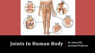
  • 1. Joints In Human Body Dr. Usha (PT) Assistant Professor
  • 2. Introduction Related Terms 1. Arthron (G. a joint). Compare with the terms arthrology, synarthrosis, diarthrosis, arthritis, arthrodesis, etc. 2. Articulatio (L a joint); articulation (NA). 3. Junctura (L a joint). 4. Syndesmology (G. syndesmosis = ligament) is the study of ligaments and related joints.
  • 3. Definition Joint is a junction between two or more bones or cartilages. It is a device to permit movements. However, immovable joints are primarily meant for growth, and may permit moulding during childbirth. There are more joints in a child than in an adult because as growth proceeds some of the bones fuse together, e.g. the ilium, ischium and pubis to form the pelvic bone; the two halves of the infant frontal bone, and of the infant mandible; the five sacral vertebrae and the four coccygeal vertebrae.
  • 4. A. Structural Classification 1. Fibrous Joints Sutures Syndesmosis Gomphosis 2. Cartilaginous Joints Primary cartilaginous joints or synchondrosis Secondary cartilaginous joints or symphysis 3. Synovial Joints Ball-and-socket or spheroidal joints Sellar or saddle joints Condylar or bicondylar joints Ellipsoid joints Hinge joints Pivot or trochoid joints Plane joints
  • 5. B. Functional Classification (According to the Degree of Mobility) 1. • Synarthrosis (immovable), like fibrous joints 2. • Amphiarthrosis (slightly movable), like cartilaginous joints 3. • Diarthrosis (freely movable), like synovial joints
  • 6. Synarthroses Fixed joints at which there is no movement. The articular surfaces are joined by tough fibrous tissue. Often the edges of the bones are dovetailed into one another as in the sutures of the skull.
  • 7. Amphiarthroses Joints at which slight movement is possible. A pad of cartilage lies between the bone surfaces, and there are fibrous ligaments to hold the bones and cartilage in place. The cartilages of such joints also act as shock absorbers, e.g. the intervertebral discs between the bodies of the vertebrae, where the cartilage is strengthened by extra collagen fibres.
  • 8. Diarthroses or Synovial Joints Freely movable joints, though at some of them the movement is restricted by the shape of the articulating surfaces and by the ligaments which hold the bones together. These ligaments are of elastic connective tissue.
  • 9. A synovial joint has a fluid-filled cavity between articular surfaces which are covered by articular cartilage. The fluid, known as synovial fluid, produced by the synovial membrane which lines the cavity except for the actual articular surfaces and covers any ligaments or tendons which pass through the joint. Synovial fluid acts as a lubricant.
  • 10. The form of the articulating surfaces controls the type of movement which takes place at any joint. The movements possible at synovial joints are: 1. Angular 2. Rotatory 3. Gliding
  • 11. • Flexion : decreasing the angle between two bones; • Extension : increasing the angle between two bones; • Abduction : moving the part away from the mid-line; • Adduction : bringing the part towards the mid-line. Angular • Rotation : turning upon an axis; • Circumduction: moving the extremity of the part round in a circle so that the whole part inscribes a cone. Rotary • one part slides on another. Gliding
  • 12. C. Regional Classification 1. • Skull type: immovable. 2. • Vertebral type: slightly movable. 3. • Limb type: freely movable.
  • 13. D. According to No. of Articulating Bones 1. Simple Joints When two bones articulate, e.g. interphalangeal joints 2. Compound Joints More than two bones articulate within one capsule, e.g. elbow joint, wrist joint 3. Complex Joint When joint cavity is divided by an intra-articular disc, e.g., temporomandibula r joint and sternoclavicular joint.
  • 14.
  • 15. 1. FIBROUS JOINTS In fibrous joints the bones are joined by fibrous tissue. These joints are either immovable or permit a slight degree of movement. These can be grouped in the following three subtypes: 1. Sututres 2. Syndesmosis 3. Gomphosis
  • 16. a. Sutures These are peculiar to skull, and are immovable. According to the shape of bony margins, the sutures can be: (i) Plane, e.g. internasal suture (ii) Serrate, e.g. interparietal suture (iii) Squamous, e.g. temporo-parietal suture (iv) Denticulate, e.g. lambdoid suture (v) Schindylesis type, e.g. between rostrum of sphenoid and upper border of vomer.
  • 17.
  • 18. Neonatal skull reveals fontanelles which are temporary in nature. At six specific points on the sutures in new born skull are membrane filled gaps called "fontanelles". These allow the underlying brain to increase in size. Anterior fontanelle is used to judge the hydration of the infant. All these fontanelles become bone by 18 months.
  • 19.
  • 20. b. Syndesmosis The bones are connected by the interosseous ligament. Example: inferior tibiofibular joint
  • 21. c. Gomphosis Also known as- Peg and socket joint. Example: root of the tooth in its bony socket
  • 22. 2. CARTILAGINOUS JOINTS In this type of joints the bones are joined by cartilage. These are of the following two types: 1. Primary cartilaginous joints 2. Secondary cartilaginous joints
  • 23. a. Primary Cartilaginous Joints Synchondrosis, or hyaline cartilage joints The bones are united by a plate of hyaline cartilage so that the joint is immovable and strong. These joints are temporary in nature because after a certain age the cartilaginous plate is replaced by bone (synostosis).
  • 24. Examples: (a) Joint between epiphysis and diaphysis of a growing long bone (b) Spheno-occipital joint (c) First chondrosternal joint (d) Costochondral joints.
  • 25. b. Secondary Cartilaginous Joints Symphyses or fibrocartilaginous joints The articular surfaces are covered by a thin layer of hyaline cartilage, and united by .a disc of fibrocartilage. These joints are permanent and persist throughout life. In this respect symphysis menti is a misnomer as it is a synostosis. Typically the secondary cartilaginous joints occur in the median plane of the body, and permit limited movements due to compressible pad of fibro-cartilage such as in the pubic symphysis and manubriosternal joints.
  • 26. The thickness of fibrocartilage is directly related to the range of movement. Secondary cartilaginous joints may represent an intermediate stage in the evolution of synovial joints. Examples: (a) Symphysis pubis (b) Manubriosternal joint (c) Intervertebral joints between the vertebral bodies
  • 27. 3. SYNOVIAL JOINTS Synovial joints are most evolved, and, therefore, most mobile type of joints Characters 1. The articular surfaces are covered with hyaline (articular) cartilage (fibrocartilage in certain membrane bones). Articular cartilage is avascular, non-nervous and elastic. Lubricated with synovial fluid, the cartilage provides slippery surfaces for free movements, like 'ice on ice'. The surface of the cartilage shows fine undulations filled with synovial fluid.
  • 28.
  • 29. 2. Between the articular surfaces there is a joint cavity filled with synovial fluid. The cavity may be partially or completely subdivided by an articular disc or meniscus. 3. The joint is surrounded by an articular capsule which is made up of a fibrous capsule lined by synovial membrane. Because of its rich nerve supply, the fibrous capsule is sensitive to stretches imposed by movements. This sets up appropriate reflexes to protect the joint from any sprain. This is called the 'watch-dog' action of the capsule.
  • 30. The fibrous capsule is often reinforced by : (a) Capsular or true ligaments representing thickenings of the fibrous capsule (b) The accessory ligaments (distinct from fibrous capsule) which may be intra or extracapsular. The synovial membrane lines whole of the interior of the joint, except for the articular surfaces covered by hyaline cartilage.
  • 31. The membrane secretes a slimy viscous fluid called the synovia or synovial fluid which lubricates the joint and nourishes the articular cartilage. The viscosity of fluid is due to hyaluronic acid secreted by cells of the synovial membrane. 4. Varying degrees of movements are always permitted by the synovial joints.
  • 32. a. Plane Synovial Joints Articular surfaces are more or less flat (plane). They permit gliding movements (translations) in various directions.
  • 33. Examples: (a) Intercarpal joints (b) Intertarsal joints (c) Joints between articular processes of vertebrae (d) Cricothyroid joint (e) Cricoarytenoid joint (f) Superior tibiofibular (g) Interchondral joint (5-9 ribs) (i) Costotransverse and Costovertebral (j) Acromioclavicular with intra- articular disc (k) Carpometacarpal (except first) (1) Tarsometatarsal (m) Intermetacarpal (n) Intermetatarsal (o) Chondrosternal (except first) (p) Sacroiliac
  • 34. b. Hinge Joints (Ginglymi) Articular surfaces are pulley-shaped. There are strong collateral ligaments. Movements are permitted in one plane around a transverse axis. Examples: (a) Elbow joint (b) Ankle joint (c) Interphalangeal joints.
  • 35. c. Pivot (Trochoid) Joints Articular surfaces comprise a central bony pivot (peg) surrounded by an osteoligamentous ring. Movements are permitted in one plane around a vertical axis. Examples: (a) Superior and inferior radio-ulnar joints (b) Median atlanto-axial joint.
  • 36.
  • 37. d. Condylar (Bicondylar) Joints Articular surfaces include two distinct condyles (convex male surfaces) fitting into reciprocally concave female surfaces (which are also, sometimes, known as condyles, such as in tibia). These joints permit movements mainly in one plane around a transverse axis, but partly in another plane (rotation) around a vertical axis.
  • 38. Examples: (a) Knee joint (b) Right and left jaw joints or temporomandibular joint
  • 39. e. Ellipsoid Joints Articular surfaces include an oval, convex, male surface fitting into an elliptical, concave female surface. Free movements are permitted around both the axes, flexion and extension around the transverse axis, and abduction and adduction around the anteroposterior axis. Combination of movements produces circumduction. Typical rotation around a third (vertical) axis does not occur.
  • 40. Examples: (a) Wrist joint (b) Metacarpophalangeal joints (c) Atlanto-occipital joints.
  • 41. f. Saddle (Sellar) Joints Articular surfaces are reciprocally concavoconvex. Movements are similar to those permitted by an ellipsoid joint, with addition of some rotation (conjunct rotation) around a third axis which, however, cannot occur independently.
  • 42. Examples: (a) First carpometacarpal joint (b) Sternoclavicular joint (c) Calcaneocuboid joint (d) Incudomalleolar joint (e) Between femur and patella.
  • 43.
  • 44. g. Ball-and-socket (Spheroidal) Joints Articular surfaces include a globular head (male surface) fitting into a cup-shaped socket (female surface). Movements occur around an indefinite number of axes which have one common centre. Flexion, extension, abduction, adduction, medial rotation, lateral rotation, and circumduction, all occur quite freely.
  • 45. Examples: (a) Shoulder joint (b) Hip joint (Fig. 3.19) (c) Talocalcaneonavicular joint (d) Incudostapedial joint Shoulder joint
  • 46.
  • 47.
  • 48. Classification And Movements Of Synovial Joints
  • 49. 1. Terminology & Definition Human Kinesiology: Study of geometry of surfaces and their associated movements. Male surface: An articulating surface which is larger in surface area and always convex in all directions. Female surface: An articulating surface which is smaller and concave in all directions. Simple joints: Joints with only two articulating surfaces, i.e. male and female. Compound joints: Joint possessing more than one pair of articulating surfaces. Degrees of freedom: Number of axes at which the bone in a joint can move. Uniaxial: Movement of bone at a joint is limited to one axis, i.e. with one degree of freedom, e.g. interphalangeal joints. Biaxial: With two degrees of freedom, e.g. wrist joint. Multi-axial: Three axes along with intermediate positions also, e.g. shoulder joint Translation: Sliding movements of one articulating surface over the other.
  • 50. 2. Movements & Mechanism of Joints A. Angular movement: Movement leading to diminution or increase in angle between two adjoining bones. They are of two types: (a) Flexion and extension: Bending and straightening respectively. (b) Abduction and adduction: Movement away and towards the median plane respectively. Circumduction: When a long bone circumscribes a conical space. B. Rotation: Bone moves around a longitudinal axis. (a) Adjunct rotation: independent rotations, e.g. locking of knee joint. (b) Conjunct rotation: rotations which accompany other movements as in 1st carpometacarpal joint.
  • 51. 3. Shape of Articular Surface The common shapes of the articular surface are: (a) Ovoid: When concave-female ovoids. When convex-male ovoids. (b) Sellar/saddle-shaped: These are convex in one plane, concave in the perpendicular plane.
  • 52. 4. Mechanical Axis of A Bone & Movement of A Bone It is a reference point around which joint mechanics can be studied and around which the most habitual conjunct rotation occurs. Spin: Simple rotation around the bone's stationary mechanical axis. Swing: Any other displacement of the bone and its mechanical axis apart from spin is termed a swing. Swing may be pure or impure (swing + element of spin). Ovoid of motion: This represents the imaginary surface which would include all possible paths of a point on the mechanical axis at some distance from its related joint. Cardinal swing: When the mechanical axis moves in the shortest pathway when bone moves.
  • 53. Arcuate swing: When the mechanical axis moves in the longest pathway along with the bony movement. Co-spin: When the effect of adjunct rotation is additive to the rotation. Anti-spin: Adjunct rotation which has a nullifying effect on rotation. Basic components of movements of the synovial joints are: (1) Spin, (2) Sliding, and (3) Rolling. 1. Spin: It occurs around a fixed mechanical axis.
  • 54. 2. Slide-. During sliding movement, the mechanical axis of the joint and both ends of a moving bone move in the same direction. The transverse axis of movement is not fixed and it undergoes gliding or translation or linear movement. 3. Rolling: In rolling movement, one end of the mechanical axis moves in a particular direction and the other end moves in opposite direction. The transverse axis of movement is almost fixed. The resultant movement is rolling along an arc. Rolling and sliding occur together in knee joint.
  • 55. Joint Positions Close packed position: When the joint surfaces become completely congruent, their area of contact is maximal and they are tightly compressed. In this position fibrous capsule and ligaments are maximally spiralized and tense; no further movement is possible; surfaces cannot be separated by disruptive forces; articular surfaces are liable to trauma
  • 56.
  • 57. Loose packed: All other positions of incongruency. Examples: Least packed position. Shoulder - semiabduction Hip - semiflexion Knee - semiflexion Ankle - plantar flexion.
  • 58. Mechanism of Lubrication of A Synovial Joint 1. Synovial fluid, secreted by synovial membrane, is sticky and viscous due to hyaluronic acid (a mucopolysaccharide). It serves the main function of lubrication of the joint, but also nourishes the articular cartilage. 2. Hyaline cartilage covering the articular surfaces possesses inherent slipperiness, like that of the ice. 3. Intra-articular fibrocartilages, articular discs or menisci, complete or incomplete, help in spreading the synovial fluid throughout the joint cavity, but particularly between the articular surfaces, e.g. temporomandibular joint
  • 59. 4. Haversian fatty pads (Haversian glands) occupy extra spaces in the joint cavity between the incongruous bony surfaces. All of them are covered with synovial membrane, and perhaps function as swabs to spread the synovial fluid. 5. Bursa is a synovial fluid filled bag in relation to joints and bones, to prevent friction. The inflammation of bursa is called bursitis.
  • 60.
  • 61. Blood Supply of Synovial Joints The articular and epiphysial branches given off by the neighbouring arteries form a periarticular arterial plexus. Numerous vessels from this plexus pierce the fibrous capsule and form a rich vascular plexus in the deeper parts of synovial membrane. The blood vessels of the synovial membrane terminate around the articular margins in a fringe of looped anastomoses termed the circulus vasculosus (circulus articularis vasculosus).
  • 62. It supplies capsule, synovial membrane, and the epiphysis. The articular cartilage is avascular. After epiphysial fusion, communications between circulus vasculosus and the end arteries of metaphysis are established, thus minimizing the chances of osteomyelitis in the metaphysis.
  • 63. Nerve Supply of Synovial Joints 1. The capsule and ligaments possess a rich nerve supply, which makes them acutely sensitive to pain. The synovial membrane has a poor nerve supply and is relatively insensitive to pain. The articular cartilage is non-nervous and totally insensitive. Articular nerves contain sensory and autonomic fibres. Some of the sensory fibres are proprioceptive in nature; these are sensitive to position and movement, and are concerned with the reflex control of posture and locomotion. Other sensory fibres are sensitive to pain.
  • 64. Autonomic fibres are vasomotor or vasosensory. The joint pain is often diffuse, and may be associated with nausea, vomiting, slowing of pulse, and fall in blood pressure. The pain commonly causes reflex contraction of muscles which fix the joint in a position of maximum comfort. Like visceral pain, the joint pain is also referred to uninvolved joints.
  • 65. 2. The principles of distribution of nerves to joints were first described by Hilton (1891). Hilton's law states that a motor nerve to the muscle acting on joint tends to give a branch to that joint (capsule) and another branch to the skin covering the joint. Each nerve innervates a specific region of the capsule, and that the part of the capsule which is rendered taut by a given muscle is innervated by the nerve supplying its antagonists. Thus the pattern of innervation is concerned with the maintenance of an efficient stability at the joint.
  • 66. Stability of Synovial Joints 1. Muscles: The tone of different groups of muscles acting on the joint is the most important and indispensable factor in maintaining the stability. Without muscles, the knee and shoulder would be unstable, and arches of the foot would collapse. 2. Ligaments: Are important in preventing any over-movement, and in guarding against sudden accidental stresses. However, they do not help against a continuous strain, because once stretched, they tend to remain elongated. In this respect the elastic ligaments (ligamenta flava and ligaments of the joints of auditory ossicles) are superior to the common type of white fibrous ligaments. 3. Bones: Help in maintaining stability only in firm type of joints, like the hip and ankle. Otherwise in most of the joints (shoulder, knee, sacroiliac, etc.) their role is negligible.