Download as PDF, PPTX


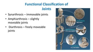
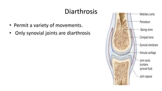
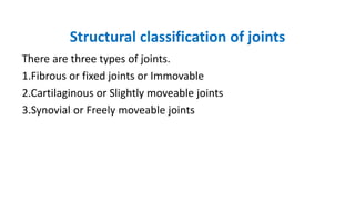
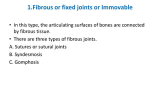
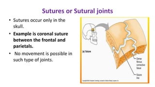
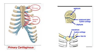
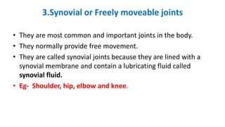
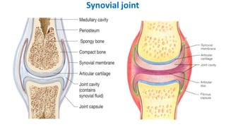
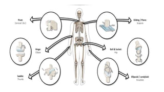
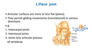
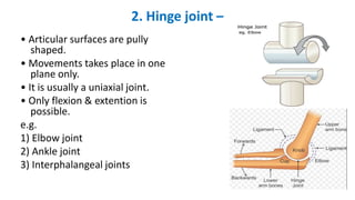
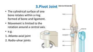
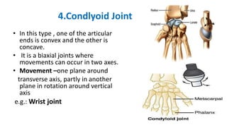
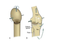
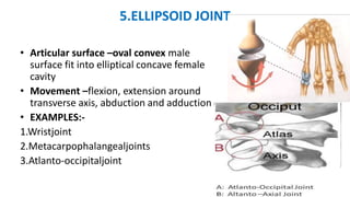
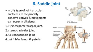
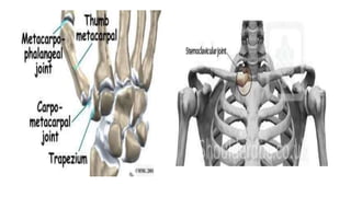
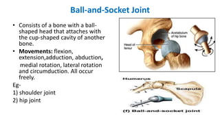


The document classifies and describes the different types of joints in the human body. It begins by defining a joint and discussing the functional classification of joints into synarthrosis (immovable), amphiarthrosis (slightly movable), and diarthrosis (freely movable). It then describes the structural classification of joints into fibrous/fixed joints, cartilaginous joints, and synovial joints. Finally, it provides details on the seven types of synovial joints: plane, hinge, pivot, condyloid, ellipsoid, saddle, and ball-and-socket.


![ONFH[AVN HIP] -TRIPLE REGIME -A NOVAL SURGICAL CONCEPT .pptx](https://cdn.slidesharecdn.com/ss_thumbnails/onfhavnhip2026koaconcalicutdrgokuldevdrmashraf-260210064517-213ec005-thumbnail.jpg?width=640&height=640&fit=bounds)


![CTEV [ clubfoot] DR ARUN LAL ,DR MOHAMED ASHRAF travancore medical college k...](https://cdn.slidesharecdn.com/ss_thumbnails/ctevclubfootdrarunlaldrmohamedashraftravancoremedicalcollegekollamkeralaindia-260208063247-18fc466c-thumbnail.jpg?width=640&height=640&fit=bounds)


