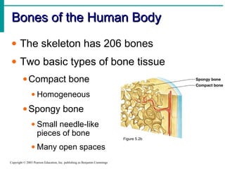
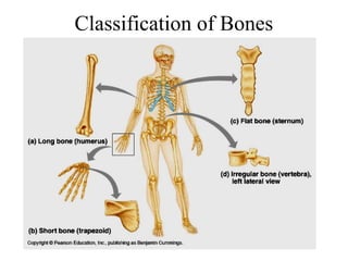
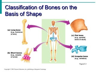
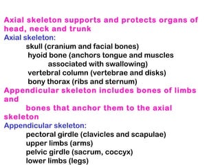
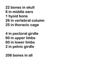
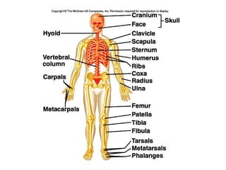
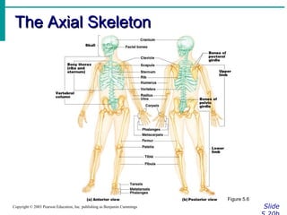
The document discusses the skeletal system and bones. It covers the structure and function of bones, classification of bones, bone tissues, bone cells, bone growth and healing, joints, and common bone diseases. The skeletal system includes bones, cartilage, ligaments, and joints. It is divided into the axial skeleton which includes the skull, vertebral column, ribs, and thoracic cage, and the appendicular skeleton which includes the limbs and their attachments.
Skeletal system. anatomy and physiology of skeletal system. appendicular skel...mamtabisht10
SKELETAL SYSTEM
bones, cartilage and ligaments are tightly joined to form a strong, flexible framework called skeletal system
anatomy and physiology of axial and appendicular skeletal system
Axial Skeleton: The axial skeleton includes the skull, spine, ribs and sternum.
Appendicular Skeleton:
The appendicular skeleton includes the appendages of the body, which are the shoulders, arms, hips, and legs.
all the stages of bone formation described in easiest way possible for better understanding including graphical representation for better understanding. description of each and very thing.
A detail account of Bones, their histological features, classification, composition, Formation, blood and nerve supply, functions, plus some interesting facts about bones.
young bone , blood supply , types of epiphysis, parts of young bone, traction epiphysis, atavastic epiphysis, aberant epiphysis, pressure epiphysis, diaphysis, metaphysis, part of long bone, internal structure of shaft, periosteum, cortex of bone, medullary cavity, epiphysial artery, metaphysial artery, periosteal artery, nutrient artery, arterial supply of short boneperi
1. Unit - 7- Skeleton anatomy by Thiru muruganthiru murugan
The Skeletal System
By Thiru murugan. M
Unit – 7: Anatomy - The Musculoskeletal system:
The Skeletal system
Anatomical positions
Bones: types, structure, growth and ossification
Axial and appendicular skeleton
Joints: classification, major joints and structure
Application and implications in nursing
The Muscular system:
Types and structure of muscles
Muscle groups: muscles of the head, neck, thorax, abdomen, pelvis, upper limb and lower limbs
Principal muscles: deltoid, biceps, triceps, respiratory, abdominal, pelvic floor muscles, gluteal muscles and vastus lateralis
Major muscles involved in nursing procedures
Skeletal system:
The human skeletal system consists of all of the bones, cartilage, tendons, and ligaments in the body
It Provide framework of the body.
Altogether, the skeleton makes up about 20% of a person's body weight. An adult's skeleton contains 206 bones.
It providing support and protection for the internal organs
The skeletal system also provides attachment points for muscles to allow movements at the joints.
Components of skeletal system:
Cartilage: This smooth and flexible substance covers the tips of your bones where they meet. It enables bones to move without friction (rubbing against each other).
Functions of Cartilage:
Model for bone growth in embryo & fetus
Provides a smooth cushion between adjacent bones
Provides firm flexible support (nose, ears, ribs & trachea)
Excellent shock absorber
Ligaments: Bands of strong connective tissue called ligaments hold bones together.
Functions of Ligaments:
Attach bones to bones
Provide stability
Tendons: Tendons are bands of tissue that connect the ends of a muscle to your bone.
Functions of Tendons:
Attach muscles to bones
Anchors muscle to bone for movement
Joints: A joint is where two or more bones in the body come together.
Anatomical position:
Anatomical position, or standard anatomical position, refers to the positioning of the body when it is standing upright and facing forward with each arm hanging on either side of the body, and the palms facing forward. The legs are parallel, with feet flat on the floor and facing forward.
Bones – types, structure, growth and ossification:
Bones:
Bone are specialized forms of strong connective tissue that forms the skeleton of the body.
It is composed of calcium phosphate and calcium carbonate.
It also serves as a storage area for calcium, playing a large role in calcium balance in the blood
The smallest bone in the human body is called the stirrup or stapes bone, located deep inside the ear & The longest bone in the human is called the femur.
Classification or types of bones:
Bones are divided into 5 types.
Long Bone
Short Bone
Flat Bone
Irregular Bone
Sesamoid Bone
1. Long Bone:
A long bone is one that is cylindrical in shape, being longer than it is wide.
Shape of a bone, not its size.
Long bones are found in: Arms (humerus, ulna, radius) & fingers (metacarpals, phalanges) and also Legs (femur, tibia, fibula),
Osteology, derived from the from Greek ὀστέον (ostéon) 'bones', and λόγος (logos) 'study', is the scientific study of bones, practised by osteologists. A subdiscipline of anatomy, anthropology, and paleontology, osteology is the detailed study of the structure of bones, skeletal elements, teeth, microbone morphology, function, disease, pathology, the process of ossification (from cartilaginous molds), and the resistance and hardness of bones (biophysics).[1]
Osteologists frequently work in the public and private sector as consultants for museums, scientists for research laboratories, scientists for medical investigations and/or for companies producing osteological reproductions in an academic context.
Osteology and osteologists should not be confused with osteopathy and its practitioners, osteopaths.
Structure of bone By M Thiru murugan.pptxthiru murugan
Structure of Bone
By,M. Thiru murugan
Structure of bone:
The basic structure of bones is bone matrix, which makes up the underlying rigid framework of bones, composed of both compact bone and spongy bone.
The bone matrix consists of tough protein fibers, mainly collagen, that become hard and rigid due to mineralization with calcium crystals.
Bone matrix is crossed by blood vessels and nerves and also contains specialized bone cells that are actively involved in metabolic processes.
Bone matrix provides bones with their basic structure. Notice the spongy bone in the middle, and the compact bone towards the outer region. The osteon is the functional unit of compact bone.
The microscopic structural unit of compact bone is called an osteon, or Haversian system.
Each osteon is composed of concentric rings of calcified matrix called lamellae (singular = lamella).
Running down the center of each osteon is the central canal, or Haversian canal, which contains blood vessels, nerves, and lymphatic vessels.
These vessels and nerves branch off at right angles through a perforating canal, also known as Volkmann’s canals, to extend to the periosteum and endosteum
Bone Cells: Bones are made of four main kinds of cells:
Osteoblasts
Osteocytes
Osteoclasts
Lining cells.
Osteoblasts: are responsible for making new bone as your body grows.
They also rebuild existing bones when they are broken. To make new bone, many osteoblasts come together in one spot then begin making a flexible material called osteoid.
Minerals are then added to osteoid, making it strong and hard. When osteoblasts are finished making bone, they become either lining cells or osteocytes.
Osteocytes: Mature bone cells are called osteocytes
Osteoclasts: Bone-destroying cells & Break down bone matrix for remodelling and release of calcium
Lining cells: are very flat bone cells.
These cover the outside surface of all bones and are also formed from osteoblasts that have finished creating bone material.
These cells play an important role in controlling the movement of molecules in and out of the bone
Bone Tissues:
Bones consist of different types of tissue, including periosteum, compact bone, spongy bone, and bone marrow.
Periosteum.
Cortical, or Compact Bone.
Cancellous, or Spongy Bone.
Bone Marrow.
1.Periosteum: The periosteum is a tough membrane that covers and protects the outside of the bone.
2.Compact bone: Below the periosteum, compact bone is white, hard, and smooth. It provides structural support and protection.
3.Spongy bone: The core, inner layer of the bone is softer than compact bone. It has small holes called pores to store marrow
4. Bone Marrow: The inside bones are filled with a soft tissue called marrow.
There are 2 types of bone marrow: red and yellow.
Red bone marrow is where all new RBC, WBC, and platelets are produced.
Red bone marrow is found in the center of flat bones such as your scapula and ribs.
Yellow marrow is made mostly of fat and is found in th
If you are Fsc student want to study the basics of support and movement, view this ppt about bones and cartilage. it will be must easier than reading long paragraphs of your textbooks. Also view my next ppts related to this topic.
Instructions for Submissions thorugh G- Classroom.pptxJheel Barad
This presentation provides a briefing on how to upload submissions and documents in Google Classroom. It was prepared as part of an orientation for new Sainik School in-service teacher trainees. As a training officer, my goal is to ensure that you are comfortable and proficient with this essential tool for managing assignments and fostering student engagement.
Palestine last event orientationfvgnh .pptxRaedMohamed3
An EFL lesson about the current events in Palestine. It is intended to be for intermediate students who wish to increase their listening skills through a short lesson in power point.
Honest Reviews of Tim Han LMA Course Program.pptxtimhan337
Personal development courses are widely available today, with each one promising life-changing outcomes. Tim Han’s Life Mastery Achievers (LMA) Course has drawn a lot of interest. In addition to offering my frank assessment of Success Insider’s LMA Course, this piece examines the course’s effects via a variety of Tim Han LMA course reviews and Success Insider comments.
Biological screening of herbal drugs: Introduction and Need for
Phyto-Pharmacological Screening, New Strategies for evaluating
Natural Products, In vitro evaluation techniques for Antioxidants, Antimicrobial and Anticancer drugs. In vivo evaluation techniques
for Anti-inflammatory, Antiulcer, Anticancer, Wound healing, Antidiabetic, Hepatoprotective, Cardio protective, Diuretics and
Antifertility, Toxicity studies as per OECD guidelines
How to Make a Field invisible in Odoo 17Celine George
It is possible to hide or invisible some fields in odoo. Commonly using “invisible” attribute in the field definition to invisible the fields. This slide will show how to make a field invisible in odoo 17.
The Roman Empire A Historical Colossus.pdfkaushalkr1407
The Roman Empire, a vast and enduring power, stands as one of history's most remarkable civilizations, leaving an indelible imprint on the world. It emerged from the Roman Republic, transitioning into an imperial powerhouse under the leadership of Augustus Caesar in 27 BCE. This transformation marked the beginning of an era defined by unprecedented territorial expansion, architectural marvels, and profound cultural influence.
The empire's roots lie in the city of Rome, founded, according to legend, by Romulus in 753 BCE. Over centuries, Rome evolved from a small settlement to a formidable republic, characterized by a complex political system with elected officials and checks on power. However, internal strife, class conflicts, and military ambitions paved the way for the end of the Republic. Julius Caesar’s dictatorship and subsequent assassination in 44 BCE created a power vacuum, leading to a civil war. Octavian, later Augustus, emerged victorious, heralding the Roman Empire’s birth.
Under Augustus, the empire experienced the Pax Romana, a 200-year period of relative peace and stability. Augustus reformed the military, established efficient administrative systems, and initiated grand construction projects. The empire's borders expanded, encompassing territories from Britain to Egypt and from Spain to the Euphrates. Roman legions, renowned for their discipline and engineering prowess, secured and maintained these vast territories, building roads, fortifications, and cities that facilitated control and integration.
The Roman Empire’s society was hierarchical, with a rigid class system. At the top were the patricians, wealthy elites who held significant political power. Below them were the plebeians, free citizens with limited political influence, and the vast numbers of slaves who formed the backbone of the economy. The family unit was central, governed by the paterfamilias, the male head who held absolute authority.
Culturally, the Romans were eclectic, absorbing and adapting elements from the civilizations they encountered, particularly the Greeks. Roman art, literature, and philosophy reflected this synthesis, creating a rich cultural tapestry. Latin, the Roman language, became the lingua franca of the Western world, influencing numerous modern languages.
Roman architecture and engineering achievements were monumental. They perfected the arch, vault, and dome, constructing enduring structures like the Colosseum, Pantheon, and aqueducts. These engineering marvels not only showcased Roman ingenuity but also served practical purposes, from public entertainment to water supply.
Synthetic Fiber Construction in lab .pptxPavel ( NSTU)
Synthetic fiber production is a fascinating and complex field that blends chemistry, engineering, and environmental science. By understanding these aspects, students can gain a comprehensive view of synthetic fiber production, its impact on society and the environment, and the potential for future innovations. Synthetic fibers play a crucial role in modern society, impacting various aspects of daily life, industry, and the environment. ynthetic fibers are integral to modern life, offering a range of benefits from cost-effectiveness and versatility to innovative applications and performance characteristics. While they pose environmental challenges, ongoing research and development aim to create more sustainable and eco-friendly alternatives. Understanding the importance of synthetic fibers helps in appreciating their role in the economy, industry, and daily life, while also emphasizing the need for sustainable practices and innovation.
Acetabularia Information For Class 9 .docxvaibhavrinwa19
Acetabularia acetabulum is a single-celled green alga that in its vegetative state is morphologically differentiated into a basal rhizoid and an axially elongated stalk, which bears whorls of branching hairs. The single diploid nucleus resides in the rhizoid.
5. Introduction
• One of the most remarkable tissues of the
human body
• Far from inert and lifeless, bones are living,
dynamic structures
• Bones serve a wide variety of very diverse
functions within us
• Noted for their strength and resiliency
during life, bones will remain after we are
long gone
10. Skeletal Cartilages
• Initially our skeleton is made up of
cartilages and fibrous membranes
• Gradually our skeletal cartilages are
replaced by bone
• Upon reaching adulthood the skeleton
becomes almost fully ossified
• Only a few cartilages remain in the adult
skeleton
11. Basic structure, type & location
• A skeletal cartilage is made of some variety
of cartilage tissue
• Each type contains a high proportion of
water which makes them resilient
• Cartilage has no nerves or blood supply
• It is surrounded by a dense tissue membrane
called a perichondrium
12. Basic structure, type & location
• There are three types of cartilage tissue:
hyaline, elastic, and fibrocartilage
• Each contains a matrix of jellylike ground
substance and fibers
14. Hyaline cartilages
• The most prevalent type of cartilage
• Its high proportion of collagen fibers give it
flexibility and resilience while providing
support
• Upon examination the tissue appears white,
frosted, and smooth
15. Elastic cartilage
• Elastic cartilage is similar to hyaline
cartilage but with more elastic fibers
• Its elastic fibers enable it to withstand
repeated bending
• Found only in the external ear and the
epiglottis
16. Fibrocartilage
• The tissue contains parallel rows
chondrocytes alternating with collagen
fibers
• Tissue is highly compressible and has great
tensile strength
• Found in thick pad-like structures like the
menisci of the knee or the discs of the
vertebral column
17. Growth of cartilage
• Cartilage grows in two ways
• Appositional growth occurs when cells in
the surrounding perichondrium secrete new
matrix next to existing cartilage tissue
(growth from the outside)
• Interstitial growth occurs when the
chondrocytes within the cartilage divide and
secrete new matrix, expanding the cartilage
(growth from within)
19. Function of Bones
• Support • Bones provide a hard
framework that
supports the body
• Bones provide support
for internal organs
20. Function of Bone
• Protection • Fused bones provide a
brain case that protects
this vital tissue
• Spinal cord is
surrounded by
vertebrae
• Rib cage protects vital
organs
21. Function of Bone
• Movement • Skeletal muscle
attached to bones use
the bones as levers to
move the body
• Arrangement of bones
and joints determine
the movements
possible
22. Function of Bones
• Mineral Storage • Bone serves as a
mineral reservoir
• Phosphate and calcium
ions can be released
into the blood steam
for distribution
• Deposition and
removal are ongoing
23. Function of Bones
• Blood cell formation • Hematopoiesis occurs
within the marrow
cavities of the long
bones
• The majority of
hematopoiesis occurs
in bones
25. Classification of Bone:
• Bones vary in shape and size
• The unique shape of each bone fulfills a
particular need
• Bones are classified by their shape as long,
short, flat, or irregular bone
• Bones differ in the distribution of compact
and spongy osseous tissues
32. Blood Vessels
• Unlike cartilage bone
is well vascularized
• Nutrient arteries
serve the diaphysis
• The nutrient artery
runs inward to supply
the bone marrow and
the spongy bony
33. Medullary cavity
• The interior of all bones
consists largely of spongy
bone
• The very center of the
bone is an open cavity or
marrow cavity
• The cavity is filled with
yellow bone marrow
34. Membranes
• Periosteum covers
outer bone surface
• Consists of dense
irregular connective
tissue & osteoblasts
• Contain nerve fiber
blood and lymph
vessels secured by
Sharpey’s fibers
• Endosteum covers
internal bone surfaces
35.
36. Short, Irregular
and Flat Bones
• Bones consist of thin
layers of compact
bones over spongy
bone
• No shaft, epiphysis or
marrow cavity
• Spongy area between is
a diploe
• Flat sandwich of bone
41. Compact Bone
• Compact bone appears very dense
• It actually contains canals and passageways
that provide access for nerves, blood vessels,
and lymphatic ducts
• The structural unit of compact bone is the
osteon or Haversian system
• Each osteon is an elongated cylinder running
parallel to the long axis of the bone
• Structurally each osteon represents a weight
bearing pillar
43. An Osteon
• Each osteon is a
group of hollow
tubes of bone
matrix
• Each matrix tube is
a lamella
• Collagen fibers in
each layer run in
opposite directions
• Resists torsion
stresses
44. An Osteon
• Running through
the core of each
osteon is the central
or Haversian canal
• The canal contains
small blood vessels
that supply the cells
of the osteon
45. Perforating (Volkmann’s) Canal
• Canals lie at
right angles to
long axis of
bone
• Connect the
vascular supply
of the
periosteum to
those of the
central canal
and medullary
cavity
53. Axial skeleton supports and protects organs of
head, neck and trunk
Axial skeleton:
skull (cranium and facial bones)
hyoid bone (anchors tongue and muscles
associated with swallowing)
vertebral column (vertebrae and disks)
bony thorax (ribs and sternum)
Appendicular skeleton includes bones of limbs
and
bones that anchor them to the axial
skeleton
Appendicular skeleton:
pectoral girdle (clavicles and scapulae)
upper limbs (arms)
pelvic girdle (sacrum, coccyx)
lower limbs (legs)
54. 22 bones in skull
6 in middle ears
1 hyoid bone
26 in vertebral column
25 in thoracic cage
4 in pectoral girdle
60 in upper limbs
60 in lower limbs
2 in pelvic girdle
206 bones in all
81. Bursitis
• Inflammation of the Bursa (fluid
filled sac surrounding the joint).
• A bursa can become inflamed from
injury, infection (rare in the
shoulder), or due to an underlying
rheumatic condition.
• Bursitis is typically identified by
localized pain or swelling,
tenderness, and pain with motion of
the tissues in the affected area.
82.
83.
84. Tendonitis
• Sometimes the tendons become inflamed
for a variety of reasons, and the action of
pulling the muscle becomes irritating. If
the normal smooth gliding motion of your
tendon is impaired, the tendon will
become inflamed and movement will
become painful. This is called
tendonitis, and literally means
inflammation of the tendon.
• The most common cause of tendonitis is
overuse.
85.
86. Carpal Tunnel Syndrome
• Any condition that causes swelling
or a change in position of the tissue
within the carpal tunnel can squeeze
and irritate the median nerve.
Irritation of the median nerve in this
manner causes tingling and
numbness of the thumb, index, and
the middle fingers, a condition
known as "carpal tunnel syndrome."
87.
88.
89. Osteoporosis
• Osteoporosis is a term that means
"porous bones." It is a skeletal disease
affecting women and men. Osteoporosis
is a condition in which bones have lost
minerals especially calcium ム making
them weaker, more brittle, and
susceptible to fractures (broken bones).
Any bone in the body can be affected by
osteoporosis, but the most common
places where fractures occur are the
back (spine), hips, and wrists.
90.
91.
92.
93. Scoliosis
• Scoliosis is an abnormal curvature of the
spine. If your child has scoliosis, the view
from behind may reveal one or more
abnormal curves.Scoliosis runs in
families, but doctors often don't know the
cause. More girls than boys have severe
scoliosis. Adult scoliosis may be a
worsening of a condition that began in
childhood, but wasn't diagnosed or
treated. In other cases, scoliosis may
result from a degenerative joint condition
in the spine.
94.
95.
96.
97.
98. Kyphosis
• With kyphosis, your spine may look
normal or you may develop a hump.
Kyphosis can occur as a result of
developmental problems; degenerative
diseases, such as arthritis of the spine;
osteoporosis with compression fractures
of the vertebrae; or trauma to the spine. It
can affect children, adolescents and
adults.
99.
100.
101. Lordosis
• A normal spine, when viewed from
behind appears straight. However, a
spine affected by lordosis shows
evidence of a curvature of the back
bones (vertebrae) in the lower back
area, giving the child a "swayback"
appearance.
102.
103.
104. Tuberculosis of the
Spine- Pott’s Disease
• As a form of extrapulmonary tuberculosis that impacts the
spine, Pott’s disease has an effect that is sometimes
described as being a sort of arthritis for the vertebrae that
make up the spinal column. More properly known as
tuberculosis spondylitis, Pott’s disease is named after Dr.
Percivall Pott, an eighteenth century surgeon who was
considered an authority in issues related to the back and
spine.Pott's disease is often experienced as a local
phenomenon that begins in the thoracic section of the
spinal column. Early signs of the presence of Pott’s
disease generally begin with back pain that may seem to
be due to simple muscle strain. However, in short order,
the symptoms will begin to multiply.
105.
106.
107.
108. Rickets
• Rickets is the softening and
weakening of bones in children,
usually because of an extreme and
prolonged vitamin D deficiency.
• Some skeletal deformities caused by
rickets may need corrective
surgery.
109.
110.
111.
112. Scurvy
• The human body lacks the ability to
synthesize and make vitamin C and
therefore depends on exogenous dietary
sources to meet vitamin C needs.
Consumption of fruits and vegetables or
diets fortified with vitamin C are essential
to avoid ascorbic acid deficiency. Even
though scurvy is uncommon, it still occurs
and can affect adults and children who
have chronic dietary vitamin C deficiency.
113.
114.
115. Gout
• Gout is a disease that results from an
overload of uric acid in the body. This
overload of uric acid leads to the
formation of tiny crystals of urate that
deposit in tissues of the body, especially
the joints. When crystals form in the joints
it causes recurring attacks of joint
inflammation (arthritis). Chronic gout can
also lead to deposits of hard lumps of uric
acid in and around the joints and may
cause joint destruction, decreased kidney
function, and kidney stones.
116.
117.
118.
119. Acromegaly
• Acromegaly is a serious condition that occurs
when the body produces too much of the
hormones that control growth. ・ The hormone
most often affected is called growth hormone, or
GH. It ハ is produced by the pituitary gland, a tiny
organ at the base of the brain. ・・ Growth
hormone ハ promotes growth of bone, cartilage,
muscle, organs, and other tissues. ・・ When
there is too much growth hormone in the body,
these tissues grow larger than normal. This
excessive growth can cause serious disease and
even premature death.
120.
121.
122.
123.
124. Poliomyelitis
• Poliomyelitis (polio) is a highly infectious disease caused by a
virus. It invades the nervous system, and can cause total
paralysis in a matter of hours. It can strike at any age, but affects
mainly children under three (over 50% of all cases). The virus
enters the body through the mouth and multiplies in the intestine.
Initial symptoms are fever, fatigue, headache, vomiting, stiffness
in the neck and pain in the limbs. One in 200 infections leads to
irreversible paralysis (usually in the legs). Amongst those
paralysed, 5%-10% die when their breathing muscles become
immobilized. Although polio paralysis is the most visible sign of
polio infection, fewer than 1% of polio infections ever result in
paralysis. Poliovirus can spread widely before cases of paralysis
are seen. As most people infected with poliovirus have no signs
of illness, they are never aware they have been infected. After
initial infection with poliovirus, the virus is shed intermittently in
faeces (excrement) for several weeks. During that time, polio can
spread rapidly through the community.
125.
126.
127.
128. Spina Bifida
• Spina bifida is a birth defect that involves
the incomplete development of the spinal
cord or its coverings. The term spina
bifida comes from Latin and literally
means "split" or "open" spine.Spina bifida
occurs at the end of the first month of
pregnancy when the two sides of
the ハ embryo's spine fail to join together,
leaving an open area. In some cases, the
spinal cord or other membranes may
push through this opening in the back.
The condition usually is ハ detected before
a baby is born and treated right away.
129.
130.
131.
132. Talipes Equinovarus-
“Clubfoot”
• Clubfoot is a deformity of the whole foot
that is present at birth. There are several
types of clubfoot that are jointly known as
'talipes', as the deformity is mostly in the
talus (a bone in the ankle). The most
common of the talipes is what is known as
"talipes equino varus" - it is so common
that the word clubfoot is commonly used
to refer to this. In talipes equino varus,
the child is born with the foot pointing
down and twisted inwards at the ankle.
133.
134.
135.
136. Sarcoma
• Osteosarcoma-The most common
type of bone cancer. It arises in
bone and is most commonly found in
children and adolescents but a rare
form occurs in adults, particularly in
patients who have been cured of
other cancers with radiation
therapy.
137.
138.
139.
140.
141.
142. Myeloma
• Multiple myeloma is a cancer in which abnormal
cells collect in the bone marrow and form
tumors. Sometimes these abnormal cells (called
myeloma cells) collect in only one bone and form
a single tumor known as a plasmacytoma.
However, in most cases, the myeloma cells
collect in many bones, forming several tumors
and causing other problems. When this happens,
the disease is called multiple myeloma.
143.
144.
145. Leukemia
• Leukemia is cancer of the blood cells. It starts in
the bone marrow, the soft tissue inside most
bones. Bone marrow is where blood cells are
made.When you are healthy, your bone marrow
makes: ・ White blood cells, which help your body
fight infection. ・ Red blood cells, which carry
oxygen to all parts of your body. ・ Platelets,
which help your blood clot.When you have
leukemia, the bone marrow starts to make a lot
of abnormal white blood cells, called leukemia
cells. They don't do the work of normal white
blood cells, they grow faster than normal cells,
and they don't stop growing when they should.
149. Joints
• Fibrous-Fibrous joints connect bones without
allowing any movement. The bones of your
skull and pelvis are held together by fibrous
joints.
• Cartilaginous-Cartilaginous joints are joints
in which the bones are attached by cartilage.
These joints allow for only a little movement,
such as in the spine or ribs.
• Synovial-Synovial joints allow for much more
movement than cartilaginous joints. Cavities
between bones in synovial joints are filled
with synovial fluid. This fluid helps lubricate
and protect the bones. Bursa sacks contain
the synovial fluid. within fixed limits
150. • A joint, or articulation, is the
place where two bones come
together.
• There are three types of joints
classified by the amount of
movement they allow:
Immovable
slightly movable
freely movable
151. Types of Joints
Hinge- A hinge joint allows extension and
retraction of an appendage. (Elbow,
Knee)
152. Ball and Socket- A ball and socket joint
allows for radial movement in almost
any direction. They are found in the
hips and shoulders. (Hip, Shoulder)
153. Gliding- In a gliding or plane joint bones
slide past each other. Mid-carpal and mid-
tarsal joints are gliding joints. (Hands,
Feet)
154. Saddle- This type of joint occurs when the
touching surfaces of two bones have both
concave and convex regions with the
shapes of the two bones complementing
one other and allowing a wide range of
movement. (Thumb)