Download to read offline

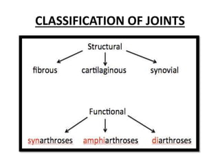
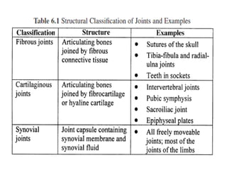


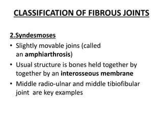


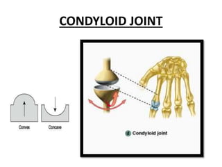


The document discusses different types of joints in the human body. It begins by explaining that joints allow movement between bones and come in many varieties. It then covers three main types of joints - fibrous joints, cartilaginous joints, and synovial joints. Synovial joints are the most mobile and complex. They are further classified into six categories based on their structure and motion: gliding, hinge, pivot, condylar, saddle, and ball and socket. Each joint type allows for specific ranges of motion.


