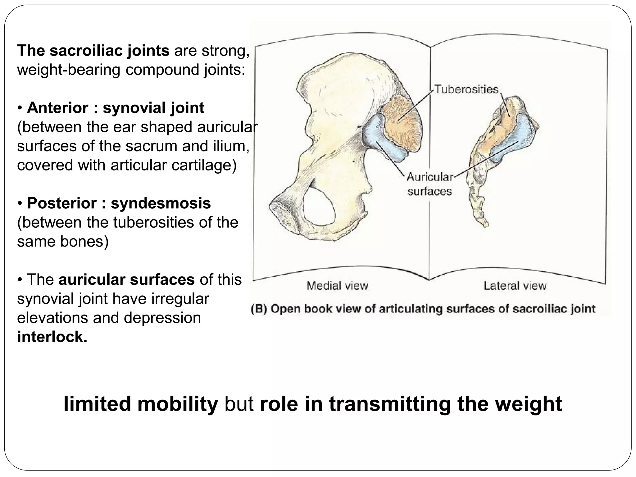
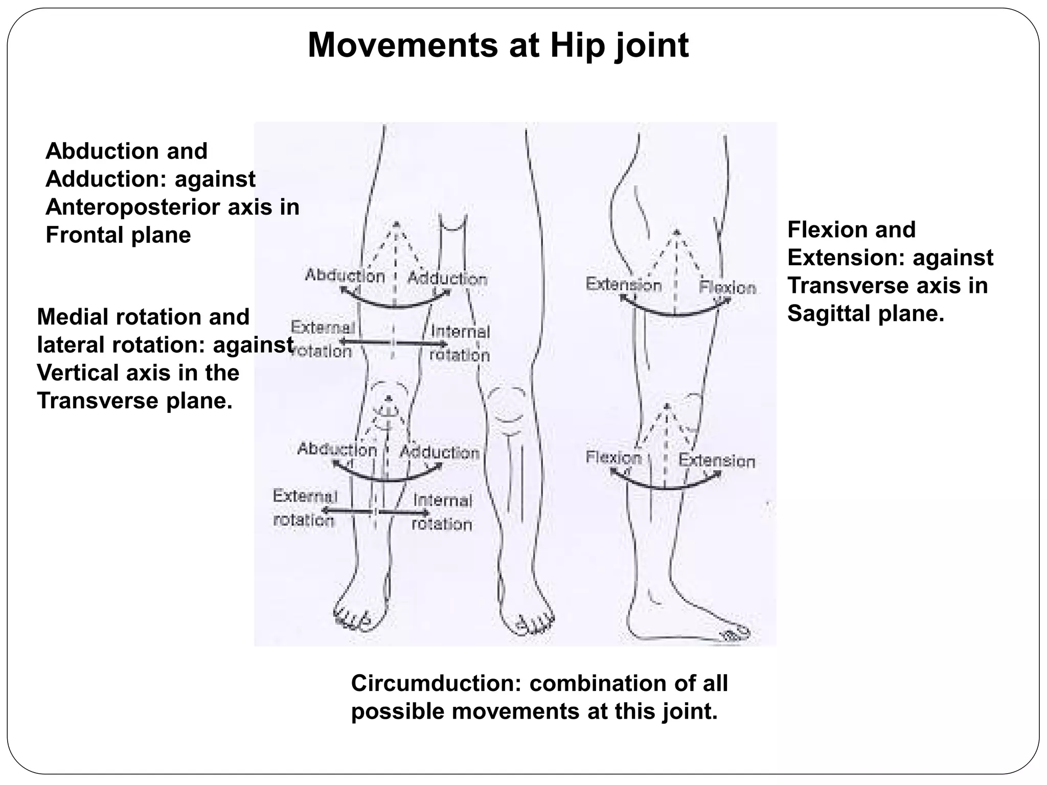
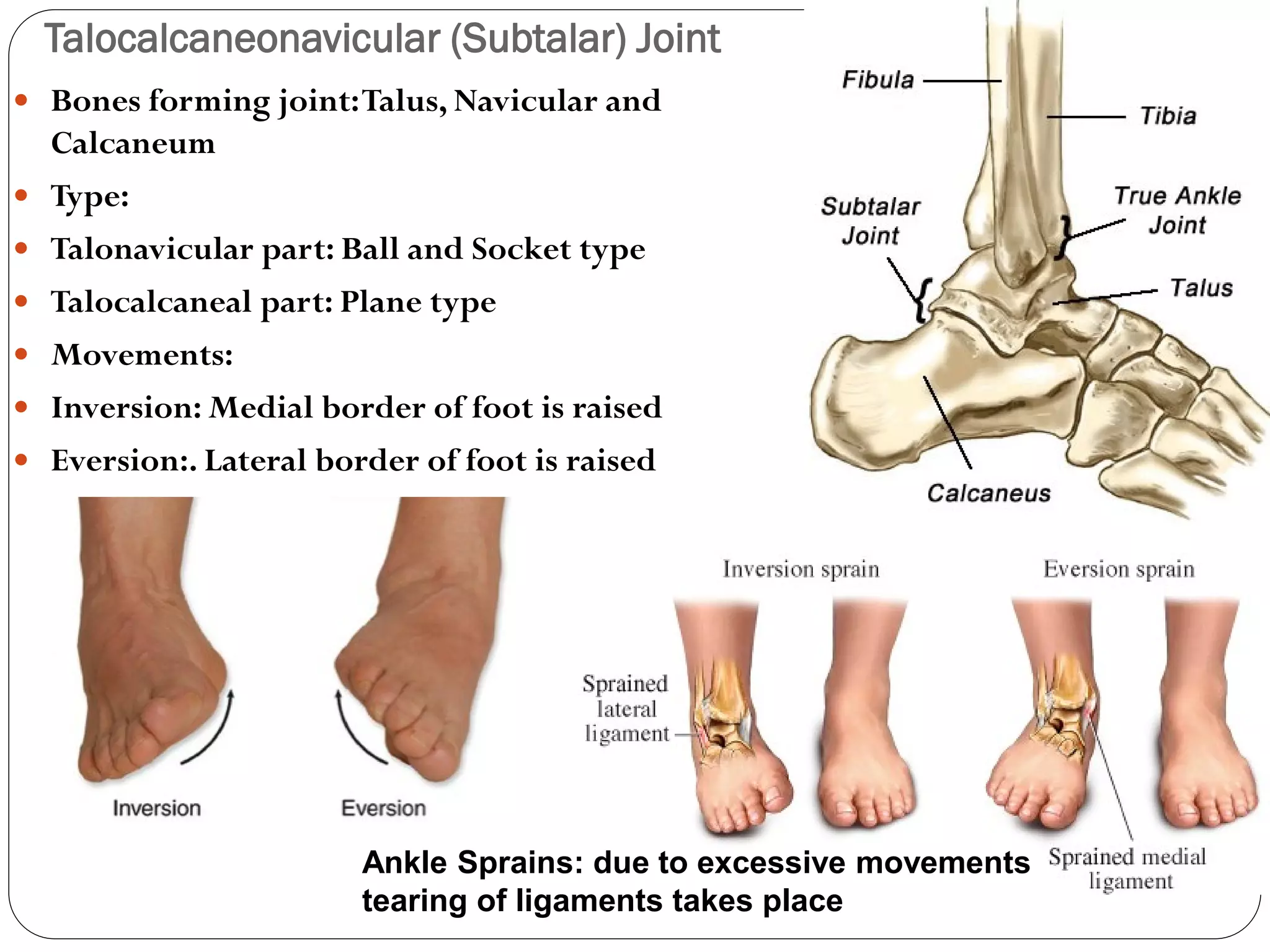
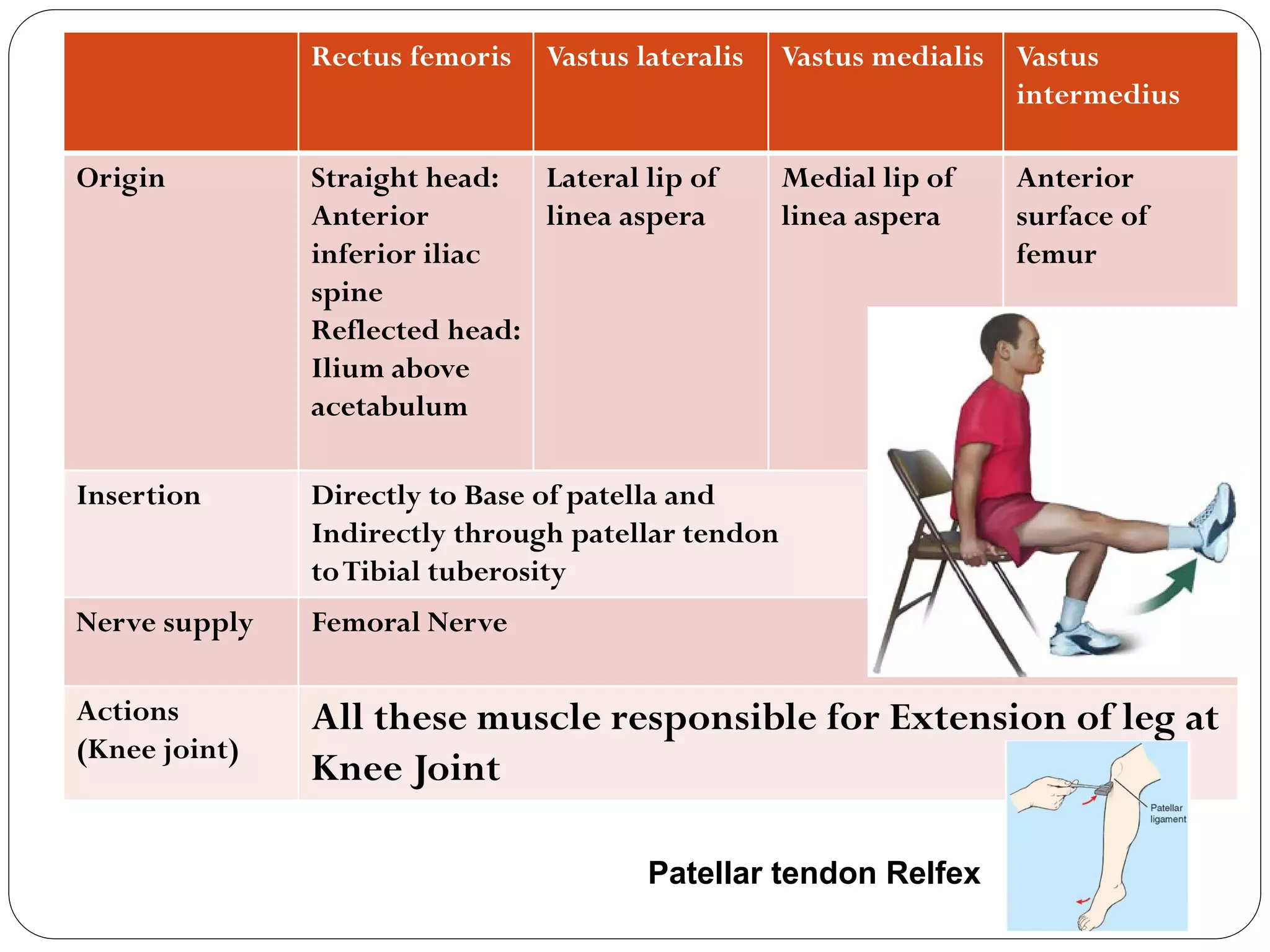
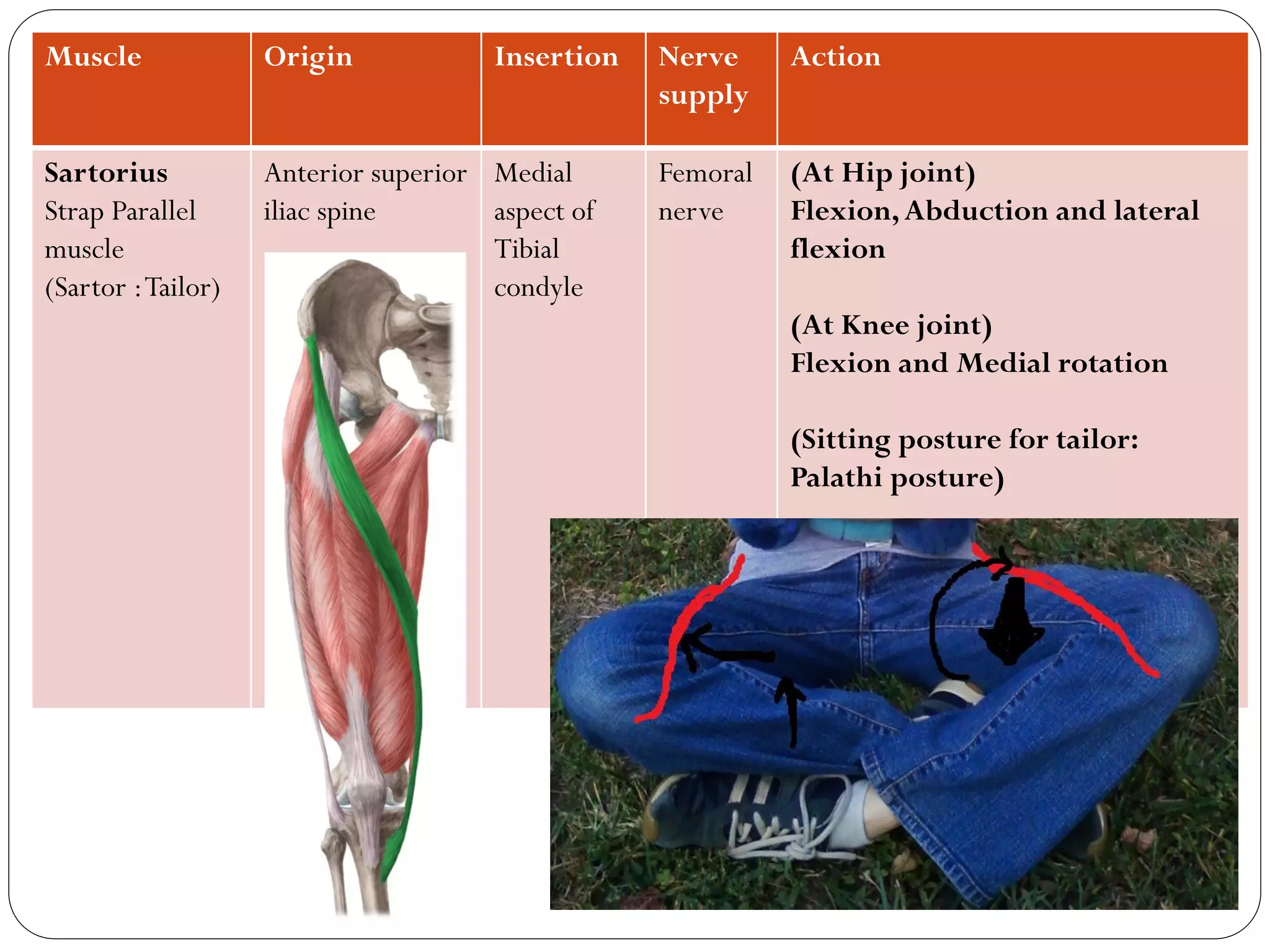
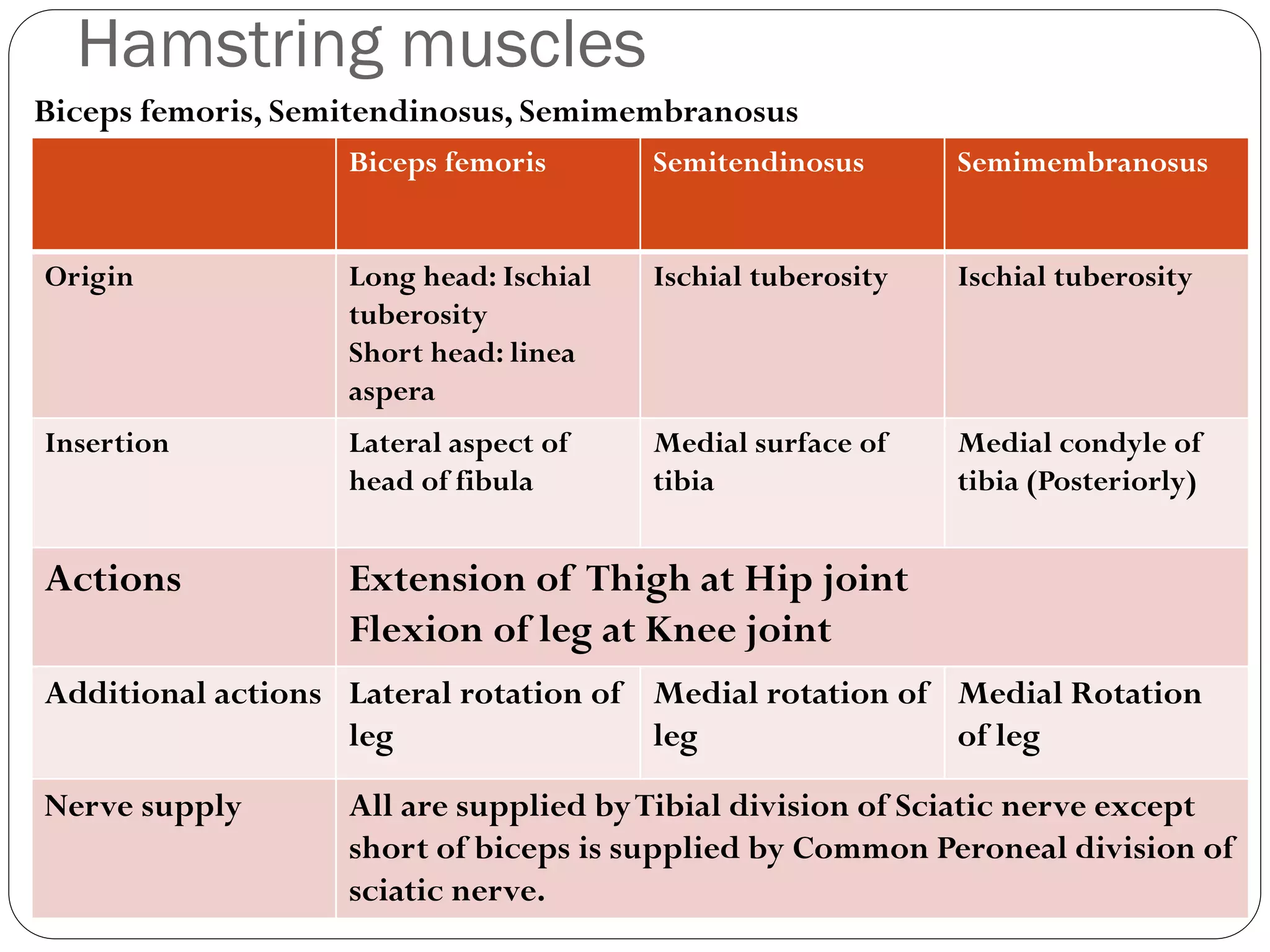
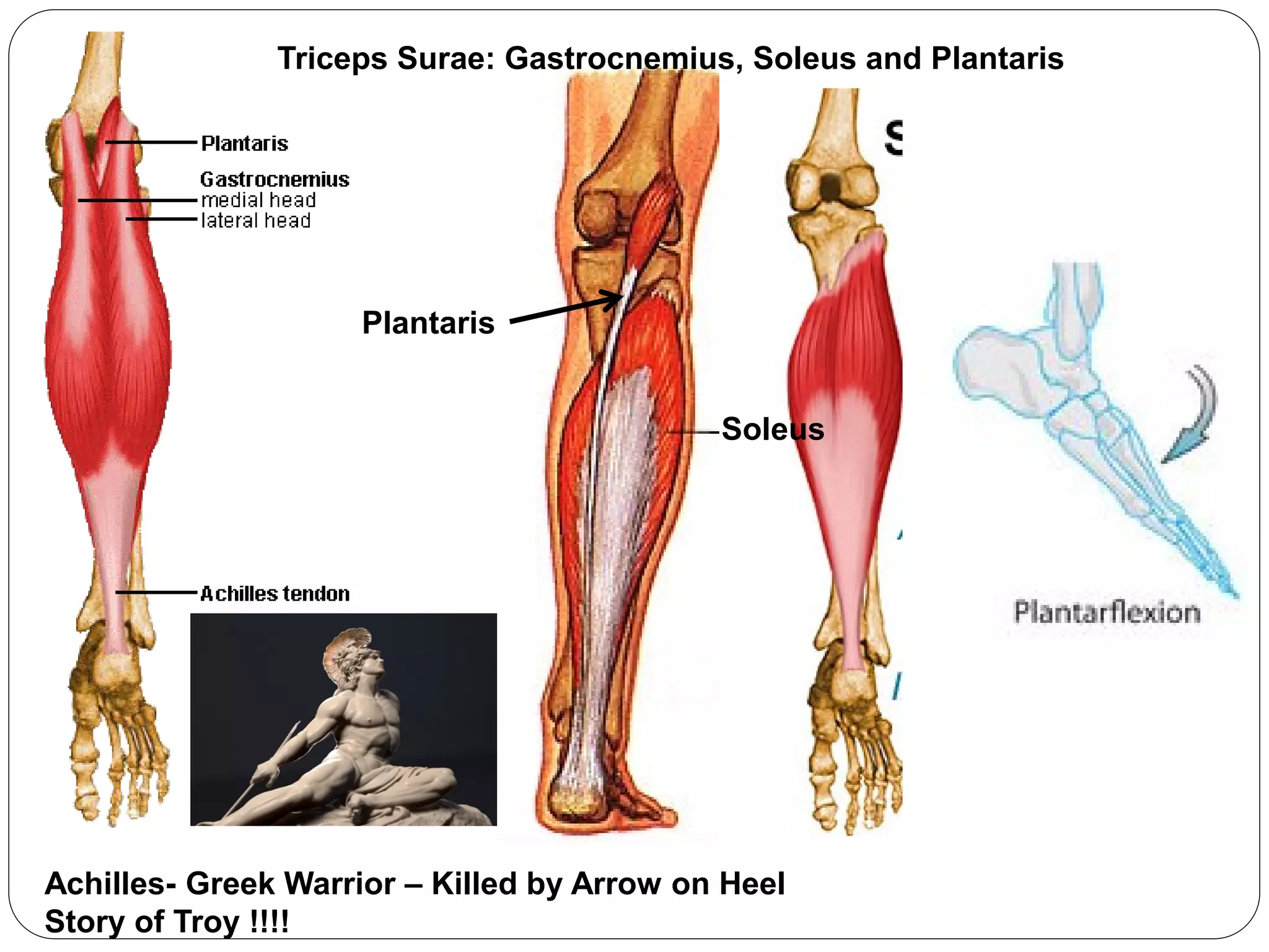
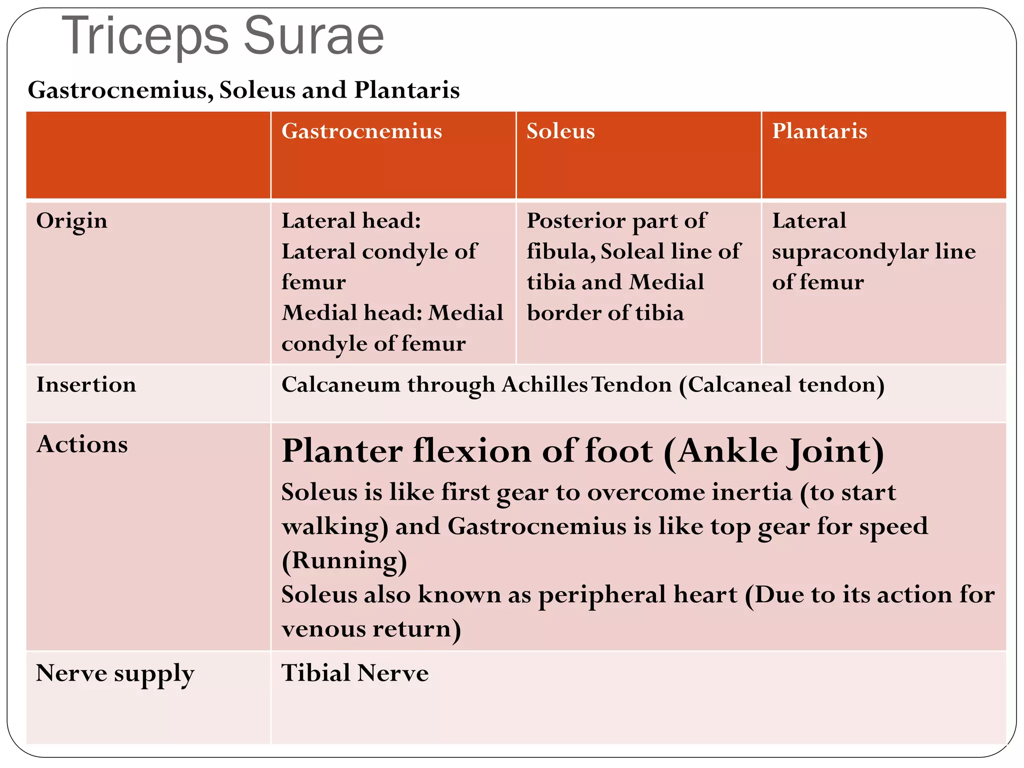
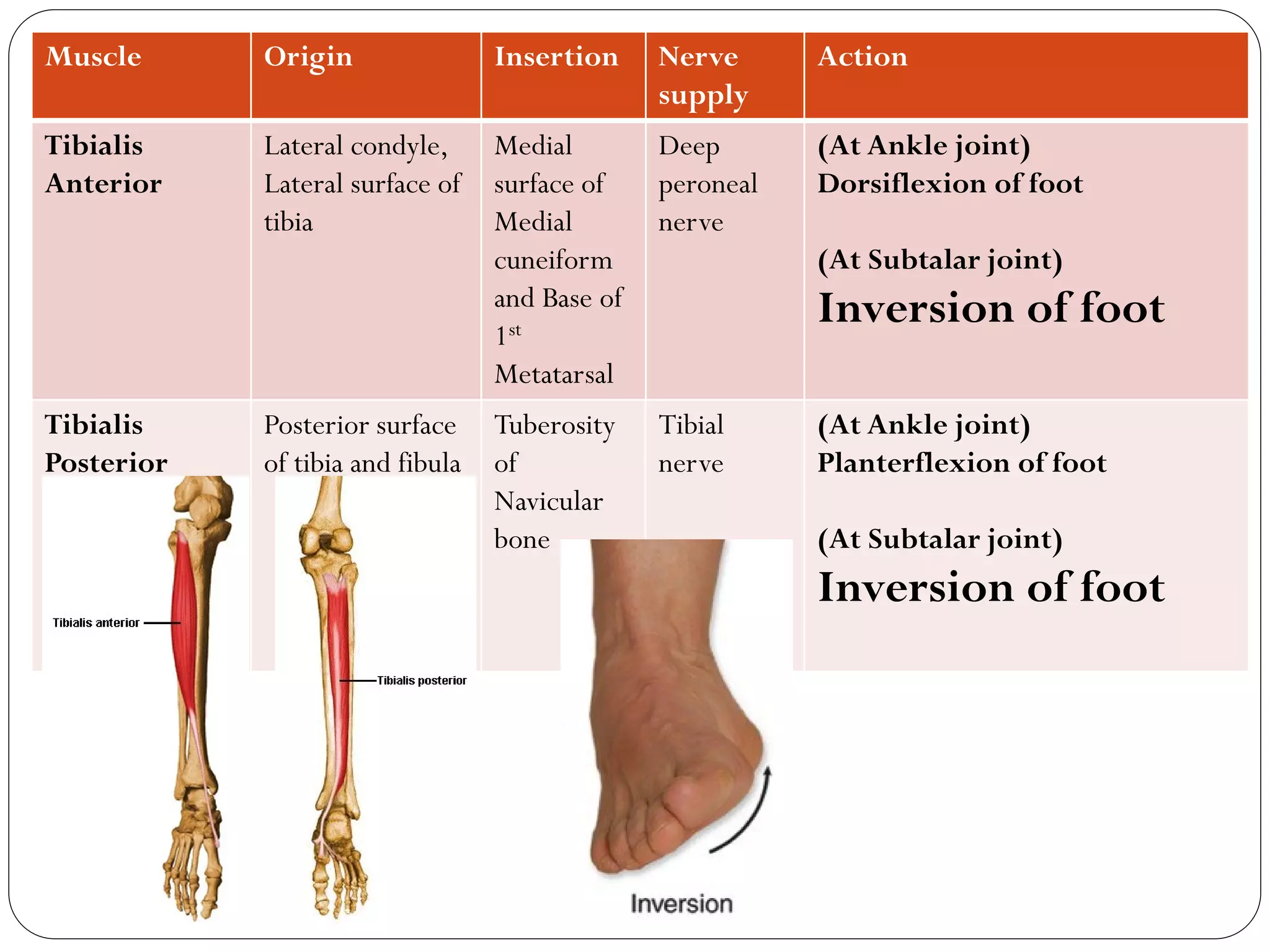
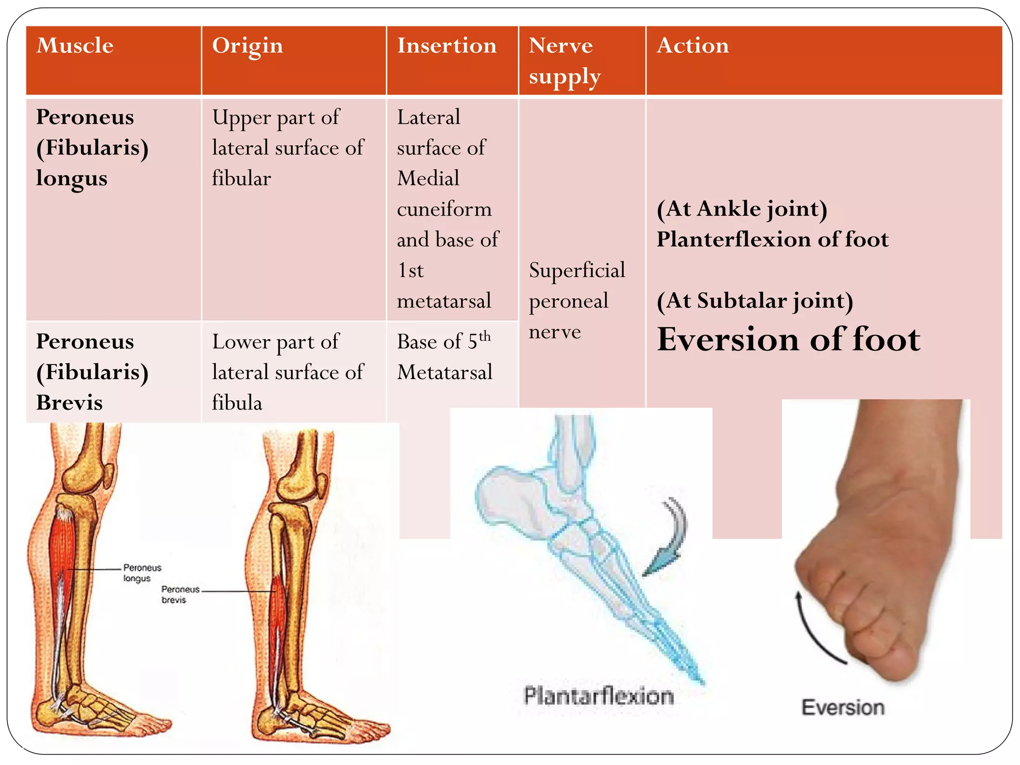
1. HIP REPLACEMENT SURGERY
Fracture-Neck Femur
My Favourite Quote:
I profess to learn and to teach anatomy not from books but from dissections,
not from the tenets of Philosophers but from the fabric of Nature !!!
- William Harvey….Founder of Systemic Circulation.
2. Bones, Joints and Muscles
Dr.B.B.Gosai
Anatomy of Lower limb-Movement Appratus
3. What we will learn today?
Bones of Lower limb: Characteristics and their relation to each other.
Bones forming Pelvis and Movements at Sacroiliac joint
Joints of Lower limb-
Hip joint, Knee andAnkle jonits and overview of small joints
Muscles of Thigh
Muscles of leg
References:
Clinical Oriented Anatomy by Keith Moore : Ch 3 & 5
Atlas of Anatomy by Gilroy: Ch 26
4. Parts of Lower limb
The gluteal region : between the trunk and free
lower limbs.
The femoral region (thigh) is the region lies
between the gluteal, abdominal, and perineal regions
proximally and the knee region distally.
The knee region: includes the prominences
(condyles) of the distal femur and proximal tibia, the
head of the fibula, and the patella.
The leg region is the part that lies between the
knee and ankle.
The ankle includes the medial and lateral
prominences (malleoli) on side of the ankle
(talocrural) joint
The foot is the distal part of the lower limb
containing the tarsus, metatarsus, and phalanges (toe
bones).
5. Hip bone Large, irregularly shaped bones, each of which develops from
the fusion of three bones, the ilium, ischium, and pubis.
All three parts converge at Acetabulum
Join with femur
to form hip joint
Hamstring
muscles
Sartorius
muscle
Rectus
femoris
muscle
Adductor
longus
muscle
6. Bones forming Pelvis: The two hip bones are joined anteriorly at the pubic
symphysis and articulate posteriorly with the sacrum at the sacroiliac joints
to form the pelvic girdle.
7. The sacroiliac joints are strong,
weight-bearing compound joints:
• Anterior : synovial joint
(between the ear shaped auricular
surfaces of the sacrum and ilium,
covered with articular cartilage)
• Posterior : syndesmosis
(between the tuberosities of the
same bones)
• The auricular surfaces of this
synovial joint have irregular
elevations and depression
interlock.
limited mobility but role in transmitting the weight
9. Head join with acetabulum of hip bone to form hip joint
Most common
site of fracture
Femur: Long bone of thigh
Muscular
attachment
Shaft
Condyles join with
condyles of tibia to
form Knee joint
Articulate
with patella
Linea
aspera
10. Condyles join with condyles
of femur to form Knee joint
Tibia and Fibula
Attachment of Patellar
ligament (Tendon of
Quadriceps muscle)
Syndesmosis
Malleoli join with talus to
form Ankle joint
Medial and thicker
Lateral and thinner
11. Bones of Foot:
7 -Tarsals
5 -Metatarsals
14 -Phalanges
Talus (Form Ankle and subtalar joint)
Calcaneum (Subtalar joint)
Cuboid, Navicular, Medial,
Intermediate and Lateral
cuneiform (Other joints of foot)
12. Hip Joint
Type: Ball and socket type of synovial joint
Tiraxial (Multiaxial joint –Movement in three axes and hence 3
degrees of freedom -3 df)
Bones forming joint (Joint partners): Acetabulum of hip bone
(deepened byAcetabular labrum) and Head of Femur.
13. Ligaments of Hip joint:
Acetabular labrum:
fibrocartilaginous ring
deepens the acetabulum to
receive large head of femur.
Pubofemoral ligament:
from pubis to femur
Iliofemorall ligament: (Y
shape) from ilium to femur
(Strongest ligament of body)
Ischiofemoral ligament:
From ischium to femur.
Anterior view
Posterior view
14. Flexion and
Extension: against
Transverse axis in
Sagittal plane.
Circumduction: combination of all
possible movements at this joint.
Movements at Hip joint
Medial rotation and
lateral rotation: against
Vertical axis in the
Transverse plane.
Abduction and
Adduction: against
Anteroposterior axis in
Frontal plane
15. Knee Joint
Type: Hinge type of synovial joint
(Primarily a Hinge type of synovial joint,
allowing flexion and extension; however,
the hinge movements are combined with
gliding and rolling and with rotation about a
vertical axis, hence some text book mention
it as Condyloid joint)
Uniaxial joint –Movement in one axis
(Transverse axis) and hence 1 degrees
of freedom -1 df)
Bones forming joint (Joint partners):
Femoral condyles articulate withTibial
condyles.Anteriorly lower end of femur
also articulate with Patella.
16. Ligaments of Knee joint:
Anteriorly: Patellar tendon
Laterally: Fibular collateral ligament
Medially: Tibial collateral ligament
Within the joint holding femur and tibia
together and form cross like arrangement
are Anterior and Posterior cruciate
ligaments.
Ligaments of Knee joint:
Menisci:
Medial meniscus: semilunar shape
Lateral meniscus: oval/round
shape
Importance:
1. Makes articular surface
congruent (Matching)
2. Divide joint in upper and lower
compartment
17.
18. Movements at Knee joint:
Flexion and Extension: against the transverse
axis in sagittal plane.
Some rotation can occur in flexed leg.
Locking of Knee: During fully extended knee, the knee passively “locks” because
of medial rotation of the femoral condyles on the tibial plateau (the “screw-home
mechanism”). This position makes the lower limb a solid column and more adapted
for weight-bearing
Unlocking of Knee: Reverse of locking and initiated by Popliteus muscle
20. Tibio-Fibular joints:
Superior Tibio-fibular joint: Plane synovial
joint
Middel tibio-fibular and Inferior Tibio-fibular
joint: Syndesmosis
Movements: Sliding/Gliding
Fibula is spare bone in lower limb as most
of the weight transmission occur through
tibia only.
Hence Fibula is taken for bone grafting in
severe fractures at other places.
21. Talocrural (Ankle) Joint
Type: Hinge type of synovial joint
Uniaxial joint –Movement in one axis
(Transverse axis) and hence 1 degrees
of freedom -1 df)
Bones forming joint (Joint partners):
Inferior end of tibia, Medial Malleolus ,
Lateral Malleolus and Body of talus.
Ligaments of Ankle:
Deltoid ligament (Medially)
Talo-fibular and calcaneo-fibular ligament
(Laterally).
Movements at Ankle joint:
Planterflexion and Dorsiflexion
(The transverse axis in sagittal plane )
Malleolar fracture
22.
23. Talocalcaneonavicular (Subtalar) Joint
Bones forming joint:Talus, Navicular and
Calcaneum
Type:
Talonavicular part: Ball and Socket type
Talocalcaneal part: Plane type
Movements:
Inversion: Medial border of foot is raised
Eversion:. Lateral border of foot is raised
Ankle Sprains: due to excessive movements
tearing of ligaments takes place
25. Rectus femoris Vastus lateralis Vastus medialis Vastus
intermedius
Origin Straight head:
Anterior
inferior iliac
spine
Reflected head:
Ilium above
acetabulum
Lateral lip of
linea aspera
Medial lip of
linea aspera
Anterior
surface of
femur
Insertion Directly to Base of patella and
Indirectly through patellar tendon
toTibial tuberosity
Nerve supply Femoral Nerve
Actions
(Knee joint)
All these muscle responsible for Extension of leg at
Knee Joint
Patellar tendon Relfex
26. Muscle Origin Insertion Nerve
supply
Action
Sartorius
Strap Parallel
muscle
(Sartor :Tailor)
Anterior superior
iliac spine
Medial
aspect of
Tibial
condyle
Femoral
nerve
(At Hip joint)
Flexion,Abduction and lateral
flexion
(At Knee joint)
Flexion and Medial rotation
(Sitting posture for tailor:
Palathi posture)
27. Muscle Origin Insertion Nerve
supply
Action
Adductor
longus
Body of Pubis
(Hip bone)
Middle
third of
linea aspera
Obturator
nerve
(At Hip joint)
Adduction of thigh
Forms boundary of Femoral
triangle
28.
29. Hamstring muscles
Biceps femoris, Semitendinosus, Semimembranosus
Biceps femoris Semitendinosus Semimembranosus
Origin Long head: Ischial
tuberosity
Short head: linea
aspera
Ischial tuberosity Ischial tuberosity
Insertion Lateral aspect of
head of fibula
Medial surface of
tibia
Medial condyle of
tibia (Posteriorly)
Actions Extension of Thigh at Hip joint
Flexion of leg at Knee joint
Additional actions Lateral rotation of
leg
Medial rotation of
leg
Medial Rotation
of leg
Nerve supply All are supplied byTibial division of Sciatic nerve except
short of biceps is supplied by Common Peroneal division of
sciatic nerve.
32. Triceps Surae
Gastrocnemius, Soleus and Plantaris
Gastrocnemius Soleus Plantaris
Origin Lateral head:
Lateral condyle of
femur
Medial head: Medial
condyle of femur
Posterior part of
fibula, Soleal line of
tibia and Medial
border of tibia
Lateral
supracondylar line
of femur
Insertion Calcaneum through AchillesTendon (Calcaneal tendon)
Actions Planter flexion of foot (Ankle Joint)
Soleus is like first gear to overcome inertia (to start
walking) and Gastrocnemius is like top gear for speed
(Running)
Soleus also known as peripheral heart (Due to its action for
venous return)
Nerve supply Tibial Nerve
33. Muscle Origin Insertion Nerve
supply
Action
Tibialis
Anterior
Lateral condyle,
Lateral surface of
tibia
Medial
surface of
Medial
cuneiform
and Base of
1st
Metatarsal
Deep
peroneal
nerve
(At Ankle joint)
Dorsiflexion of foot
(At Subtalar joint)
Inversion of foot
Tibialis
Posterior
Posterior surface
of tibia and fibula
Tuberosity
of
Navicular
bone
Tibial
nerve
(At Ankle joint)
Planterflexion of foot
(At Subtalar joint)
Inversion of foot
34. Muscle Origin Insertion Nerve
supply
Action
Peroneus
(Fibularis)
longus
Upper part of
lateral surface of
fibular
Lateral
surface of
Medial
cuneiform
and base of
1st
metatarsal
Superficial
peroneal
nerve
(At Ankle joint)
Planterflexion of foot
(At Subtalar joint)
Eversion of footPeroneus
(Fibularis)
Brevis
Lower part of
lateral surface of
fibula
Base of 5th
Metatarsal