UTERUS_Nursing.pptx
•Download as PPTX, PDF•
0 likes•427 views

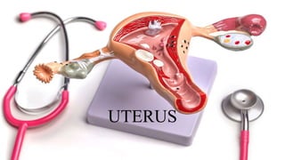
Once the egg has left the ovary it can be fertilized and implant itself in the lining of the uterus. The main function of the uterus is to nourish the developing fetus prior to birth.

Recommended
More Related Content
What's hot
What's hot (20)
Similar to UTERUS_Nursing.pptx
Similar to UTERUS_Nursing.pptx (20)
More from ABHIJIT BHOYAR
More from ABHIJIT BHOYAR (20)
Recently uploaded
Recently uploaded (20)
UTERUS_Nursing.pptx
- 1. UTERUS
- 2. INTRODUCTION • The uterus is a hollow muscular organ located in the female pelvis between the bladder and rectum • In layman’s language, the uterus is called the womb. • It is also called hystera, on which word hysterectomy is based.
- 3. DEFINITION • Uterus is a child-bearing organ in females, situated in the pelvis between bladder and rectum. Though hollow it is thick walled and firm, and can be palpated bimanually during a PV (per vaginal) examination.
- 4. Conti.. • It is the organ which protects and provides nutrition to a fertilized ovum, enabling it to grow into a fully formed fetus. • At the time of childbirth or parturition, contractions of muscle in the wall of the organ result in expulsion of the fetus from the uterus.
- 5. SIZE AND SHAPE • The uterus is pyriform in shape. • It is about 7.5 cm long, 5 cm broad, and 2.5 cm thick. It weighs 30 to 40 grams
- 6. Parts of Uterus A fundus Body with two surfaces, anterior or vesicles and posterior or intestinal Two lateral borders Cervix The uterus comprises:
- 7. Conti.. • It is divisible into an upper expanded part called the body and a lower cylindrical part called the cervix. • The junction of these two parts is marked by a circular constriction called the isthmus. • Part of uterus above the opening of fallopian tube is called the fundus. • The body forms the upper two-thirds of the organ, and the cervix forms the lower one- third
- 8. Fundus • The fundus is formed by the free upper end of the uterus. • Fundus lies above the openings of the uterine tubes. • It is convex like a dome. • It is covered with peritoneum and is directed forward when the bladder is empty. • The fertilized oocyte is usually implanted in the posterior wall of the fundus or upper part of body of uterus
- 9. Conti.. • The anterior or vesical surface of the body is flat and related to the urinary bladder. • It is covered with peritoneum and forms the posterior or superior wall of the uterovesical pouch. • The posterior or intestinal surface is convex and is related to coils of the terminal ileum and to the sigmoid colon. • It is covered with peritoneum and forms the anterior wall of the rectouterine pouch
- 10. Conti.. • Each lateral border is rounded and convex. • It provides attachment to the broad ligament of the uterus which connects it to the lateral pelvic wall. • The uterine tube opens into the uterus at the upper end of this border. • This end of the border gives attachment to the round ligament of the uterus, antero-inferior to the tube; and to the ligament of the ovary postero-inferior to the tube. • The uterine artery ascends along the lateral border of the uterus between the two layers of the broad ligament
- 11. Conti.. • In sagittal section, the cavity of the body of the uterus is seen as a mere slit because the uterus is compressed anteroposteriorly. • In coronal section, the cavity is seen to be triangular in shape, the apex being directed downwards. At the apex, the cavity becomes continuous with the canal of the cervix. The junction is called the internal os. • The superolateral angles of the cavity receive the openings of the right and left uterine tubes
- 12. Layers of uterus • uterus consists of three layers: • Perimetrium: The outermost, protective layer. • Myometrium: The highly muscular middle layer. This is what expands during pregnancy and contracts to push your baby out. • Endometrium: The inner layer or lining of your uterus (uterine lining). This layer of your uterus is shed during your menstrual cycle.
- 13. LIGAMENTS OF UTERUS Peritoneal Ligaments 1. The anterior ligament consists of the uterovesical fold of peritoneum. 2. The posterior ligament consists of the rectovaginal fold forming rectovaginal pouch of peritoneum 3. The right and left broad ligaments are folds of peritoneum Fibromuscular Ligaments 1. Round ligaments of the uterus 2. Transverse cervical ligaments 3. Uterosacral ligaments
- 16. VENUS SUPPLY OF UTERUS
- 17. SUPPORTS OF THE UTERUS • The uterus is a mobile organ which undergoes extensive changes in size and shape during the reproductive period of life. • It is supported and prevented from sagging down by a number of factors which are chiefly muscular and fibromuscular.
- 18. Primary Supports Muscular or active supports 1. Pelvic diaphragm 2. Perineal body 3. Distal urethral sphincter mechanism Fibromuscular or mechanical supports 1. Uterine axis 2. Pubocervical ligaments 3. Transverse cervical ligaments of Mackenrodt 4. Uterosacral ligaments 5. Round ligaments of uterus
- 19. Secondary Supports These are of doubtful value and are formed by peritoneal ligaments. 1. Broad ligaments 2. Vesicouterine pouch and fold of peritoneum 3. Rectovaginal or rectouterine pouch and fold of peritoneum
- 21. Pelvic Diaphragm • The pelvic diaphragmsupports the pelvic viscera and resists any rise in the intra- abdominal pressure. • The pubococcygeus part of the levator ani is partly inserted into the perineal body between the vagina and the rectum. • Some of these fibres also form a supporting sling and a sphincter for the vagina, and so indirectly for the uterus and the urinary bladder.
- 22. Conti.. • If the pubococcygeus is torn during parturition, the support to the vagina is lost, and the latter tends to sink into the vestibule along with the uterus, thus causing prolapse of the uterus. • The efficacy of the levator ani as a support is also lost when the perineal muscles are torn. They normally fix the perineal body, and make it an anchor for the levator ani
- 23. Perineal Body • It is a fibromuscular node to which ten muscles are attached. • It acts as an anchor for the pelvic diaphragm, and thus maintains the integrity of the pelvic floor. • The muscles are two superficial transversus perinei, two deep transversus perinei, two pubococcygeus part of levator ani, two bulbospongiosus and single sphincter ani externus and unstriped fibres of longitudinal muscle coat of the anal canal
- 24. Urethral Sphincter Mechanism • The urogenital diaphragm does not exist as sphincter urethrae is within the wall of the urethra. • The urethral sphincter mechanism exists. • In addition, there are compressor urethrae and sphincter urethrae vaginalis. • Since these are inserted into vagina, they support the uterus indirectly
- 25. Uterine Axis • The anteverted position of the uterus itself prevents the organ from sagging down through the vagina. • Any rise in intra-abdominal pressure tends to push the uterus against the bladder and pubic symphysis, which further accentuates anteversion. • The angle of anteversion is maintained by the uterosacral and round ligaments
- 26. Pubocervical Ligaments • These ligaments connect the cervix to the posterior surface of pubis. • They are derived from the endopelvic fascia, and correspond to the medial and lateral puboprostatic ligaments in the male
- 27. Transverse Cervical Ligaments of Mackenrodt • These are also known by various other names like lateral cervical ligaments; cardinal ligaments; paracervical ligaments; retinacula uterine sustentaculum of Bonny
- 28. Uterosacral Ligaments • These are also condensations of the endopelvic fascia. They connect the cervix to the periosteum of the sacrum and are enclosed within rectouterine folds ofperitoneum (which form the lateral boundaries of the rectouterine pouch). • The uterosacral ligaments keep the cervix braced backwards against the forward pull of the round ligaments. The two ligaments form a couple that maintains the uterine axis
- 29. Round Ligaments of Uterus • The round ligaments are two fibromuscular flat bands, 10 to 12 cm long, which lie between the two layers of the broad ligament, anteroinferior to the uterine tube. • Each ligament begins at the lateral angle of the uterus, runs forwards and laterally, passes through the deep inguinal ring, traverses the inguinal canal and merges with the areolar tissue of the labium majus after breaking up into thin filaments.
- 30. Function What does a uterus do? • The three main jobs of your uterus are: • Pregnancy: Your uterus stretches to grow your baby during pregnancy. It can also contract to help push your baby out of your vagina. • Fertility: Your uterus is where a fertilized egg implants during conception and where your baby grows. • Menstrual cycle: Your uterine lining is where blood and tissue come from during menstruation.
- 31. What are the common conditions of the uterus? • Uterine fibroids: Small, noncancerous tumors in uterus. • Uterine polyps: Growths in the endometrial lining of uterus. • Uterine cancer: Cancer of your uterus, such as endometrial cancer or uterine sarcoma. • Endometriosis: A condition when uterine lining grows in places other than uterus. • Pelvic inflammatory disease: An infection of reproductive organs. • Uterine prolapse: A condition where uterus slips out of position. • Infertility: The inability to get pregnant.