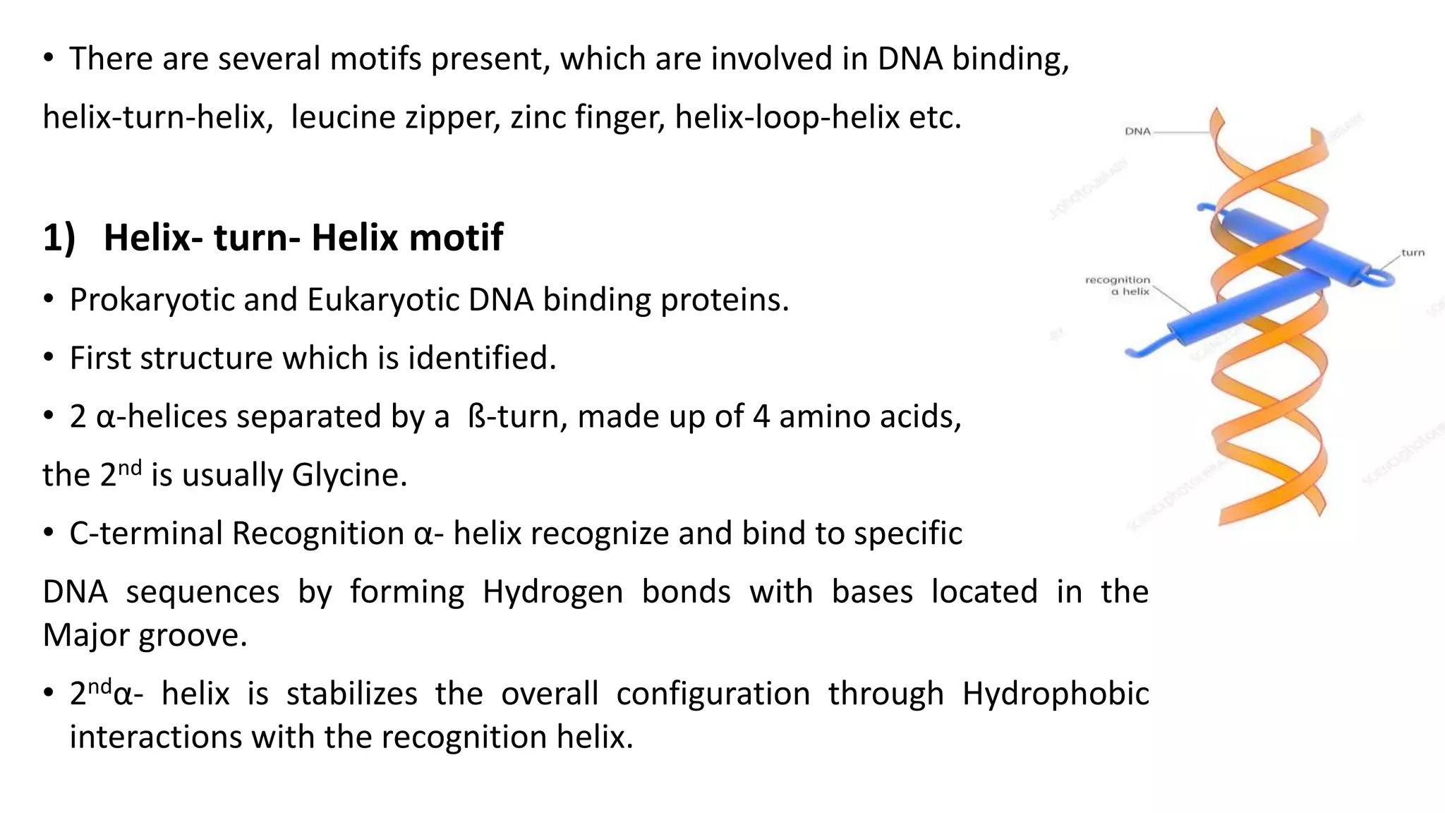
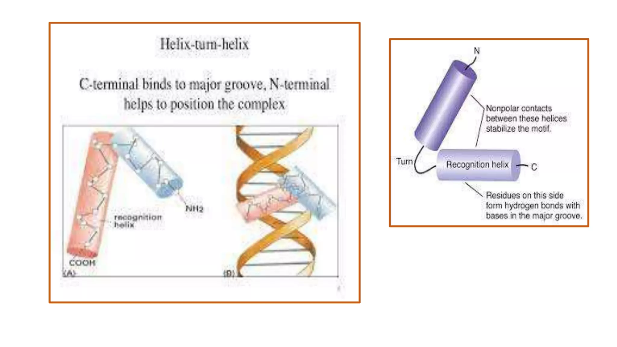
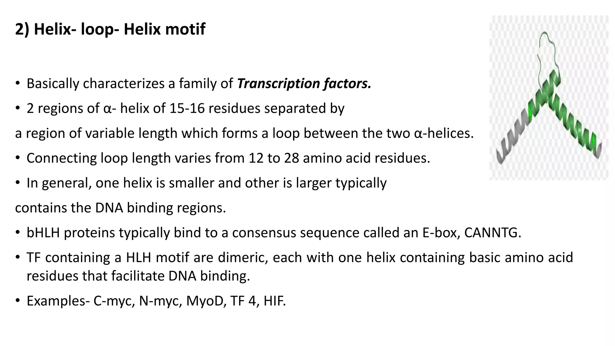
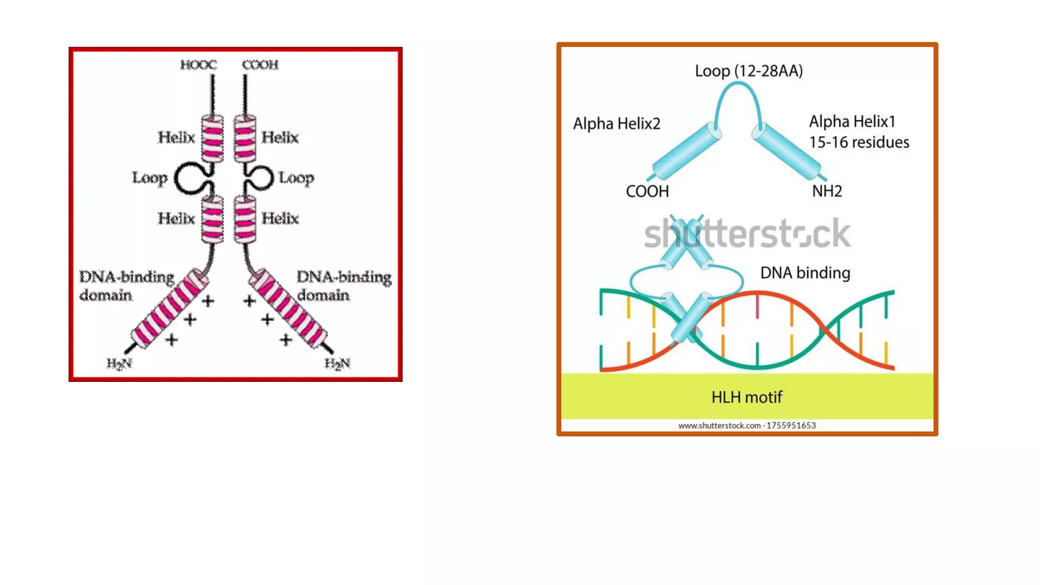
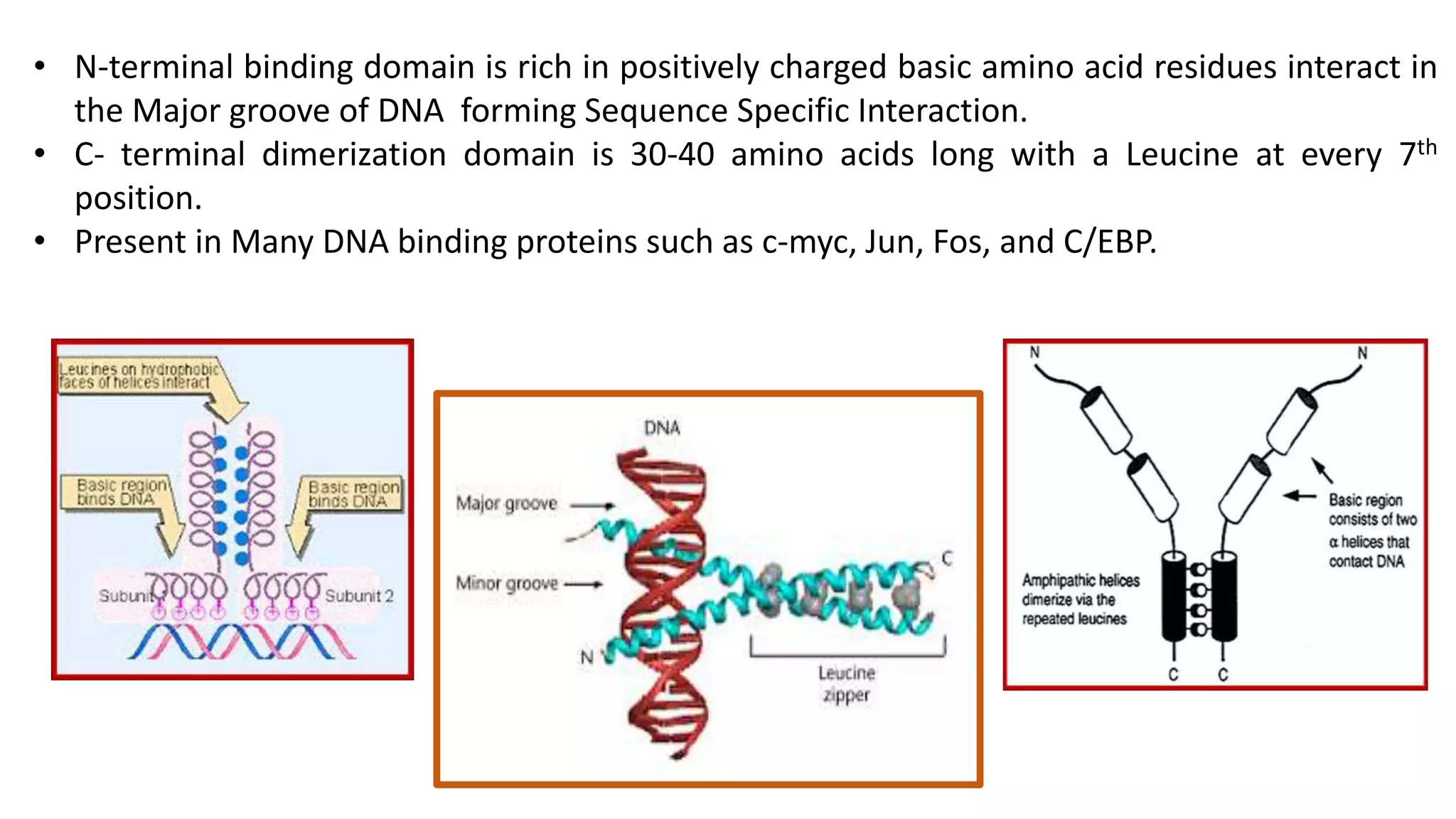
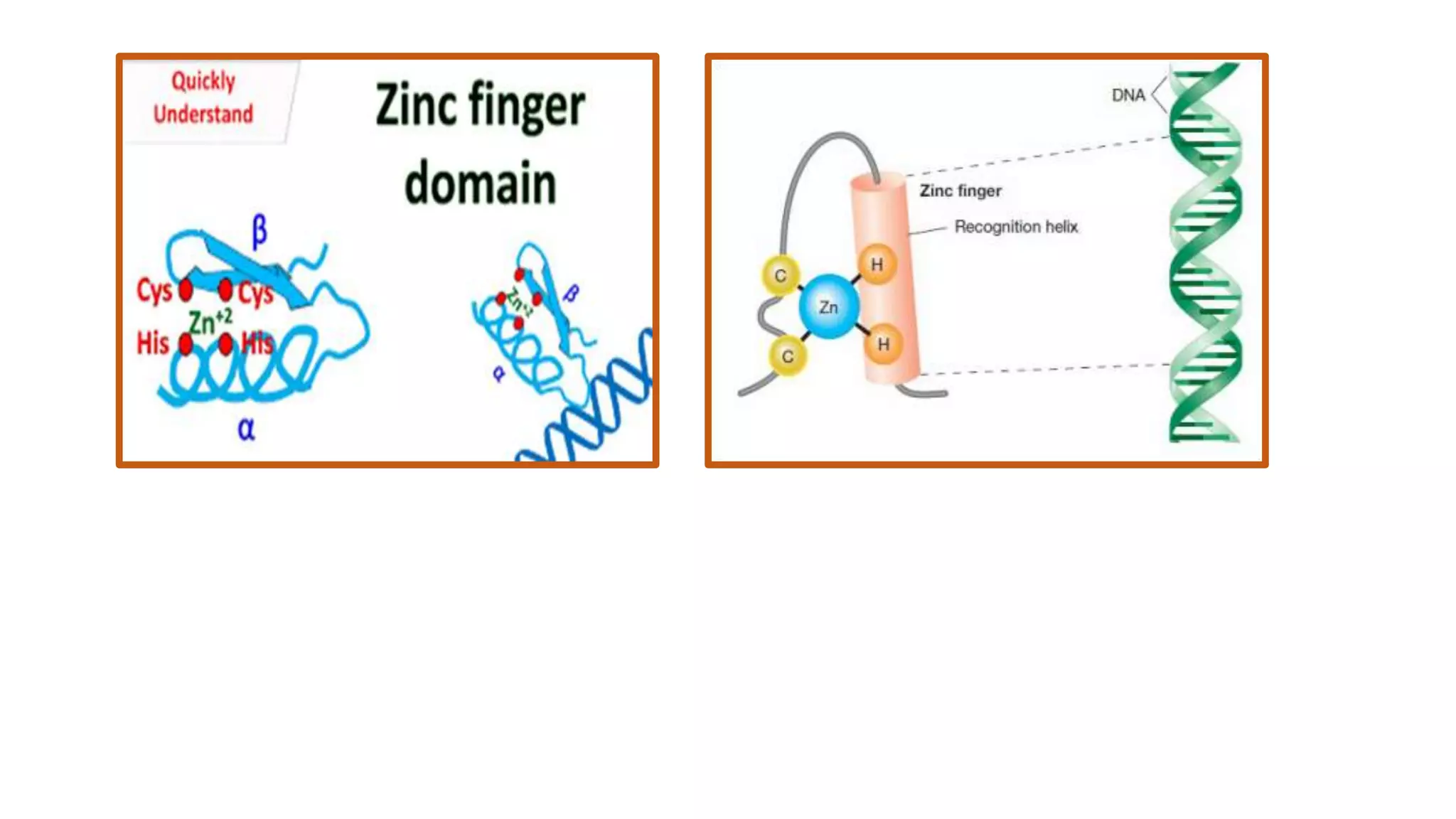
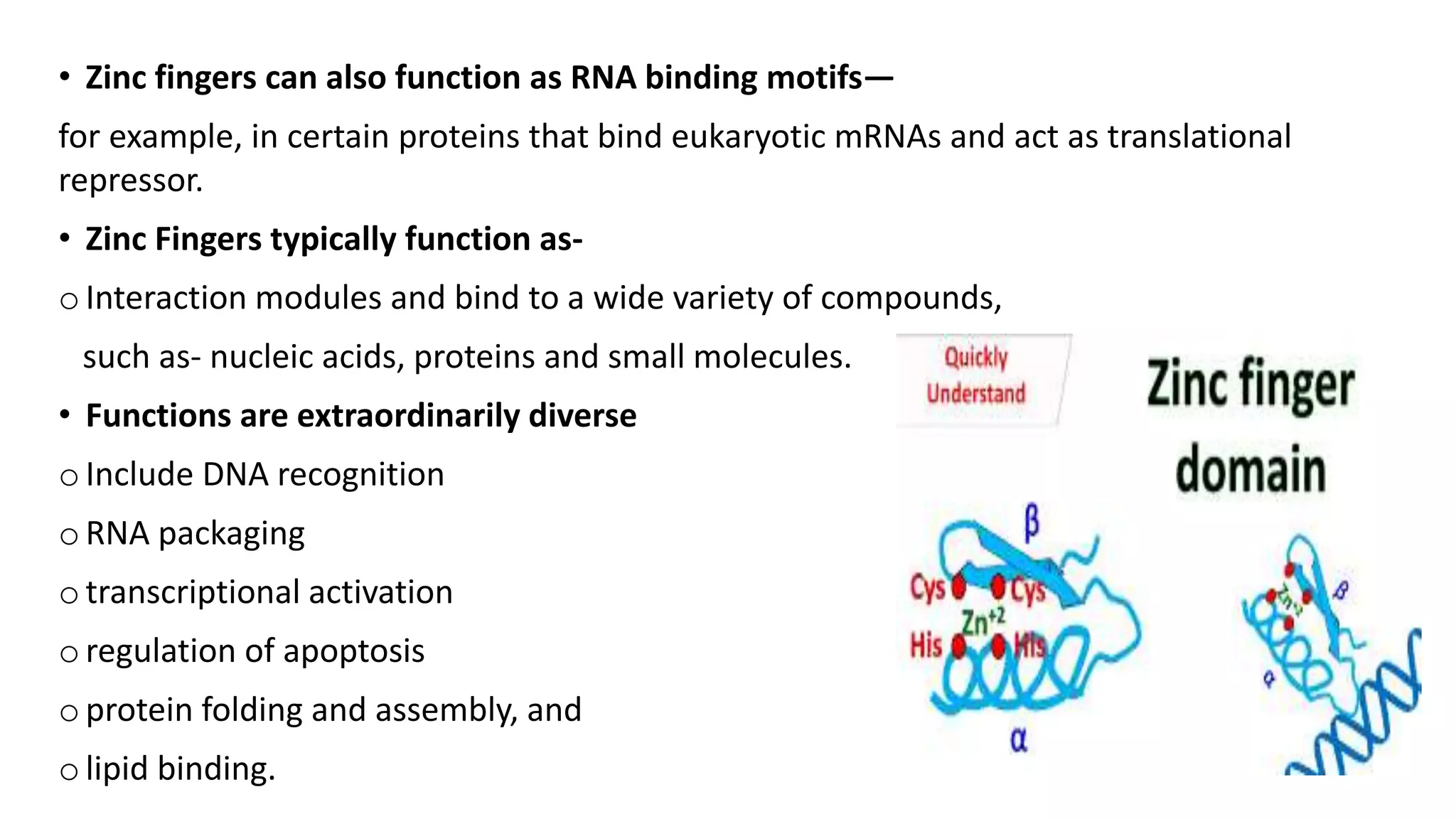
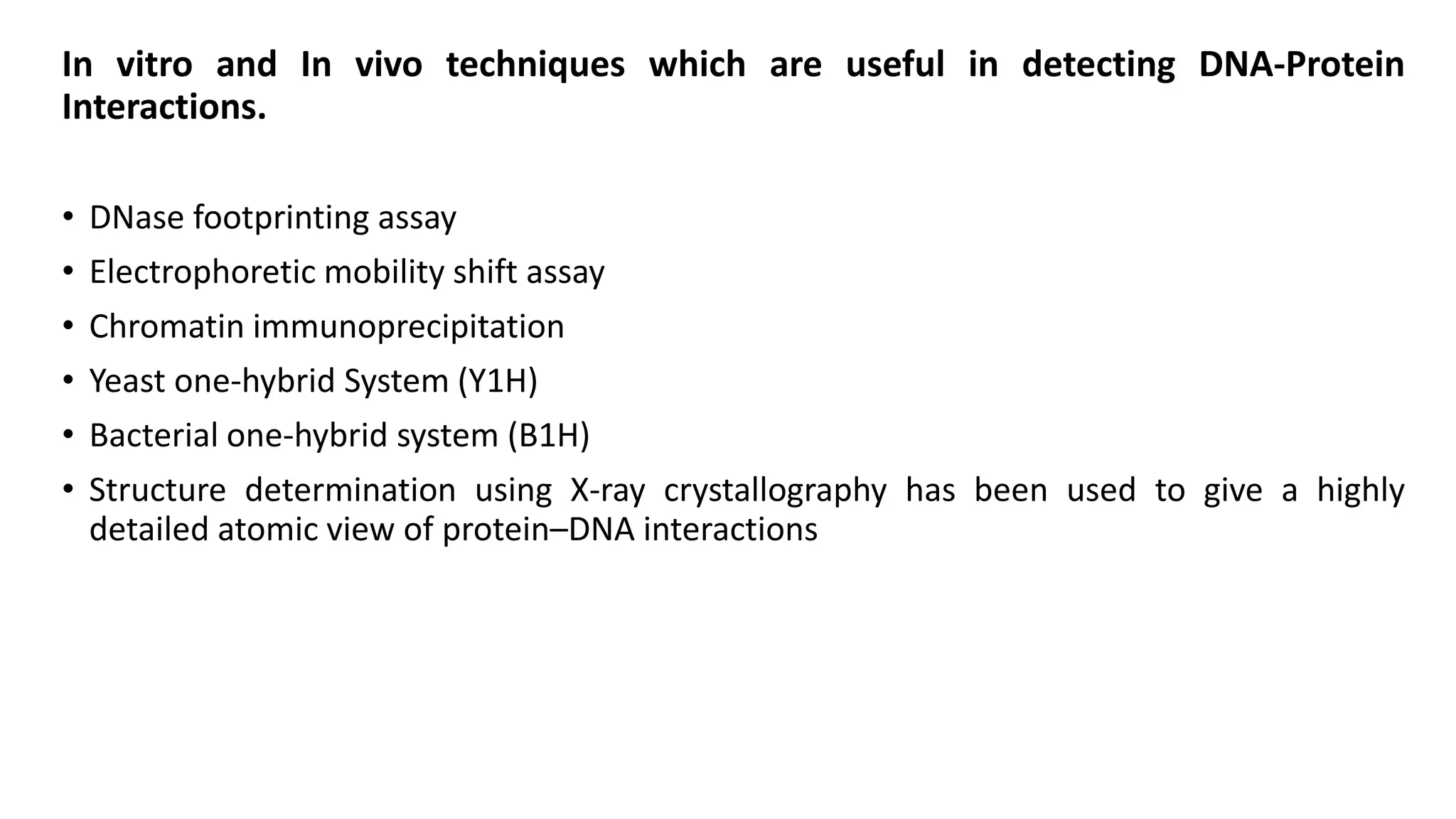
DNA – Protein Interaction
Presented by:
Shwetali N. Prajapati
Ph.D
Animal Biotechnology
• DNA-Protein interactions play very vital roles in any living cell.
• Controls replication, transcription, recombination, DNA repair etc.
• There are several types of proteins found in a cell. But only those proteins interact
with DNA, which have the DNA binding domains.
• Each DNA binding domain has at least one motif, which is a conserved amino acid
sequence of this protein, which can potentially recognize a double stranded or a
single stranded DNA.
There are mainly two broad types of DNA protein interactions :
1) Sequence specific DNA binding
• A DNA binding protein binds to a DNA on a site having a specific nucleotide sequence.
• Frequently involve DNA major groove.
• Interaction maintained by Hydrogen bonds, Ionic Interaction, van der waals forces.
• Example :- In case of transcription. The transcription factors are a special kind of DNA
binding proteins. They can only recognize a specific DNA sequence.
2) Sequence non-specific DNA binding
• The DNA binding protein can bind to a DNA in a random position on the DNA.
• In replication
During replication the DNA double strand is melted by helicase enzyme, and a replication
fork is made. A special kind of protein called single strand binding protein or SSB binds to
the melted single strand of DNA and stabilizes the system by preventing them to be re-
natured.
• There are several motifs present, which are involved in DNA binding,
helix-turn-helix, leucine zipper, zinc finger, helix-loop-helix etc.
1) Helix- turn- Helix motif
• Prokaryotic and Eukaryotic DNA binding proteins.
• First structure which is identified.
• 2 α-helices separated by a ß-turn, made up of 4 amino acids,
the 2nd is usually Glycine.
• C-terminal Recognition α- helix recognize and bind to specific
DNA sequences by forming Hydrogen bonds with bases located in the
Major groove.
• 2ndα- helix is stabilizes the overall configuration through Hydrophobic
interactions with the recognition helix.
2) Helix- loop- Helix motif
• Basically characterizes a family of Transcription factors.
• 2 regions of α- helix of 15-16 residues separated by
a region of variable length which forms a loop between the two α-helices.
• Connecting loop length varies from 12 to 28 amino acid residues.
• In general, one helix is smaller and other is larger typically
contains the DNA binding regions.
• bHLH proteins typically bind to a consensus sequence called an E-box, CANNTG.
• TF containing a HLH motif are dimeric, each with one helix containing basic amino acid
residues that facilitate DNA binding.
• Examples- C-myc, N-myc, MyoD, TF 4, HIF.
3) Leucine zipper motif
• Leucine zipper motif mediates both DNA- binding and
dimerization.
• The leucine zipper is a stretch of amino acids rich in leucine
residues that provide a dimerization motif.
• Region of the alpha-helix- containing the leucines which
line up- is called a ZIP domain, and leucines from each ZIP domain can weakly interact
with leucines from other ZIP domains, reversibly holding their alpha-helices together
(dimerization). When these alpha helices dimerize, the zipper is formed.
• 2 distinct right handed α-helices participate in the formation of homo or hetero dimer
structure.
• N-terminal binding domain is rich in positively charged basic amino acid residues interact in
the Major groove of DNA forming Sequence Specific Interaction.
• C- terminal dimerization domain is 30-40 amino acids long with a Leucine at every 7th
position.
• Present in Many DNA binding proteins such as c-myc, Jun, Fos, and C/EBP.
4) Zinc Finger motif
• Zinc- coordinated DNA binding motifs are called zinc finger motifs.
• Conserved pairs of Cys and His residues bind a Zn2+ atom
• In eukaryotes cys2-His2 zinc finger which was first discovered
in a transcription factor called TFIII A that regulates the transcription
of genes for 5S r-RNA.
• Derived from the kind of loop it generates when a covalent bond forms between a single
zinc metal ion with 2 cysteine on one side and 2 Histidine on the other side, either side of
the polypeptide or it can be between 4 cys two each on either sides. Such a structure
produces tetrahedral form.
• A stem loop structure(the finger) binds to DNA in the major groove.
• Often occure as tandem repeats with two, three or more fingers.
• Example- Sp1, TFIII-A and ADR 1.
• Zinc fingers can also function as RNA binding motifs—
for example, in certain proteins that bind eukaryotic mRNAs and act as translational
repressor.
• Zinc Fingers typically function as-
oInteraction modules and bind to a wide variety of compounds,
such as- nucleic acids, proteins and small molecules.
• Functions are extraordinarily diverse
oInclude DNA recognition
oRNA packaging
otranscriptional activation
oregulation of apoptosis
oprotein folding and assembly, and
olipid binding.
• Sequence non-specific DNA binding
• non-specific protein–DNA interactions occur with much lower affinity.
• Protein and DNA involve only weak interactions, primarily electrostatic in nature, between
the protein and the negatively charged DNA backbone.
• non-specific interactions are formed through basic residues in the histones making ionic
bonds to the acidic sugar-phosphate backbone of the DNA, and are therefore largely
independent of the base sequence.
• Chemical modifications of these basic amino acid residues
include methylation, phosphorylation and acetylation. These chemical changes alter the
strength of the interaction between the DNA and the histones, making the DNA more or
less accessible to transcription factors and changing the rate of transcription.
• Other non-specific DNA-binding proteins in chromatin include the high-mobility group
(HMG) proteins, which bind to bent or distorted DNA.
In vitro and In vivo techniques which are useful in detecting DNA-Protein
Interactions.
• DNase footprinting assay
• Electrophoretic mobility shift assay
• Chromatin immunoprecipitation
• Yeast one-hybrid System (Y1H)
• Bacterial one-hybrid system (B1H)
• Structure determination using X-ray crystallography has been used to give a highly
detailed atomic view of protein–DNA interactions
DNA protein interaction.pptx

DNA protein interaction.pptx

  • 1.
    DNA – ProteinInteraction Presented by: Shwetali N. Prajapati Ph.D Animal Biotechnology
  • 2.
    • DNA-Protein interactionsplay very vital roles in any living cell. • Controls replication, transcription, recombination, DNA repair etc. • There are several types of proteins found in a cell. But only those proteins interact with DNA, which have the DNA binding domains. • Each DNA binding domain has at least one motif, which is a conserved amino acid sequence of this protein, which can potentially recognize a double stranded or a single stranded DNA.
  • 3.
    There are mainlytwo broad types of DNA protein interactions : 1) Sequence specific DNA binding • A DNA binding protein binds to a DNA on a site having a specific nucleotide sequence. • Frequently involve DNA major groove. • Interaction maintained by Hydrogen bonds, Ionic Interaction, van der waals forces. • Example :- In case of transcription. The transcription factors are a special kind of DNA binding proteins. They can only recognize a specific DNA sequence.
  • 4.
    2) Sequence non-specificDNA binding • The DNA binding protein can bind to a DNA in a random position on the DNA. • In replication During replication the DNA double strand is melted by helicase enzyme, and a replication fork is made. A special kind of protein called single strand binding protein or SSB binds to the melted single strand of DNA and stabilizes the system by preventing them to be re- natured.
  • 5.
    • There areseveral motifs present, which are involved in DNA binding, helix-turn-helix, leucine zipper, zinc finger, helix-loop-helix etc. 1) Helix- turn- Helix motif • Prokaryotic and Eukaryotic DNA binding proteins. • First structure which is identified. • 2 α-helices separated by a ß-turn, made up of 4 amino acids, the 2nd is usually Glycine. • C-terminal Recognition α- helix recognize and bind to specific DNA sequences by forming Hydrogen bonds with bases located in the Major groove. • 2ndα- helix is stabilizes the overall configuration through Hydrophobic interactions with the recognition helix.
  • 7.
    2) Helix- loop-Helix motif • Basically characterizes a family of Transcription factors. • 2 regions of α- helix of 15-16 residues separated by a region of variable length which forms a loop between the two α-helices. • Connecting loop length varies from 12 to 28 amino acid residues. • In general, one helix is smaller and other is larger typically contains the DNA binding regions. • bHLH proteins typically bind to a consensus sequence called an E-box, CANNTG. • TF containing a HLH motif are dimeric, each with one helix containing basic amino acid residues that facilitate DNA binding. • Examples- C-myc, N-myc, MyoD, TF 4, HIF.
  • 9.
    3) Leucine zippermotif • Leucine zipper motif mediates both DNA- binding and dimerization. • The leucine zipper is a stretch of amino acids rich in leucine residues that provide a dimerization motif. • Region of the alpha-helix- containing the leucines which line up- is called a ZIP domain, and leucines from each ZIP domain can weakly interact with leucines from other ZIP domains, reversibly holding their alpha-helices together (dimerization). When these alpha helices dimerize, the zipper is formed. • 2 distinct right handed α-helices participate in the formation of homo or hetero dimer structure.
  • 10.
    • N-terminal bindingdomain is rich in positively charged basic amino acid residues interact in the Major groove of DNA forming Sequence Specific Interaction. • C- terminal dimerization domain is 30-40 amino acids long with a Leucine at every 7th position. • Present in Many DNA binding proteins such as c-myc, Jun, Fos, and C/EBP.
  • 11.
    4) Zinc Fingermotif • Zinc- coordinated DNA binding motifs are called zinc finger motifs. • Conserved pairs of Cys and His residues bind a Zn2+ atom • In eukaryotes cys2-His2 zinc finger which was first discovered in a transcription factor called TFIII A that regulates the transcription of genes for 5S r-RNA. • Derived from the kind of loop it generates when a covalent bond forms between a single zinc metal ion with 2 cysteine on one side and 2 Histidine on the other side, either side of the polypeptide or it can be between 4 cys two each on either sides. Such a structure produces tetrahedral form. • A stem loop structure(the finger) binds to DNA in the major groove. • Often occure as tandem repeats with two, three or more fingers. • Example- Sp1, TFIII-A and ADR 1.
  • 13.
    • Zinc fingerscan also function as RNA binding motifs— for example, in certain proteins that bind eukaryotic mRNAs and act as translational repressor. • Zinc Fingers typically function as- oInteraction modules and bind to a wide variety of compounds, such as- nucleic acids, proteins and small molecules. • Functions are extraordinarily diverse oInclude DNA recognition oRNA packaging otranscriptional activation oregulation of apoptosis oprotein folding and assembly, and olipid binding.
  • 14.
    • Sequence non-specificDNA binding • non-specific protein–DNA interactions occur with much lower affinity. • Protein and DNA involve only weak interactions, primarily electrostatic in nature, between the protein and the negatively charged DNA backbone. • non-specific interactions are formed through basic residues in the histones making ionic bonds to the acidic sugar-phosphate backbone of the DNA, and are therefore largely independent of the base sequence. • Chemical modifications of these basic amino acid residues include methylation, phosphorylation and acetylation. These chemical changes alter the strength of the interaction between the DNA and the histones, making the DNA more or less accessible to transcription factors and changing the rate of transcription. • Other non-specific DNA-binding proteins in chromatin include the high-mobility group (HMG) proteins, which bind to bent or distorted DNA.
  • 15.
    In vitro andIn vivo techniques which are useful in detecting DNA-Protein Interactions. • DNase footprinting assay • Electrophoretic mobility shift assay • Chromatin immunoprecipitation • Yeast one-hybrid System (Y1H) • Bacterial one-hybrid system (B1H) • Structure determination using X-ray crystallography has been used to give a highly detailed atomic view of protein–DNA interactions