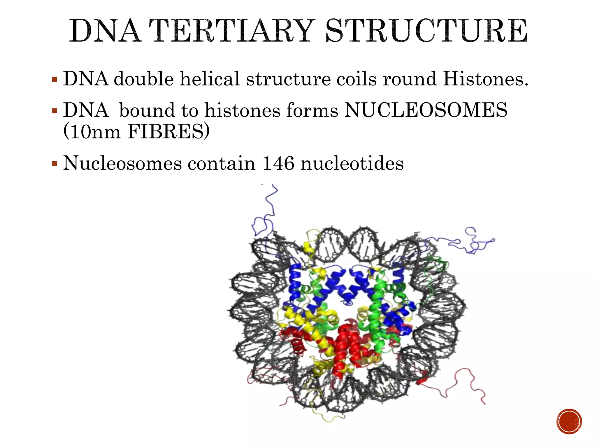
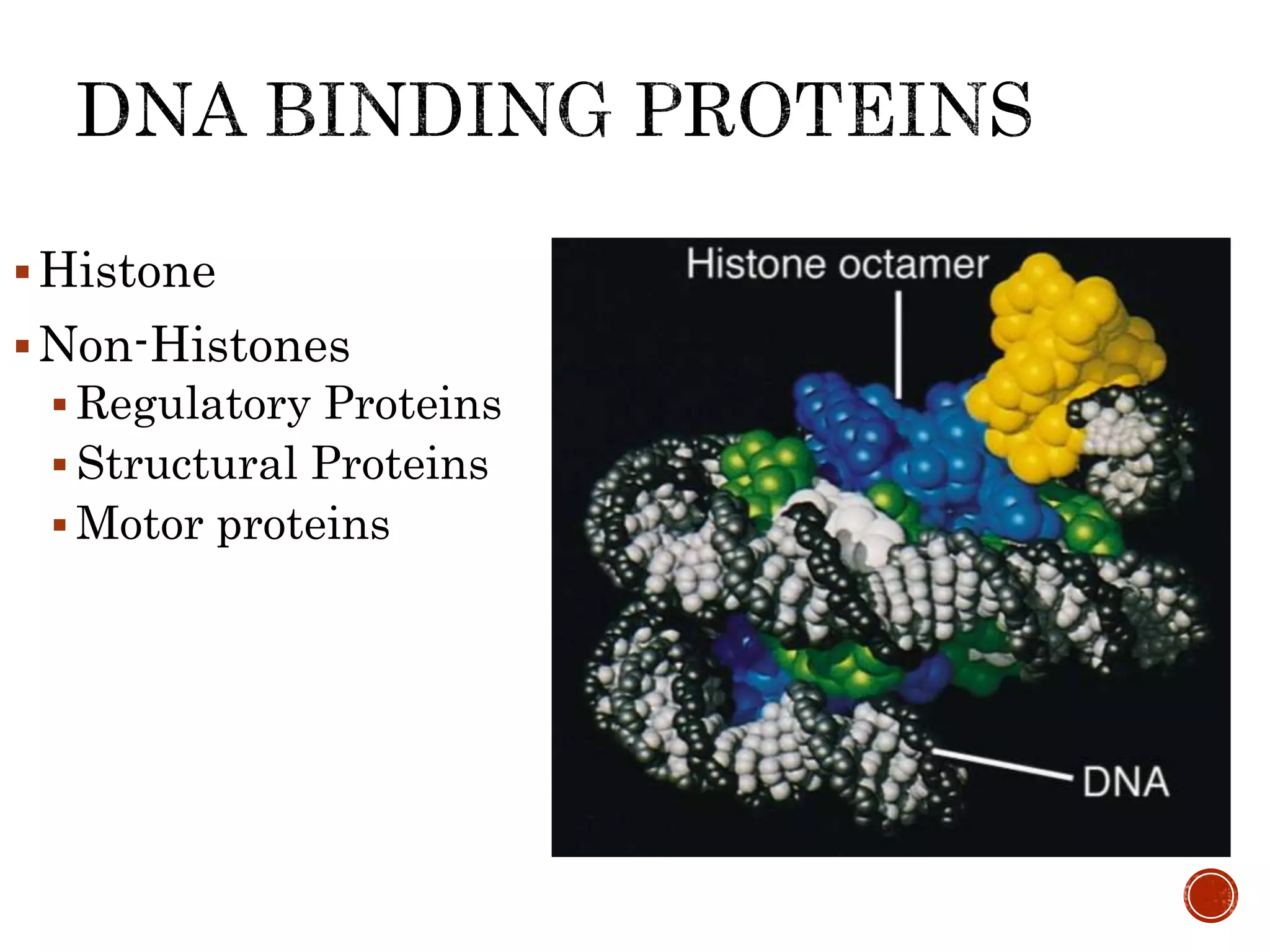
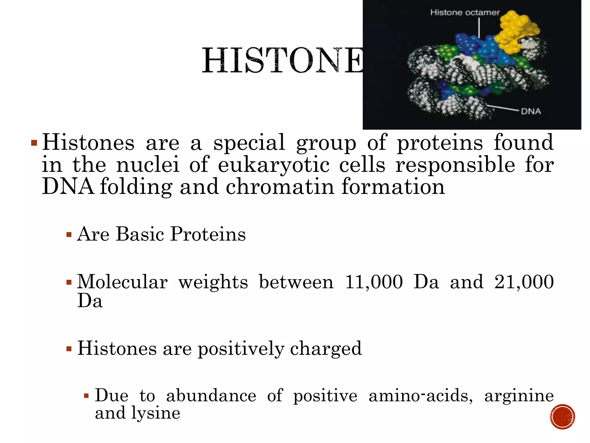
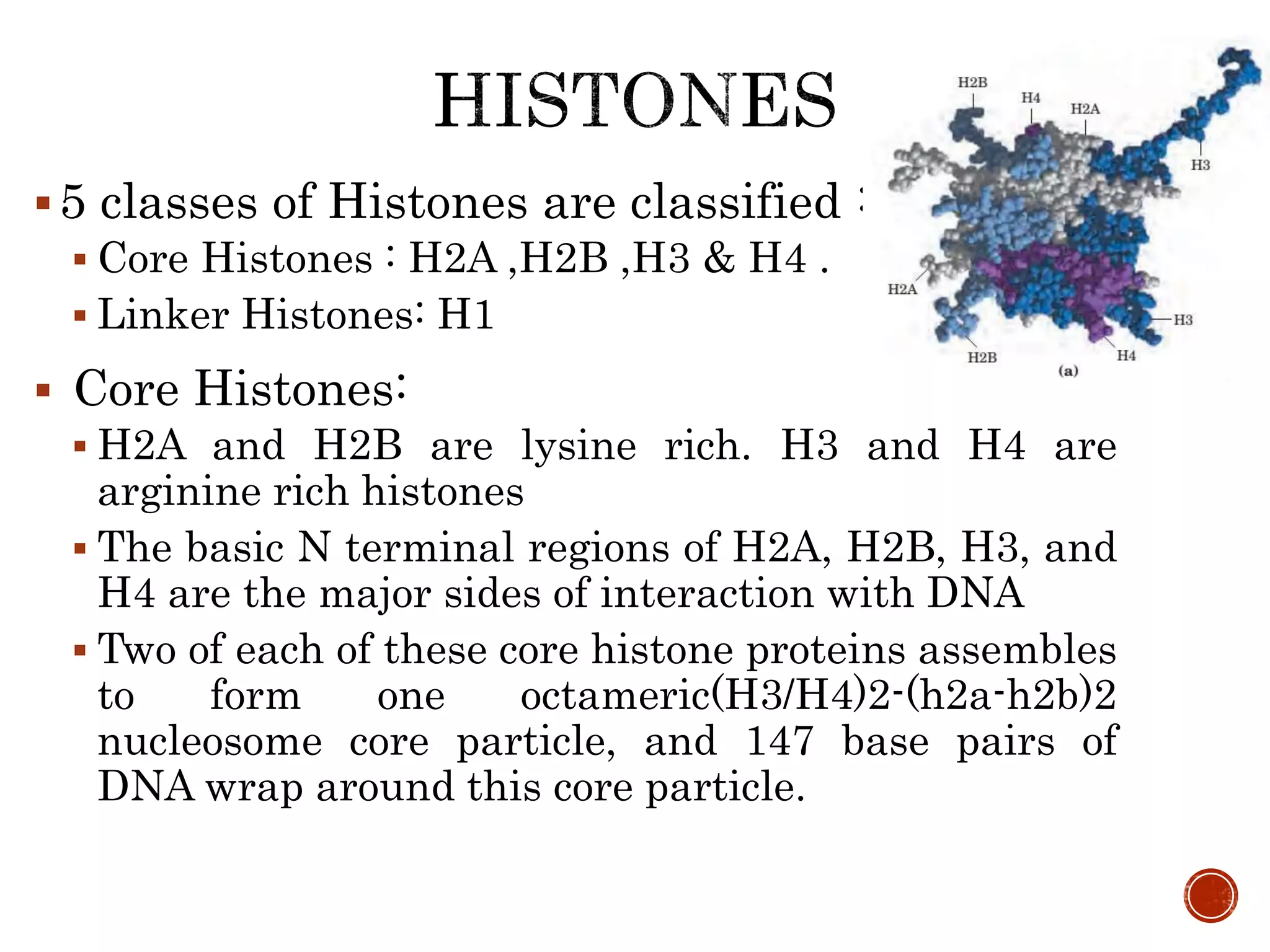
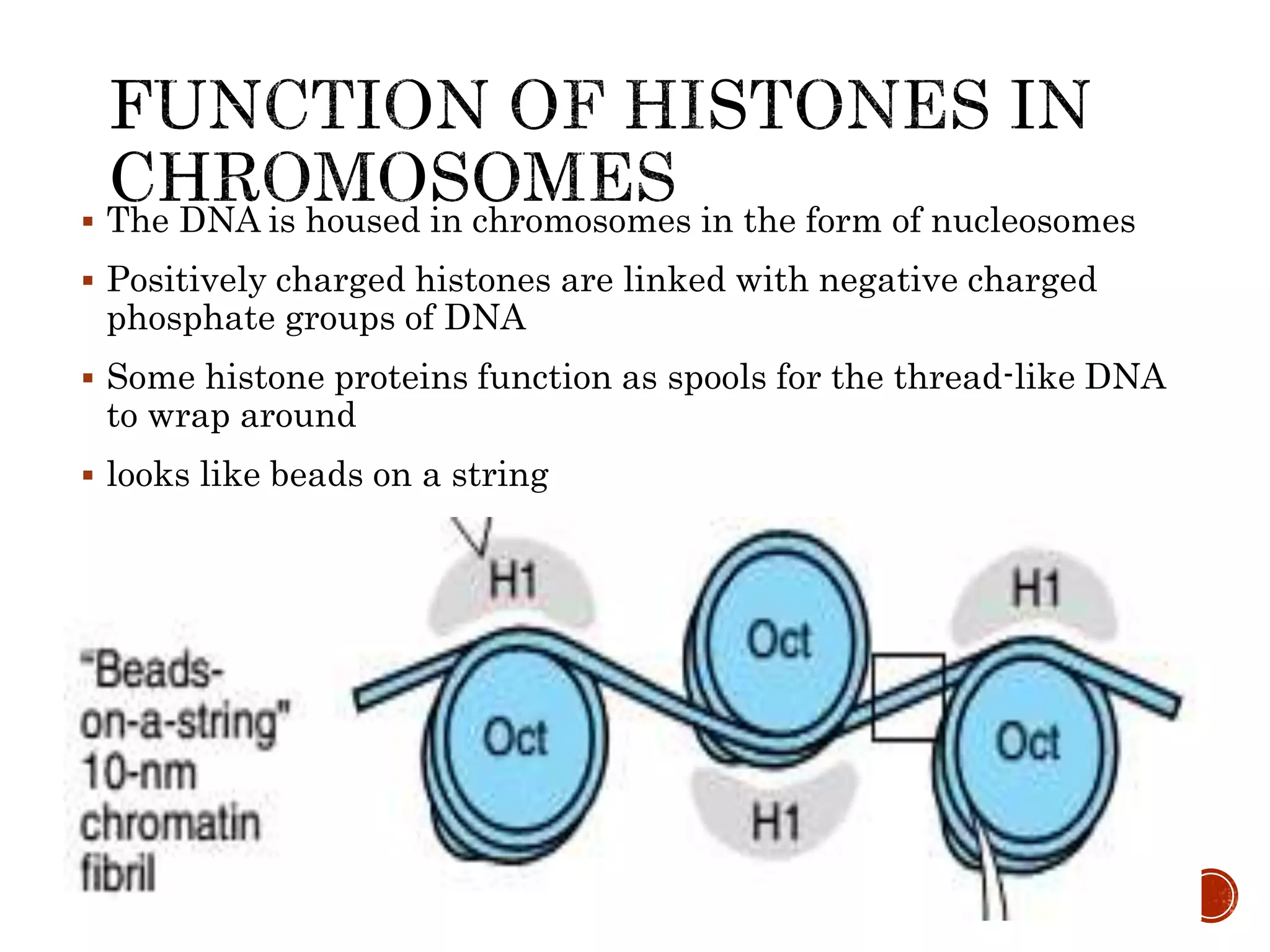
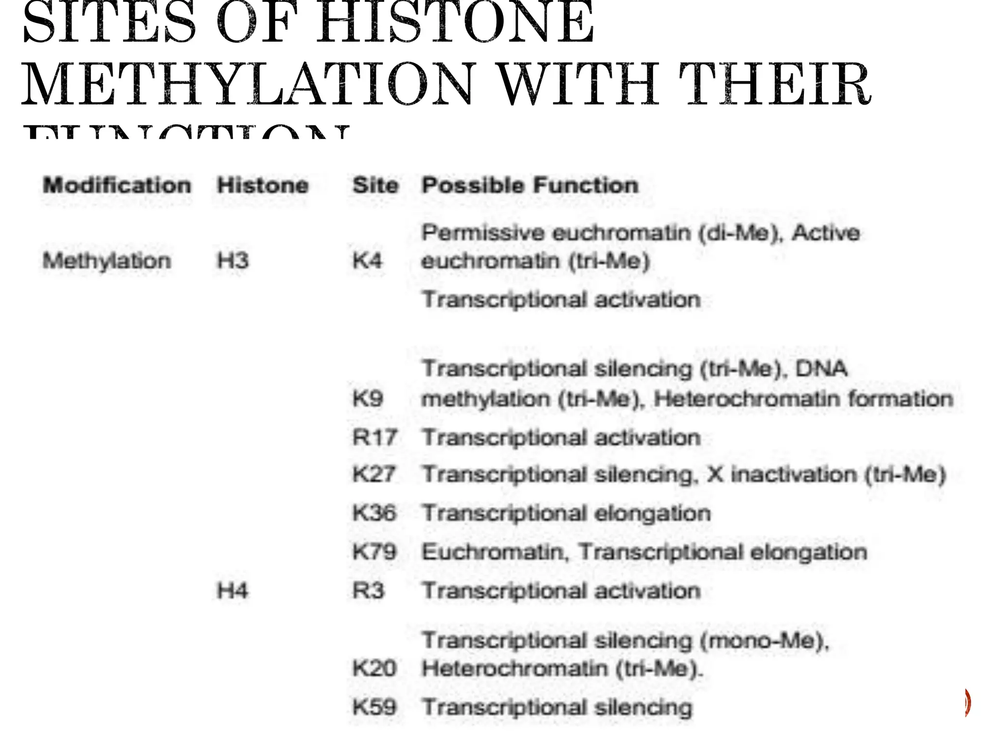
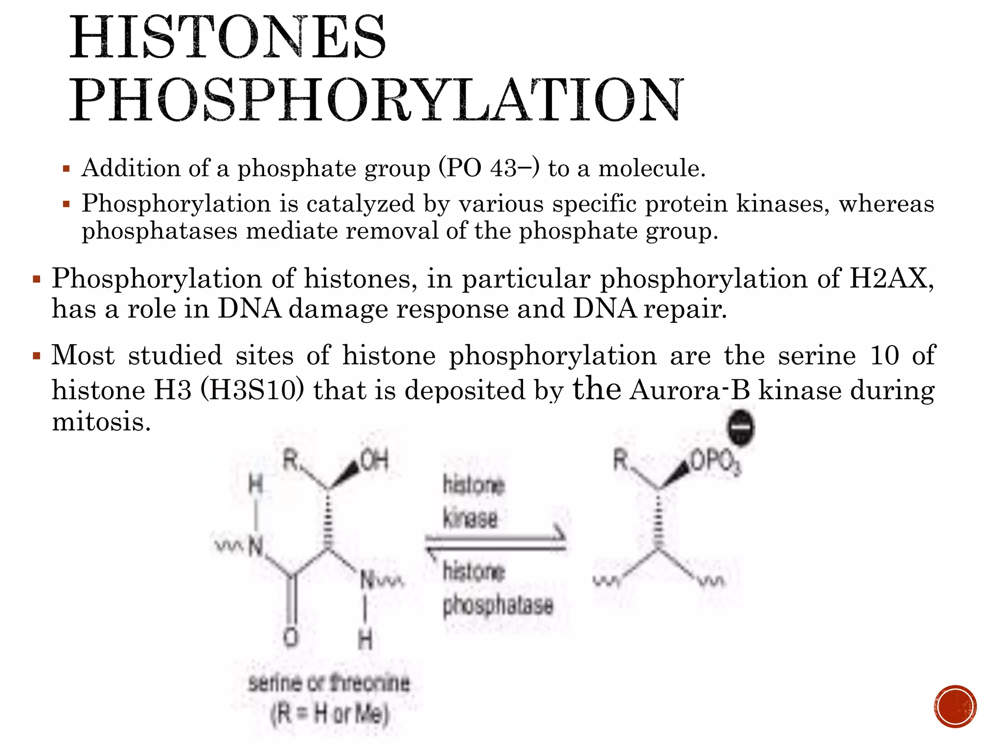
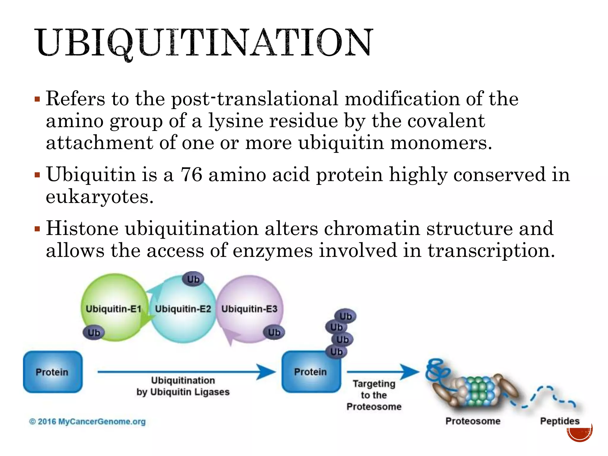
DNA BINDING
PROTEINS
Hari Sharan Makaju
M.Sc. Clinical Biochemistry
1st year
2076/4/12
DNA is a polymer of deoxyribonucleoside
monophosphates covalently linked by 3′→5′–
phosphodiester bonds
Found in chromosomes, mitochondria and
chloroplasts
Carries the genetic information
The Primary structure of
DNA is Sequence
 Two anti-parallel polynucleotide
chains wound around the same axis.
 Sugar-phosphate chains wrap
around the periphery.
 Bases (A, T, C and G) occupy the
core, forming complementary A –T
and G –C
 The double helical structure of DNA
was proposed by lames Watson and
FrancisCrick in 1953
 The DNA double helix is held
together mainly by- Hydrogen bonds
Property A-DNA B-DNA Z-DNA
Helix Right Right Left
Base pair
per turn
11 10.4 12
Pitch (Each
turn)
2.46 3.40 4.56
Rise per
base pair
along axis
0.23 0.34 1.84
Diameter 2.55 2.37 1.84
Major
Groove
Present Present Absent
Minor
Groove
Present Present Deep cleft
 DNA double helical structure coils round Histones.
 DNA bound to histones forms NUCLEOSOMES
(10nm FIBRES)
 Nucleosomes contain 146 nucleotides
Histone
Non-Histones
 Regulatory Proteins
 Structural Proteins
 Motor proteins
Histones are a special group of proteins found
in the nuclei of eukaryotic cells responsible for
DNA folding and chromatin formation
 Are Basic Proteins
 Molecular weights between 11,000 Da and 21,000
Da
 Histones are positively charged
 Due to abundance of positive amino-acids, arginine
and lysine
Histones have five major classes : H1, H2A,
H2B, H3 and H4
 Histones are characterized
Central nonpolar domain, forms a globular
structure
 N-terminal and C-terminal regions that
contain most of the basic amino acids
 The C-terminal end is primarily responsible for histone-
DNA and histone-histone interactions.
 The N-terminal tails stand as targets of post-transational
modifications (PTMs),
5 classes of Histones are classified :
 Core Histones : H2A ,H2B ,H3 & H4 .
 Linker Histones: H1
 Core Histones:
 H2A and H2B are lysine rich. H3 and H4 are
arginine rich histones
 The basic N terminal regions of H2A, H2B, H3, and
H4 are the major sides of interaction with DNA
 Two of each of these core histone proteins assembles
to form one octameric(H3/H4)2-(h2a-h2b)2
nucleosome core particle, and 147 base pairs of
DNA wrap around this core particle.
Linker histone :
 Binds the nucleosome at the starting and ending
sites of the DNA, thus locking the DNA into place
and help in the formation of higher order
structure
H1
Not part of the nucleosome core particle.
Binds to the linker DNA and is referred to
as a linker histone.
H1 is half as abundant as the other
histones, which is consistent with the
finding that only one molecule of H1 can
associate with a nucleosome.
H2A
 H2A packages DNA molecules into chromatin, the
packaging process will effect gene expression.
 H2A plays a major role in determining the overall
structure of chromatin. Inadvertently, H2A has been
found to regulate gene expression.
H2B
 Involved with the structure of the nucleosomes of the
'beads on a string' structure.
H3
 Featuring a main globular domain and a long N-
terminal tail.
 Its sequence variants and variable modification states
are thought to play a role in the dynamic and long term
regulation of Genes
H4
 Structural component of the nucleosome,
 Subject to covalent modification ,including acetylation
and methylation, which may alter expression of genes .
 The DNA is housed in chromosomes in the form of nucleosomes
 Positively charged histones are linked with negative charged
phosphate groups of DNA
 Some histone proteins function as spools for the thread-like DNA
to wrap around
 looks like beads on a string
Each type of histone has variant forms
 Because certain amino acid side chains are
enzymatically modified by
 Acetylation
 Methylation,
 Phosphorylation
 ADP-Ribosylation,
 Unibiquitination
 Sumoylation
Such modifications affect:
The net electric charge, shape, and other
properties of histones
The structural and functional properties of
the chromatin
 They play a role in the regulation of
transcription
 Adds acetyl groups group to the Lysine amino acid of
the histone tails
 Enzymes:
 Histone acetyl transferases (HATs)
 Reduces positive charge and weakens interaction of
histones with DNA
 Facilitates transcription by making DNA more
accessible to RNA polymerase II
Removes acetyl groups from histone tails
Enzyme:
Histone deacetylases (HDACs)
Increases interaction of DNA and
histones
Represses transcription
 Addition of an Methyl functional group to Lysine or Arginine of
the histone tail.
 Enzymes
 "histone methyltransferase”
 Methylation can result in activation or repression of
expression .
 Activation (H3K4, H3K36, H3K79)
 Trimethylation of histone H3 at lysine 4 (H3K4) is an
universal active mark for transcription.
 Repression (H3K9, H3K27, H4K20)
 Dimethylation of histone H3 at lysine 9 (H3K9) and at
27 (H3K27) are the universal signal for transcriptional
silencing.
 Addition of a phosphate group (PO 43−) to a molecule.
 Phosphorylation is catalyzed by various specific protein kinases, whereas
phosphatases mediate removal of the phosphate group.
 Phosphorylation of histones, in particular phosphorylation of H2AX,
has a role in DNA damage response and DNA repair.
 Most studied sites of histone phosphorylation are the serine 10 of
histone H3 (H3S10) that is deposited by the Aurora-B kinase during
mitosis.
 Refers to the post-translational modification of the
amino group of a lysine residue by the covalent
attachment of one or more ubiquitin monomers.
 Ubiquitin is a 76 amino acid protein highly conserved in
eukaryotes.
 Histone ubiquitination alters chromatin structure and
allows the access of enzymes involved in transcription.
 Addition of an ADP-ribose moiety onto a protein using NAD+ as
a substrate.
 Mono ADP-ribosylation is mediated by ADP ribosyl
transferases (ART) and the enzymes responsible for the Poly-
ADP-ribosylation are the poly ADP ribose polymerases
(PARPs).
 PARP1 prefers to linker histone H1 while PARP2 prefers core
histones
Addition of a “Small Ubiquitin-related MOdifier
protein” (SUMO) of ~100 amino acids.
Histone sumoylation was first reported in 2003,
Shiio et al.
 Found that H4 can be modified by SUMO and
 They suggested that this modification leads to the
repression of transcriptional activity
The putative sumoylation sites were identified
as K6/7
 To a lesser extent K16/17of H2B, K126 of H2A,
 All four lysine in the N-terminal tail of H4.
Apart from histones, there are many other
special proteins which
will interact at specific regions of DNA.
The protein–DNA interactions are mainly
mediated by 3 motifs :–
 Helix-turn-helix
 Zinc finger
 Leucine zipper motifs.
Only small regions of the protein make direct
contact with the DNA
The rest of the proteins are involved in other
activities, like dimerization, ligand-binding,
interaction with coactivators and corepressors,
etc.
DNA sequence-specificity of
DNA binding proteins
 Sequence-specific
interactions
 Frequently involve DNA
major groove
 The protein-DNA
interactions are maintained
by hydrogen bonds, ionic
interactions and van der
Waals forces.
 Non-specific interactions
 Interactions with DNA
phosphate backbones
 Comprises about 20 amino acids in two short α-helical
segments
 Each seven to nine amino acid residues long,
 Separated by a β-turn
 One of the two α-helical segments is called the
recognition helix,
 Because it usually contains many of the amino acids that
interact with the DNA in a sequence-specific way.
 When bound to DNA, the recognition helix is positioned
in or nearly in the major groove.
Based on their structure and the spatial arrangement of their
helices.
 Di-helical
 Simplest helix-turn-helix motif.
 Example: Homeodomain
 Tri-helical
 Example: Transcriptional activator Myb
 Tetra-helica
 Example TetR repressors.
 Multihelical versions with additional helices also occur.
 Winged helix-turn-helix
 Formed by a 3-helical bundle and a 3- or 4-strand beta-sheet(wing).
 Example :
 transcription factor ETS
 Scaffold arranged in the order α1-β1-β2-α2-α3-β3-β4 where the third helix is
the DNA recognition helix
Organism Regulatory
protein
E coli lac repressor, Cap
Phages λcI, cro, and 434
repressors
Mammals homeobox proteins
pit-1,
Oct1, Oct2
DNA-binding domain of
the Lac repressor
Homeobox
protein
 Very common in eukaryotes
 About 30 amino acid residues form an
elongated loop held together at the base by a
single Zn2+ion
 α-helix plus two antiparallel β-sheets
 And a Zn2+ ion that is coordinated by
cysteines or histidines
 α-helix makes sequence-specific contacts
along the major groove.
 Zinc Finger Proteins may have more than one
Zn finger per protein.
 A Zn2+ ion coordinated by 4 Cys or 2 Cys and 2 His
residues.
 Often occur as tandem repeats with two, three,
or more fingers.
Structure of the six-finger TFIIIA–DNA
complex
 Some zinc fingers contain the amino
acid residues that are important in
sequence discrimination.
 Zinc fingers can also function as
RNA binding motifs—for example, in
certain proteins that bind eukaryotic
mRNAs and act as translational
repressor
 Zinc fingers designed to bind
targeted DNA sequences with
ultimate goal of therapeutics
Fig. Three zinc fingers (gray)
of the regulatory protein
Zif268, complexed with DNA
(blue and white)
Zinc Fingers typically function as
 Interaction modules and bind to a wide variety
of compounds, such as
 nucleic acids, proteins and small molecules.
Functions are extraordinarily diverse
 Include DNA recognition,
 RNA packaging,
 transcriptional activation,
 regulation of apoptosis,
 protein folding and assembly, and
 lipid binding.
 DNA interaction
 First “finger” binds DNA
 Second “finger” involved in dimerisation
 Binds to neighboring “major grooves” on same side of DNA
 Extensive phosphate contact and recognition helix docked
into the groove
 specificity determined by 3 aa in recognition helix
 Classical Zinc finger (C2H2 )
 Gag-knuckle
 Treble-clef
 Zinc ribbon
 Zn2/Cys6
 TAZ2 domain
Organism Regulatory
protein
E coli Gene 32 protein
Yeast Gal 4
Xenopus TFIII A
Mammals Steroid receptor
family Sp1
 Contain leucine residues every 7th position in
an α-helix.
 Form homo- or heterodimers with coiled coil
structure (blue region)
 Although researchers initially thought the
Leu residues interdigitated (hence the name
“zipper”)
 The basic region with arginine and lysine
residues bind to the major groove of DNA
 The basic amino acids interact with the
phosphate backbone of DNA through
electrostatic interactions and also the DNA
bases through hydrogen bonding.
 Leucine zippers also function as dimers to regulate gene
transcription
 Example
Organism Regulatory Protein
Yeast GCN4
Mammals C/EBP, Fos, Jun, Fra-1,
cAMP response
element-binding
protein (CREB),
c-myc, n-myc, I-myc
proto-
oncogene JUN (purp
le) binding as a
homodimer to DNA.
 In vitro and In vivo techniques which are useful in
detecting DNA-Protein Interactions.
 Electrophoretic mobility shift assay
 Widespread technique to identify protein–DNA
interactions.
 DNase footprinting assay
 to identify the specific site of binding of a protein
to DNA.
 Chromatin immunoprecipitation :
 to identify the sequence of the DNA fragments
which bind to a known transcription factor.
Yeast one-hybrid System (Y1H)
to identify which protein binds to a particular
DNA fragment.
Bacterial one-hybrid system (B1H)
to identify which protein binds to a particular
DNA fragment.
Structure determination using X-ray
crystallography has been used to give a highly
detailed atomic view of protein–DNA
interactions
 Robert k. Murray, D.K.Granner ,P.A.Mayes & Victor W.Rodwell Harpers
illustrated biochemistry 26th edition
 Lippincot - Marks' Basic Medical Biochemistry A Clinical Approach
 Thomas M.Devlin , textbook of Biochemistry with clinical correlation 5th
edition
 Lehninger Principle of Biochemistry 4th edition
 https://en.wikipedia.org/wiki/zinc finger
 https://en.wikipedia.org/wiki/Helix-turn-helix
 https://www.mycancergenome.org/content/pathways/protein-degradation-
ubiquitination/
 https://epigenie.com/key-epigenetic-players/chromatin-modifying-and-
dna-binding-proteins/zinc-finger-proteins/
 Gregory R.Dressler,Epigenetics, Development, and the Kidney, J Am Soc
Nephrol 19: 2060 –2067, 2008. doi: 10.1681/ASN.2008010119
Dna binding proteins

Dna binding proteins

  • 1.
    DNA BINDING PROTEINS Hari SharanMakaju M.Sc. Clinical Biochemistry 1st year 2076/4/12
  • 2.
    DNA is apolymer of deoxyribonucleoside monophosphates covalently linked by 3′→5′– phosphodiester bonds Found in chromosomes, mitochondria and chloroplasts Carries the genetic information
  • 3.
    The Primary structureof DNA is Sequence
  • 5.
     Two anti-parallelpolynucleotide chains wound around the same axis.  Sugar-phosphate chains wrap around the periphery.  Bases (A, T, C and G) occupy the core, forming complementary A –T and G –C  The double helical structure of DNA was proposed by lames Watson and FrancisCrick in 1953  The DNA double helix is held together mainly by- Hydrogen bonds
  • 6.
    Property A-DNA B-DNAZ-DNA Helix Right Right Left Base pair per turn 11 10.4 12 Pitch (Each turn) 2.46 3.40 4.56 Rise per base pair along axis 0.23 0.34 1.84 Diameter 2.55 2.37 1.84 Major Groove Present Present Absent Minor Groove Present Present Deep cleft
  • 8.
     DNA doublehelical structure coils round Histones.  DNA bound to histones forms NUCLEOSOMES (10nm FIBRES)  Nucleosomes contain 146 nucleotides
  • 9.
    Histone Non-Histones  Regulatory Proteins Structural Proteins  Motor proteins
  • 10.
    Histones are aspecial group of proteins found in the nuclei of eukaryotic cells responsible for DNA folding and chromatin formation  Are Basic Proteins  Molecular weights between 11,000 Da and 21,000 Da  Histones are positively charged  Due to abundance of positive amino-acids, arginine and lysine
  • 11.
    Histones have fivemajor classes : H1, H2A, H2B, H3 and H4  Histones are characterized Central nonpolar domain, forms a globular structure  N-terminal and C-terminal regions that contain most of the basic amino acids  The C-terminal end is primarily responsible for histone- DNA and histone-histone interactions.  The N-terminal tails stand as targets of post-transational modifications (PTMs),
  • 13.
    5 classes ofHistones are classified :  Core Histones : H2A ,H2B ,H3 & H4 .  Linker Histones: H1  Core Histones:  H2A and H2B are lysine rich. H3 and H4 are arginine rich histones  The basic N terminal regions of H2A, H2B, H3, and H4 are the major sides of interaction with DNA  Two of each of these core histone proteins assembles to form one octameric(H3/H4)2-(h2a-h2b)2 nucleosome core particle, and 147 base pairs of DNA wrap around this core particle.
  • 15.
    Linker histone : Binds the nucleosome at the starting and ending sites of the DNA, thus locking the DNA into place and help in the formation of higher order structure
  • 16.
    H1 Not part ofthe nucleosome core particle. Binds to the linker DNA and is referred to as a linker histone. H1 is half as abundant as the other histones, which is consistent with the finding that only one molecule of H1 can associate with a nucleosome.
  • 17.
    H2A  H2A packagesDNA molecules into chromatin, the packaging process will effect gene expression.  H2A plays a major role in determining the overall structure of chromatin. Inadvertently, H2A has been found to regulate gene expression. H2B  Involved with the structure of the nucleosomes of the 'beads on a string' structure.
  • 18.
    H3  Featuring amain globular domain and a long N- terminal tail.  Its sequence variants and variable modification states are thought to play a role in the dynamic and long term regulation of Genes H4  Structural component of the nucleosome,  Subject to covalent modification ,including acetylation and methylation, which may alter expression of genes .
  • 19.
     The DNAis housed in chromosomes in the form of nucleosomes  Positively charged histones are linked with negative charged phosphate groups of DNA  Some histone proteins function as spools for the thread-like DNA to wrap around  looks like beads on a string
  • 20.
    Each type ofhistone has variant forms  Because certain amino acid side chains are enzymatically modified by  Acetylation  Methylation,  Phosphorylation  ADP-Ribosylation,  Unibiquitination  Sumoylation
  • 21.
    Such modifications affect: Thenet electric charge, shape, and other properties of histones The structural and functional properties of the chromatin  They play a role in the regulation of transcription
  • 22.
     Adds acetylgroups group to the Lysine amino acid of the histone tails  Enzymes:  Histone acetyl transferases (HATs)  Reduces positive charge and weakens interaction of histones with DNA  Facilitates transcription by making DNA more accessible to RNA polymerase II
  • 23.
    Removes acetyl groupsfrom histone tails Enzyme: Histone deacetylases (HDACs) Increases interaction of DNA and histones Represses transcription
  • 26.
     Addition ofan Methyl functional group to Lysine or Arginine of the histone tail.  Enzymes  "histone methyltransferase”
  • 27.
     Methylation canresult in activation or repression of expression .  Activation (H3K4, H3K36, H3K79)  Trimethylation of histone H3 at lysine 4 (H3K4) is an universal active mark for transcription.  Repression (H3K9, H3K27, H4K20)  Dimethylation of histone H3 at lysine 9 (H3K9) and at 27 (H3K27) are the universal signal for transcriptional silencing.
  • 29.
     Addition ofa phosphate group (PO 43−) to a molecule.  Phosphorylation is catalyzed by various specific protein kinases, whereas phosphatases mediate removal of the phosphate group.  Phosphorylation of histones, in particular phosphorylation of H2AX, has a role in DNA damage response and DNA repair.  Most studied sites of histone phosphorylation are the serine 10 of histone H3 (H3S10) that is deposited by the Aurora-B kinase during mitosis.
  • 31.
     Refers tothe post-translational modification of the amino group of a lysine residue by the covalent attachment of one or more ubiquitin monomers.  Ubiquitin is a 76 amino acid protein highly conserved in eukaryotes.  Histone ubiquitination alters chromatin structure and allows the access of enzymes involved in transcription.
  • 33.
     Addition ofan ADP-ribose moiety onto a protein using NAD+ as a substrate.  Mono ADP-ribosylation is mediated by ADP ribosyl transferases (ART) and the enzymes responsible for the Poly- ADP-ribosylation are the poly ADP ribose polymerases (PARPs).  PARP1 prefers to linker histone H1 while PARP2 prefers core histones
  • 34.
    Addition of a“Small Ubiquitin-related MOdifier protein” (SUMO) of ~100 amino acids. Histone sumoylation was first reported in 2003, Shiio et al.  Found that H4 can be modified by SUMO and  They suggested that this modification leads to the repression of transcriptional activity The putative sumoylation sites were identified as K6/7  To a lesser extent K16/17of H2B, K126 of H2A,  All four lysine in the N-terminal tail of H4.
  • 36.
    Apart from histones,there are many other special proteins which will interact at specific regions of DNA. The protein–DNA interactions are mainly mediated by 3 motifs :–  Helix-turn-helix  Zinc finger  Leucine zipper motifs. Only small regions of the protein make direct contact with the DNA The rest of the proteins are involved in other activities, like dimerization, ligand-binding, interaction with coactivators and corepressors, etc.
  • 37.
    DNA sequence-specificity of DNAbinding proteins  Sequence-specific interactions  Frequently involve DNA major groove  The protein-DNA interactions are maintained by hydrogen bonds, ionic interactions and van der Waals forces.  Non-specific interactions  Interactions with DNA phosphate backbones
  • 39.
     Comprises about20 amino acids in two short α-helical segments  Each seven to nine amino acid residues long,  Separated by a β-turn  One of the two α-helical segments is called the recognition helix,  Because it usually contains many of the amino acids that interact with the DNA in a sequence-specific way.  When bound to DNA, the recognition helix is positioned in or nearly in the major groove.
  • 40.
    Based on theirstructure and the spatial arrangement of their helices.  Di-helical  Simplest helix-turn-helix motif.  Example: Homeodomain  Tri-helical  Example: Transcriptional activator Myb  Tetra-helica  Example TetR repressors.  Multihelical versions with additional helices also occur.  Winged helix-turn-helix  Formed by a 3-helical bundle and a 3- or 4-strand beta-sheet(wing).  Example :  transcription factor ETS  Scaffold arranged in the order α1-β1-β2-α2-α3-β3-β4 where the third helix is the DNA recognition helix
  • 41.
    Organism Regulatory protein E colilac repressor, Cap Phages λcI, cro, and 434 repressors Mammals homeobox proteins pit-1, Oct1, Oct2 DNA-binding domain of the Lac repressor Homeobox protein
  • 42.
     Very commonin eukaryotes  About 30 amino acid residues form an elongated loop held together at the base by a single Zn2+ion  α-helix plus two antiparallel β-sheets  And a Zn2+ ion that is coordinated by cysteines or histidines  α-helix makes sequence-specific contacts along the major groove.  Zinc Finger Proteins may have more than one Zn finger per protein.
  • 43.
     A Zn2+ion coordinated by 4 Cys or 2 Cys and 2 His residues.  Often occur as tandem repeats with two, three, or more fingers.
  • 44.
    Structure of thesix-finger TFIIIA–DNA complex
  • 45.
     Some zincfingers contain the amino acid residues that are important in sequence discrimination.  Zinc fingers can also function as RNA binding motifs—for example, in certain proteins that bind eukaryotic mRNAs and act as translational repressor  Zinc fingers designed to bind targeted DNA sequences with ultimate goal of therapeutics Fig. Three zinc fingers (gray) of the regulatory protein Zif268, complexed with DNA (blue and white)
  • 46.
    Zinc Fingers typicallyfunction as  Interaction modules and bind to a wide variety of compounds, such as  nucleic acids, proteins and small molecules. Functions are extraordinarily diverse  Include DNA recognition,  RNA packaging,  transcriptional activation,  regulation of apoptosis,  protein folding and assembly, and  lipid binding.
  • 47.
     DNA interaction First “finger” binds DNA  Second “finger” involved in dimerisation  Binds to neighboring “major grooves” on same side of DNA  Extensive phosphate contact and recognition helix docked into the groove  specificity determined by 3 aa in recognition helix
  • 48.
     Classical Zincfinger (C2H2 )  Gag-knuckle  Treble-clef  Zinc ribbon  Zn2/Cys6  TAZ2 domain
  • 49.
    Organism Regulatory protein E coliGene 32 protein Yeast Gal 4 Xenopus TFIII A Mammals Steroid receptor family Sp1
  • 50.
     Contain leucineresidues every 7th position in an α-helix.  Form homo- or heterodimers with coiled coil structure (blue region)  Although researchers initially thought the Leu residues interdigitated (hence the name “zipper”)  The basic region with arginine and lysine residues bind to the major groove of DNA  The basic amino acids interact with the phosphate backbone of DNA through electrostatic interactions and also the DNA bases through hydrogen bonding.
  • 51.
     Leucine zippersalso function as dimers to regulate gene transcription  Example Organism Regulatory Protein Yeast GCN4 Mammals C/EBP, Fos, Jun, Fra-1, cAMP response element-binding protein (CREB), c-myc, n-myc, I-myc proto- oncogene JUN (purp le) binding as a homodimer to DNA.
  • 52.
     In vitroand In vivo techniques which are useful in detecting DNA-Protein Interactions.  Electrophoretic mobility shift assay  Widespread technique to identify protein–DNA interactions.  DNase footprinting assay  to identify the specific site of binding of a protein to DNA.  Chromatin immunoprecipitation :  to identify the sequence of the DNA fragments which bind to a known transcription factor.
  • 53.
    Yeast one-hybrid System(Y1H) to identify which protein binds to a particular DNA fragment. Bacterial one-hybrid system (B1H) to identify which protein binds to a particular DNA fragment. Structure determination using X-ray crystallography has been used to give a highly detailed atomic view of protein–DNA interactions
  • 54.
     Robert k.Murray, D.K.Granner ,P.A.Mayes & Victor W.Rodwell Harpers illustrated biochemistry 26th edition  Lippincot - Marks' Basic Medical Biochemistry A Clinical Approach  Thomas M.Devlin , textbook of Biochemistry with clinical correlation 5th edition  Lehninger Principle of Biochemistry 4th edition  https://en.wikipedia.org/wiki/zinc finger  https://en.wikipedia.org/wiki/Helix-turn-helix  https://www.mycancergenome.org/content/pathways/protein-degradation- ubiquitination/  https://epigenie.com/key-epigenetic-players/chromatin-modifying-and- dna-binding-proteins/zinc-finger-proteins/  Gregory R.Dressler,Epigenetics, Development, and the Kidney, J Am Soc Nephrol 19: 2060 –2067, 2008. doi: 10.1681/ASN.2008010119