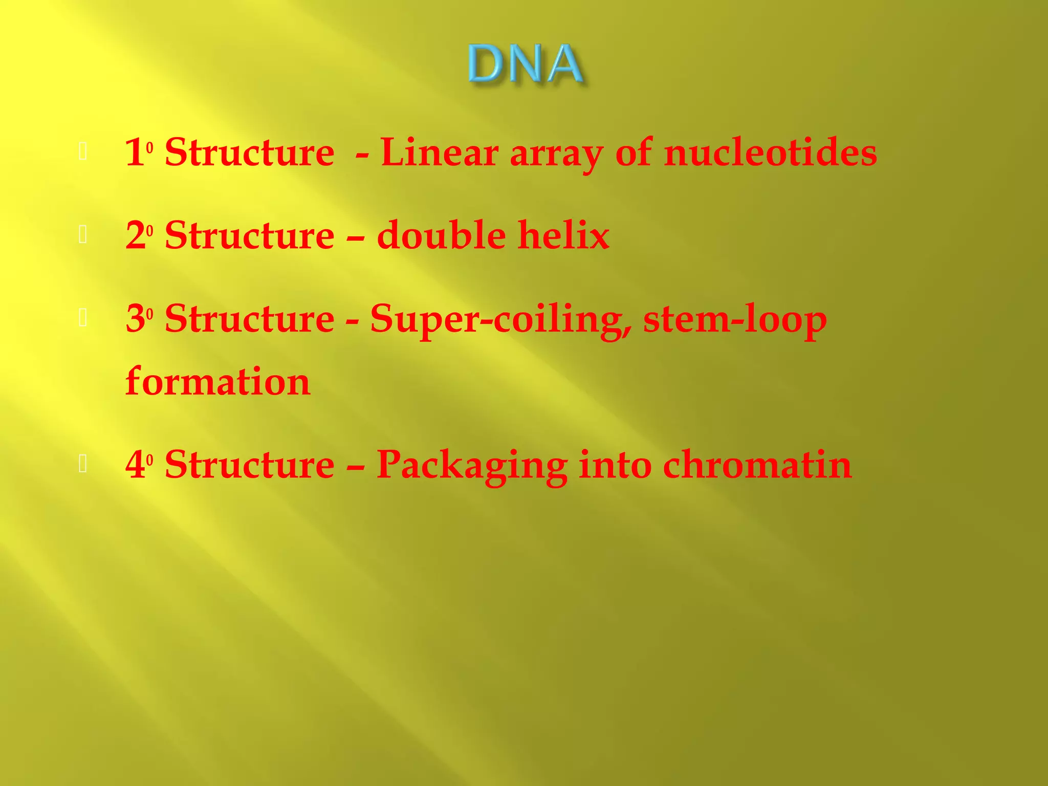
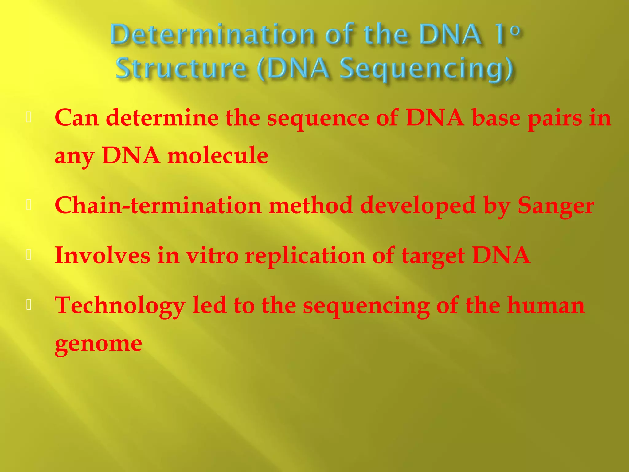
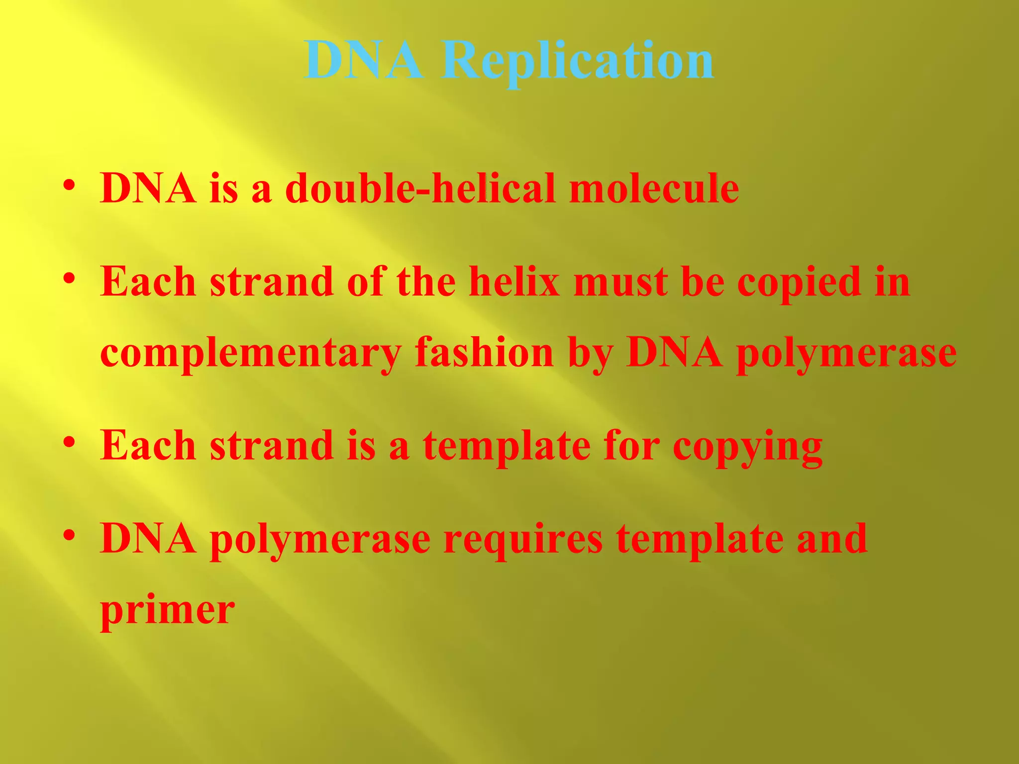
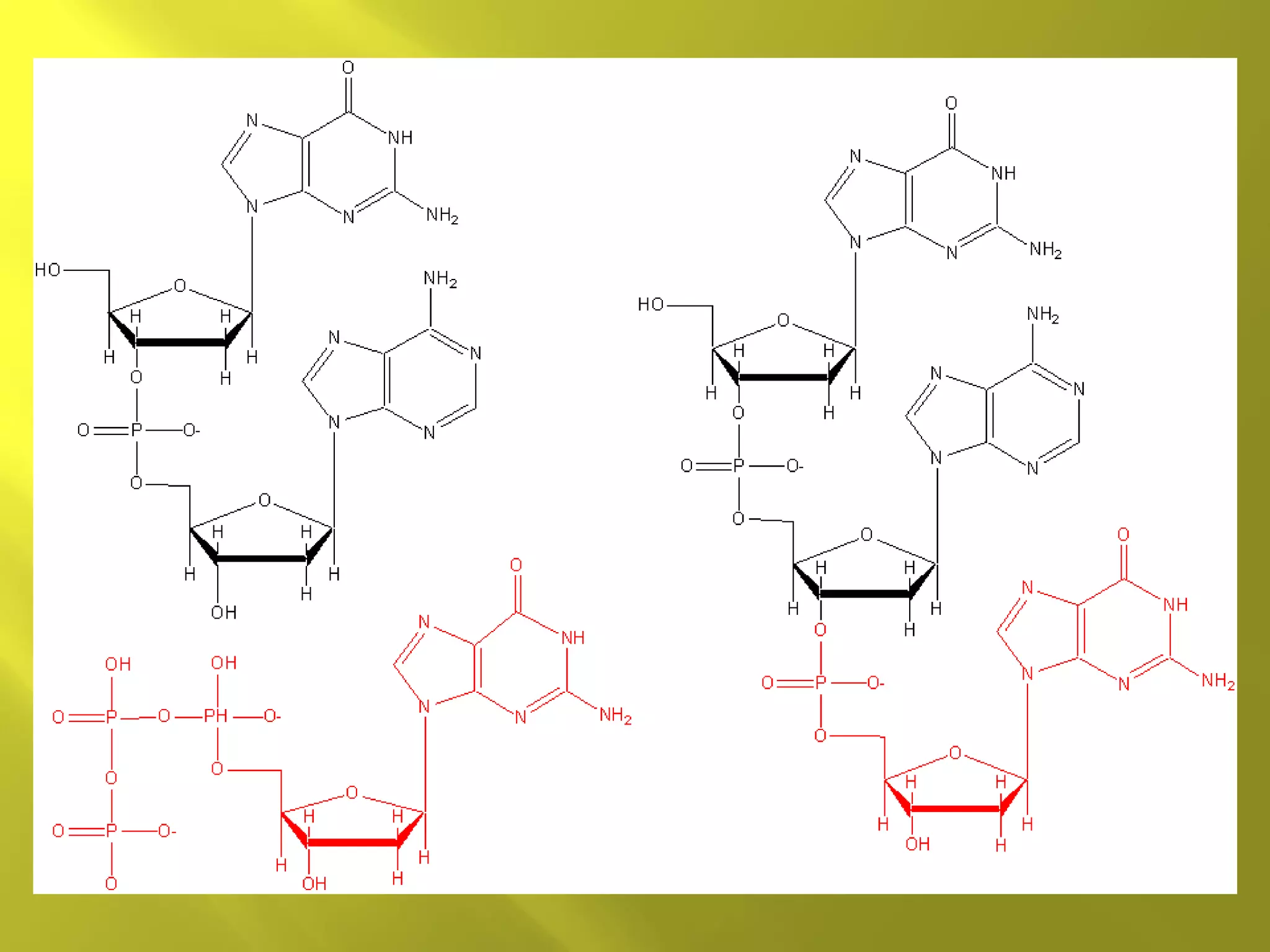
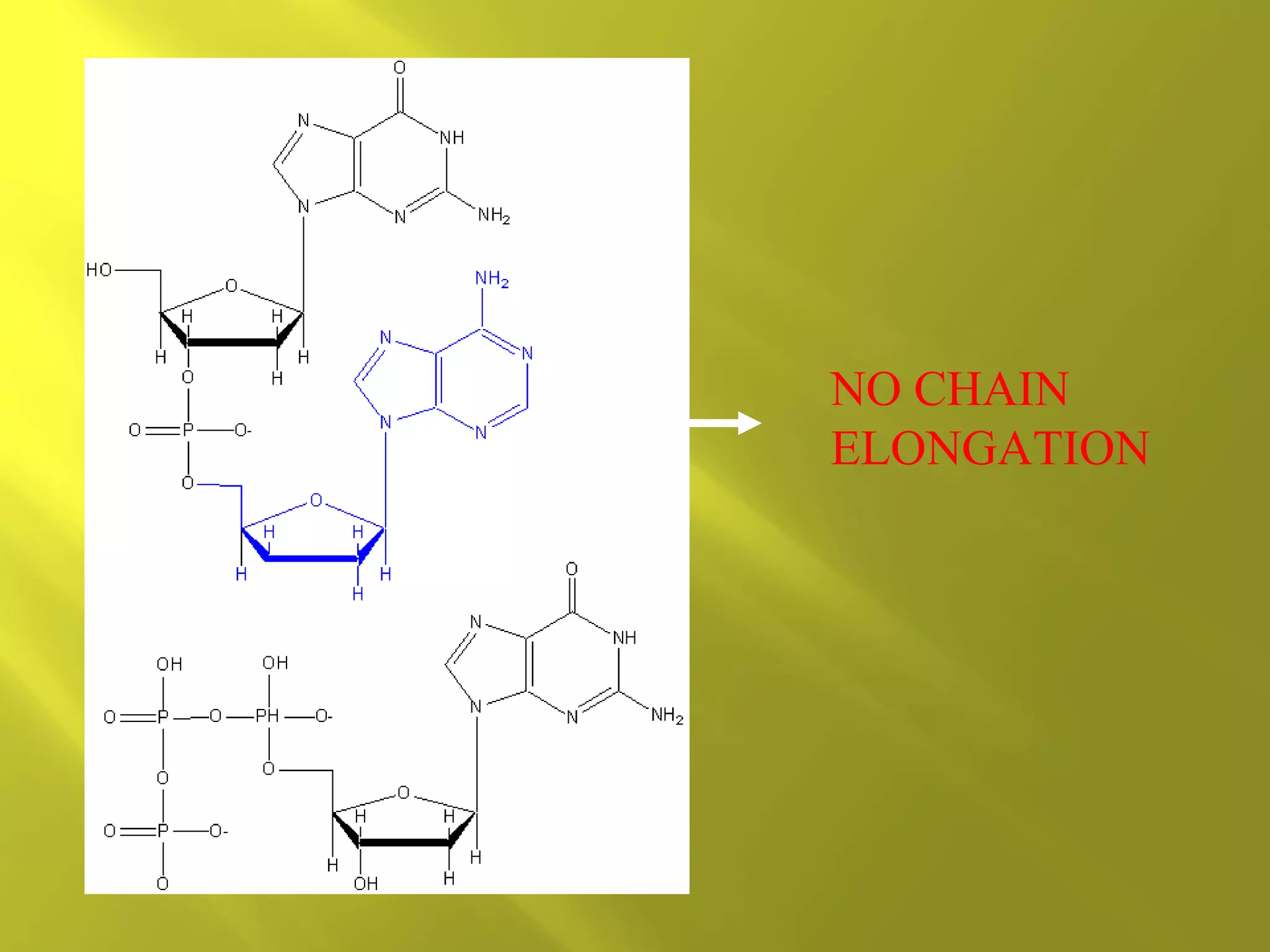
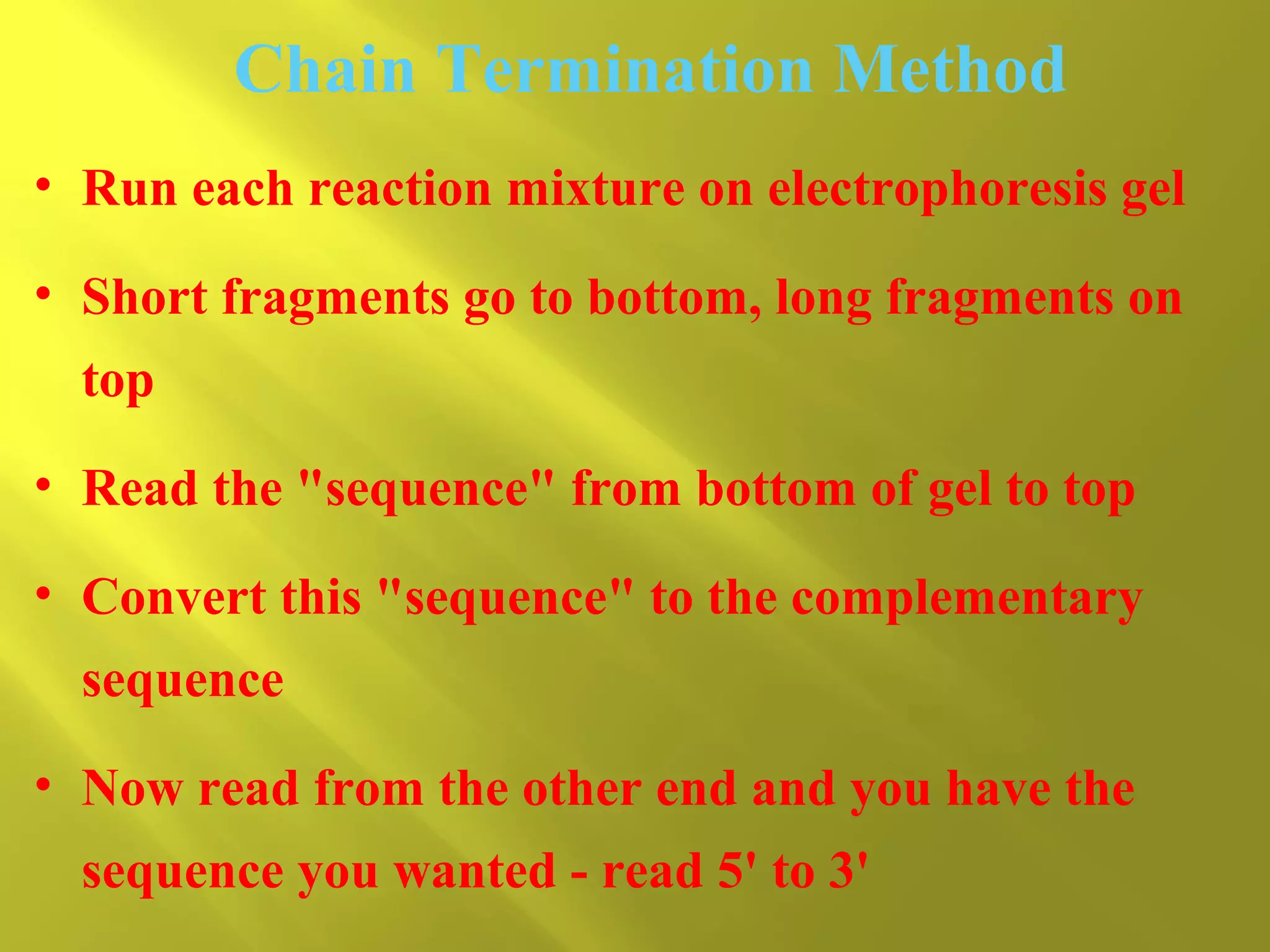
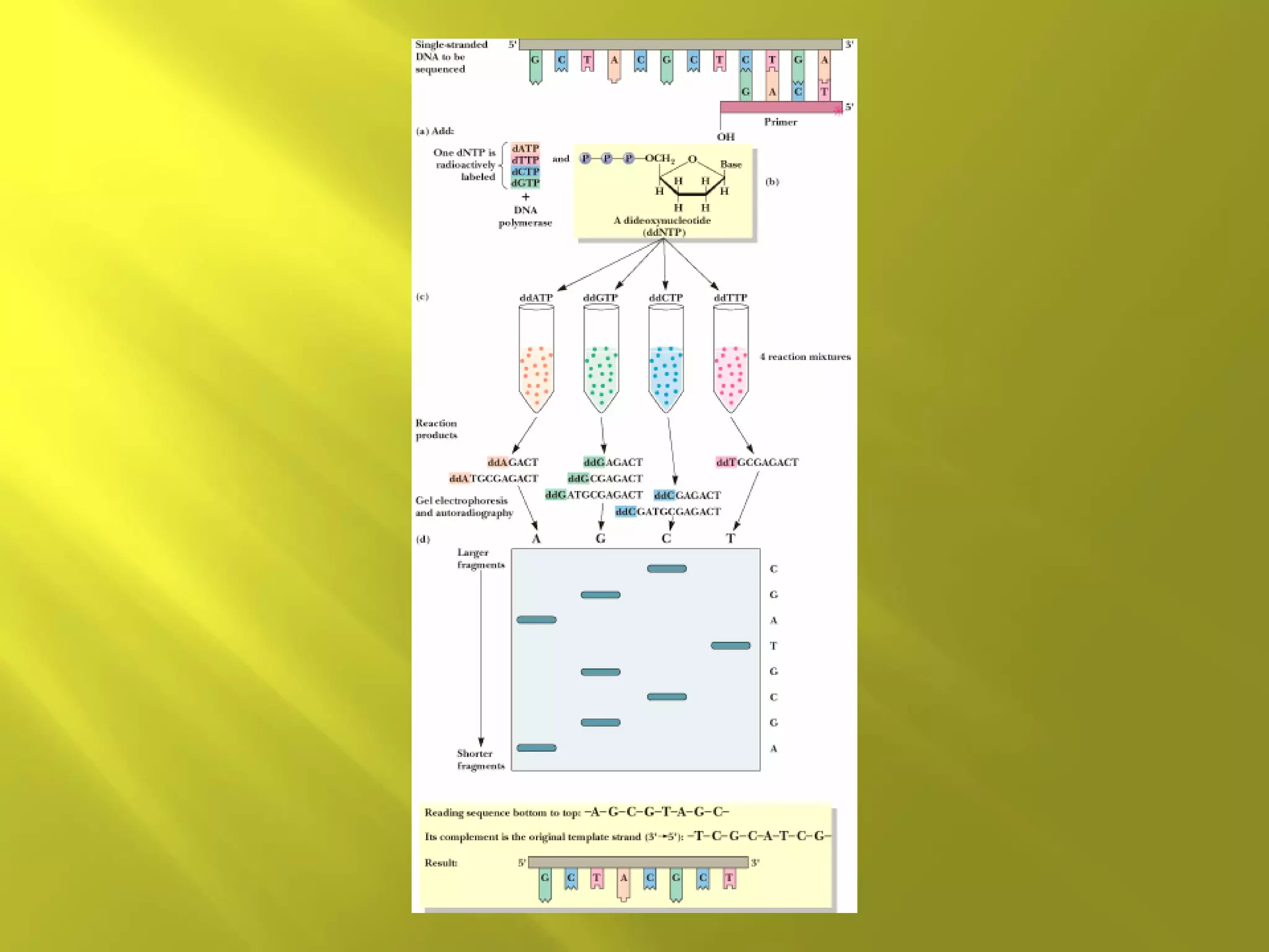
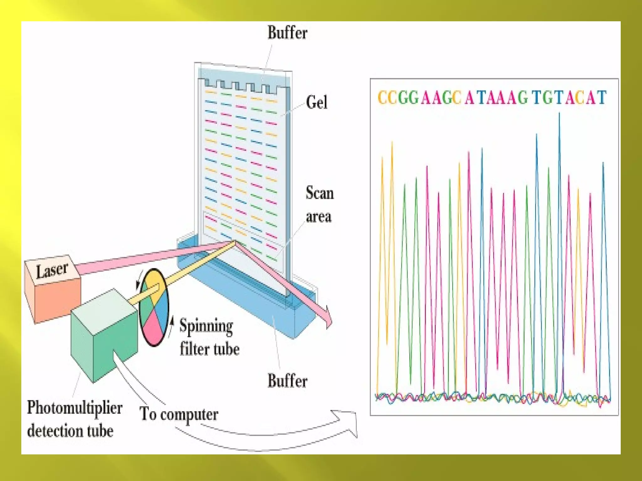
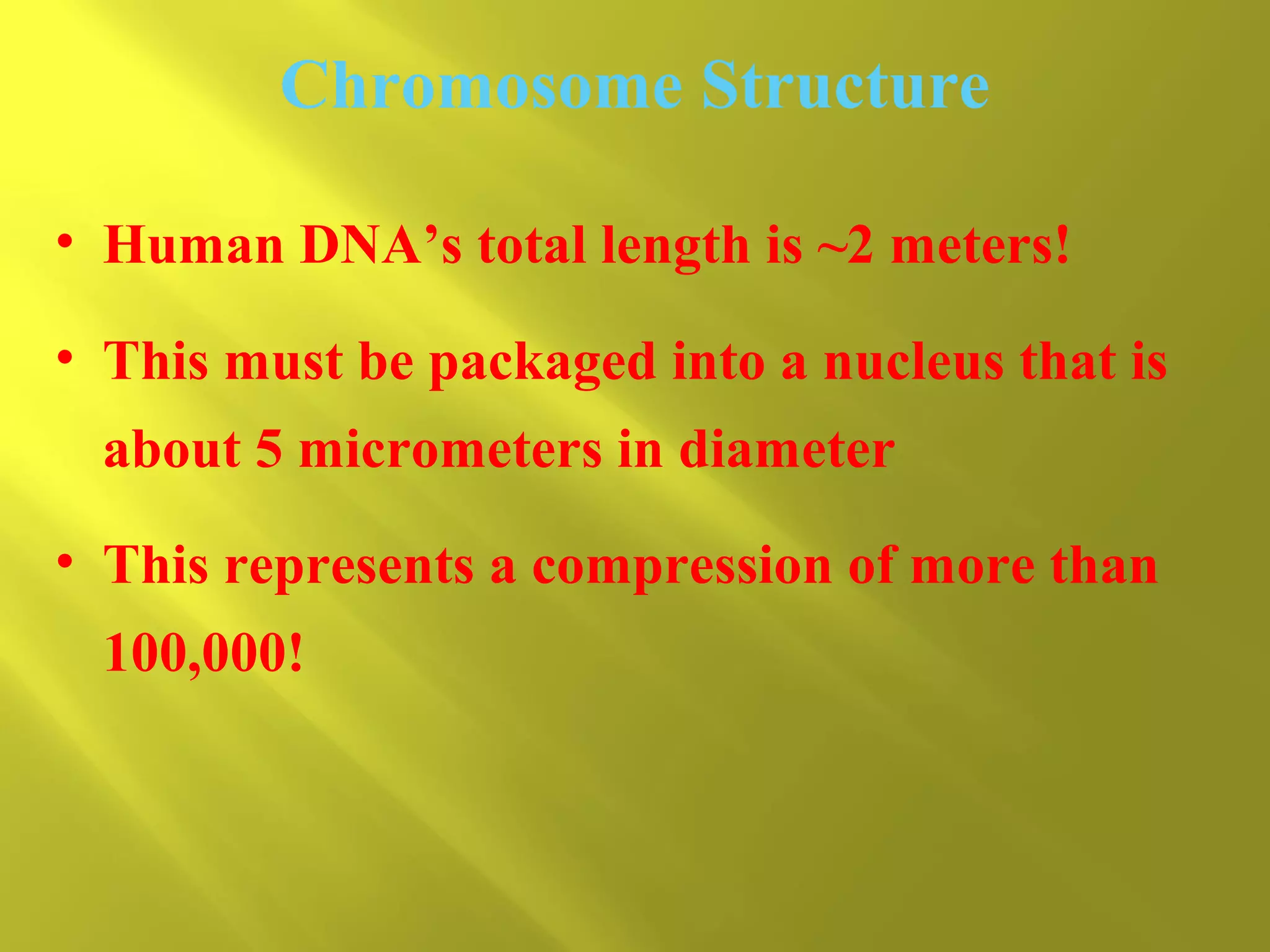
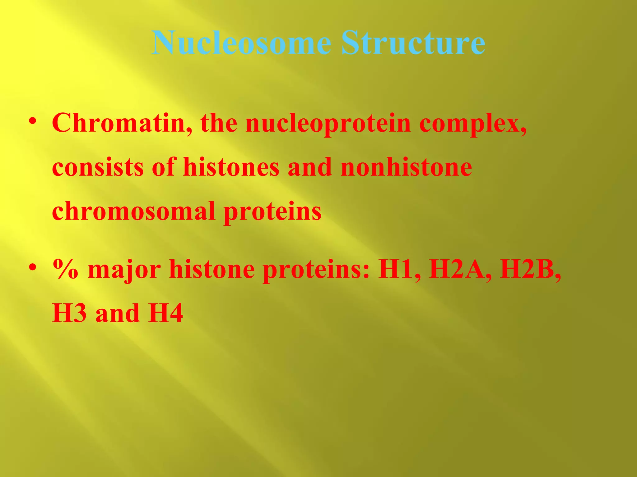
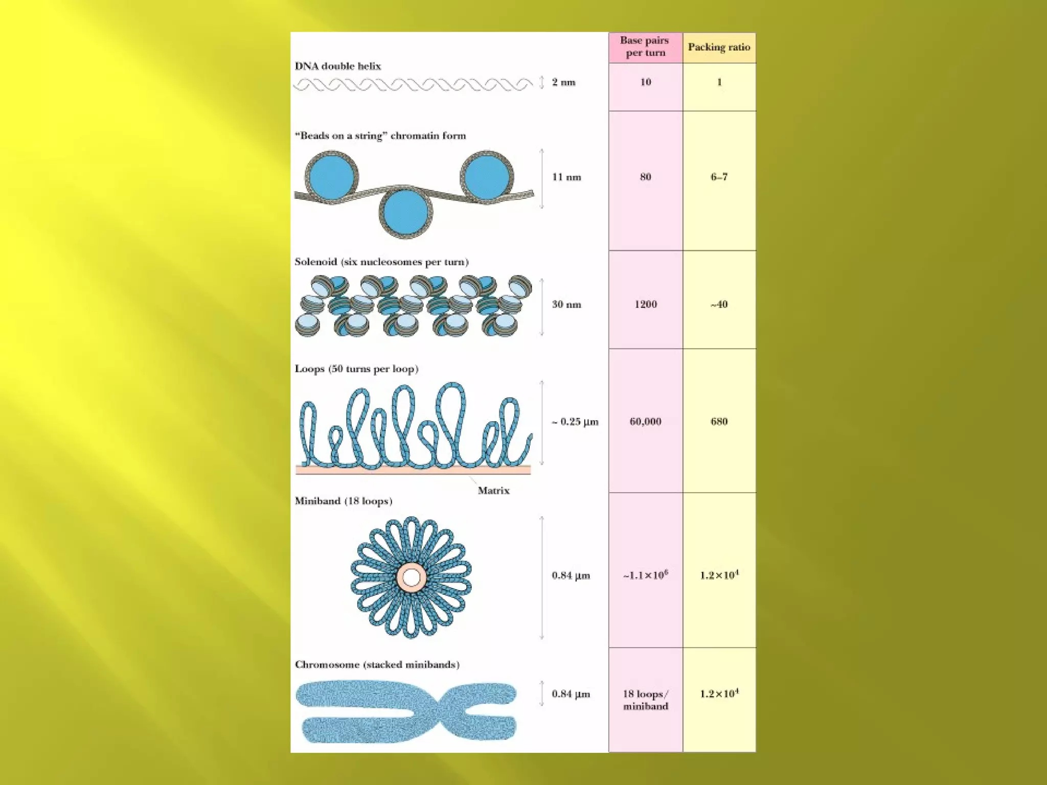


1o Structure - Linear array of nucleotides



2o Structure – double helix



3o Structure - Super-coiling, stem-loop
formation



4o Structure – Packaging into chromatin


Can determine the sequence of DNA base pairs in
any DNA molecule



Chain-termination method developed by Sanger



Involves in vitro replication of target DNA



Technology led to the sequencing of the human
genome
DNA Replication
• DNA is a double-helical molecule
• Each strand of the helix must be copied in
complementary fashion by DNA polymerase
• Each strand is a template for copying
• DNA polymerase requires template and
primer


Primer: an oligonucleotide that pairs with
the end of the template molecule to form
dsDNA



DNA polymerases add nucleotides in 5'-3'
direction
Chain Termination Method
• Based on DNA polymerase reaction
• 4 separate rxns
• Each reaction mixture contains dATP, dGTP,
dCTP and dTTP


Each reaction also contains a small amount of
one dideoxynucleotide (ddATP, ddGTP, ddCTP
and ddTTP).



Each of the 4 dideoxynucleotides are labeled
with a different fluorescent dye.



Dideoxynucleotides missing 3’-OH group. Once
incorporated into the DNA chain, chain
elongation stops)
Chain Termination Method
• Most of the time, the polymerase uses
normal nucleotides and DNA molecules grow
normally
• Occasionally, the polymerase uses a
dideoxynucleotide, which adds to the chain
and then prevents further growth in that
molecule


Random insertion of dd-nucleotides leaves
(optimally) at least a few chains terminated
at every occurrence of a given nucleotide
NO CHAIN
ELONGATION
Chain Termination Method
• Run each reaction mixture on electrophoresis gel
• Short fragments go to bottom, long fragments on
top
• Read the "sequence" from bottom of gel to top
• Convert this "sequence" to the complementary
sequence
• Now read from the other end and you have the
sequence you wanted - read 5' to 3'


DNA is double stranded with antiparallel
strands



Right hand double helix



Three different helical forms (A, B and Z
DNA.
Comparison of A, B, Z DNA
• A: right-handed, short and broad, 2.3 A, 11
bp per turn
• B: right-handed, longer, thinner, 3.32 A, 10
bp per turn
• Z: left-handed, longest, thinnest, 3.8 A, 12
bp per turn
A-DNA

B-DNA

Z-DNA
Z-DNA
• Found in G:C-rich
regions of DNA
• G goes to syn
conformation
• C stays anti but
whole C nucleoside
(base and sugar)
flips 180 degrees


Double Strand DNA can be denatured by
heat (get strand separation)



Can determine degree of denturation by
measuring absorbance at 260 nm.


Conjugated double bonds in bases absorb
light at 260 nm.



Base stacking causes less absorbance.



Increased single strandedness causes
increase in absorbance


Melting temperature related to G:C and
A:T content.



3 H-bonds of G:C pair require higher
temperatures to denture than 2 H-bonds of
A:T pair.


Super coiling



Cruciform structures
Supercoils
• In duplex DNA, ten bp per turn of helix
(relaxed form)
• DNA helix can be over-wound.
• Over winding of DNA helix can be
compensated by supercoiling


Supercoiling prevalent in circular DNA
molecules and within local regions of long
linear DNA strands



Enzymes called topoisomerases or gyrases can
introduce or remove supercoils



In vivo most DNA is negatively supercoiled.



Therefore, it is easy to unwind short regions of
the molecule to allow access for enzymes
Each super coil compensates for one + or –
turn of the double helix
Cruciforms occur in palindromic regions
of DNA
Can form intrachain base pairing
Negative supercoiling may promote
cruciforms


In chromosomes, DNA is tightly associated
with proteins
Chromosome Structure
• Human DNA’s total length is ~2 meters!
• This must be packaged into a nucleus that is
about 5 micrometers in diameter
• This represents a compression of more than
100,000!


It is made possible by wrapping the DNA
around protein spools called nucleosomes
and then packing these in helical filaments
Nucleosome Structure
• Chromatin, the nucleoprotein complex,
consists of histones and nonhistone
chromosomal proteins
• % major histone proteins: H1, H2A, H2B,
H3 and H4


Histone octamers are major part of the
“protein spools”



Nonhistone proteins are regulators of gene
expression
•4 major histone (H2A, H2B, H3, H4) proteins for
octomer
•200 base pair long DNA strand winds around the
octomer
•146 base pair DNA “spacer separates individual
nucleosomes
•H1 protein involved in higher-order chromatin
structure.
•W/O H1, Chromatin looks like beads on string
Solenoid Structure of Chromatin


Single stranded molecule



Chemically less stable than DNA



presence of 2’-OH makes RNA more
susceptible to hydrolytic attack (especially form
bases)



Prone to degradation by Ribonucleases (Rnases)


Has secondary structure. Can form
intrachain base pairing (i.e.cruciform
structures).



Multiple functions


Ribosomal RNA (rRNA) – integral part of
ribosomes (very abundant)



Transfer RNA (tRNA) – carries activated amino
acids to ribosomes.


Messenger RNA (mRNA) – endcodes
sequences of amino acids in proteins.



Catalytic RNA (Ribozymes) – catalzye
cleavage of specific RNA species.
(a)

(b)
Structure of nucleic acid

Structure of nucleic acid

  • 3.
     1o Structure -Linear array of nucleotides  2o Structure – double helix  3o Structure - Super-coiling, stem-loop formation  4o Structure – Packaging into chromatin
  • 4.
     Can determine thesequence of DNA base pairs in any DNA molecule  Chain-termination method developed by Sanger  Involves in vitro replication of target DNA  Technology led to the sequencing of the human genome
  • 5.
    DNA Replication • DNAis a double-helical molecule • Each strand of the helix must be copied in complementary fashion by DNA polymerase • Each strand is a template for copying • DNA polymerase requires template and primer
  • 6.
     Primer: an oligonucleotidethat pairs with the end of the template molecule to form dsDNA  DNA polymerases add nucleotides in 5'-3' direction
  • 8.
    Chain Termination Method •Based on DNA polymerase reaction • 4 separate rxns • Each reaction mixture contains dATP, dGTP, dCTP and dTTP
  • 9.
     Each reaction alsocontains a small amount of one dideoxynucleotide (ddATP, ddGTP, ddCTP and ddTTP).  Each of the 4 dideoxynucleotides are labeled with a different fluorescent dye.  Dideoxynucleotides missing 3’-OH group. Once incorporated into the DNA chain, chain elongation stops)
  • 10.
    Chain Termination Method •Most of the time, the polymerase uses normal nucleotides and DNA molecules grow normally • Occasionally, the polymerase uses a dideoxynucleotide, which adds to the chain and then prevents further growth in that molecule
  • 11.
     Random insertion ofdd-nucleotides leaves (optimally) at least a few chains terminated at every occurrence of a given nucleotide
  • 13.
  • 14.
    Chain Termination Method •Run each reaction mixture on electrophoresis gel • Short fragments go to bottom, long fragments on top • Read the "sequence" from bottom of gel to top • Convert this "sequence" to the complementary sequence • Now read from the other end and you have the sequence you wanted - read 5' to 3'
  • 17.
     DNA is doublestranded with antiparallel strands  Right hand double helix  Three different helical forms (A, B and Z DNA.
  • 18.
    Comparison of A,B, Z DNA • A: right-handed, short and broad, 2.3 A, 11 bp per turn • B: right-handed, longer, thinner, 3.32 A, 10 bp per turn • Z: left-handed, longest, thinnest, 3.8 A, 12 bp per turn
  • 19.
  • 20.
    Z-DNA • Found inG:C-rich regions of DNA • G goes to syn conformation • C stays anti but whole C nucleoside (base and sugar) flips 180 degrees
  • 21.
     Double Strand DNAcan be denatured by heat (get strand separation)  Can determine degree of denturation by measuring absorbance at 260 nm.
  • 22.
     Conjugated double bondsin bases absorb light at 260 nm.  Base stacking causes less absorbance.  Increased single strandedness causes increase in absorbance
  • 24.
     Melting temperature relatedto G:C and A:T content.  3 H-bonds of G:C pair require higher temperatures to denture than 2 H-bonds of A:T pair.
  • 26.
  • 27.
    Supercoils • In duplexDNA, ten bp per turn of helix (relaxed form) • DNA helix can be over-wound. • Over winding of DNA helix can be compensated by supercoiling
  • 28.
     Supercoiling prevalent incircular DNA molecules and within local regions of long linear DNA strands  Enzymes called topoisomerases or gyrases can introduce or remove supercoils  In vivo most DNA is negatively supercoiled.  Therefore, it is easy to unwind short regions of the molecule to allow access for enzymes
  • 29.
    Each super coilcompensates for one + or – turn of the double helix
  • 30.
    Cruciforms occur inpalindromic regions of DNA Can form intrachain base pairing Negative supercoiling may promote cruciforms
  • 33.
     In chromosomes, DNAis tightly associated with proteins
  • 34.
    Chromosome Structure • HumanDNA’s total length is ~2 meters! • This must be packaged into a nucleus that is about 5 micrometers in diameter • This represents a compression of more than 100,000!
  • 35.
     It is madepossible by wrapping the DNA around protein spools called nucleosomes and then packing these in helical filaments
  • 36.
    Nucleosome Structure • Chromatin,the nucleoprotein complex, consists of histones and nonhistone chromosomal proteins • % major histone proteins: H1, H2A, H2B, H3 and H4
  • 37.
     Histone octamers aremajor part of the “protein spools”  Nonhistone proteins are regulators of gene expression
  • 38.
    •4 major histone(H2A, H2B, H3, H4) proteins for octomer •200 base pair long DNA strand winds around the octomer •146 base pair DNA “spacer separates individual nucleosomes •H1 protein involved in higher-order chromatin structure. •W/O H1, Chromatin looks like beads on string
  • 40.
  • 42.
     Single stranded molecule  Chemicallyless stable than DNA  presence of 2’-OH makes RNA more susceptible to hydrolytic attack (especially form bases)  Prone to degradation by Ribonucleases (Rnases)
  • 43.
     Has secondary structure.Can form intrachain base pairing (i.e.cruciform structures).  Multiple functions
  • 44.
     Ribosomal RNA (rRNA)– integral part of ribosomes (very abundant)  Transfer RNA (tRNA) – carries activated amino acids to ribosomes.
  • 45.
     Messenger RNA (mRNA)– endcodes sequences of amino acids in proteins.  Catalytic RNA (Ribozymes) – catalzye cleavage of specific RNA species.
  • 46.