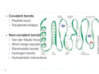
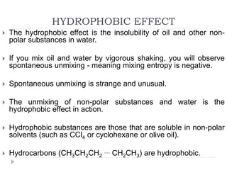
2. Covalent bonds
Peptide bond
Disulphide bridges
Non-covalent bonds
Van der Waals forces
Short range repulsion
Electrostatic bonds
Hydrogen bonds
Hydrophobic interactions
3. COVALENT BONDS
Covalent interactions (bonds) hold atoms together within
molecules.
Covalent bonds are stronger than molecular interactions.
Covalent bonds break and/or form during chemical reactions.
Bonds break during the oxidation of sugar to form carbon
dioxide and water, which is a chemical reaction called fire.
By contrast, covalent bonds remain intact and unchanged when
(a) ice melts, (b) water boils, (c) proteins unfold, (d) RNA
unfolds, (e) DNA strands separate, and (f) membranes
disassemble. These processes are not chemical reactions.
The processes (a-f) of melting, boiling, unfolding, strand
4. COVALENT BONDS
Peptide bonds between Amino Acids (C-N). Can be
broken down into individual amino acids by hydrolysis
with 6M acid/alkali, or by proteases/ proteolytic
enzymes.
Disulphide bridges form between cysteine to form
cystine. (Cysteine has -SH which forms disulphide
bridge -S-S- with another HS-). Bridges are broken
down by reduction with β-mercapto ethanol to form
cysteines once again.
5. MOLECULAR INTERACTONS
Molecular Interactions are attractive or repulsive forces between
molecules and between non-bonded atoms.
Molecular interactions are important in diverse fields of protein
folding, drug design, material science, sensors, nanotechnology,
separations, and origin of life. Molecular interactions are also
known as non-covalent interactions or intermolecular
interactions.
Molecular interactions are not bonds.
All molecular interactions are fundamentally electrostatic in
nature and can described by some variation of Coulombs Law.
6. Native states. In biological systems (i) proteins fold into globular
structures called native states, (ii) ribosomal and transfer RNAs
also fold into native globular structures, (iii) DNA forms double
stranded helices, (iv) phospholipids form membranes, and (v)
proteins assemble with DNA, RNA, membranes and with other
proteins.
These native states and assemblies are stabilized by molecular
interactions of enormous number and complexity. Native states
are destabilized by their low conformational entropy.
Denatured states. When you unfold a protein or an RNA
(denature them) or separate two strands of DNA (melt it), or
disassemble and melt the ribosome, then interior regions
become exposed to the surroundings, which are mostly water
plus some ions.
Molecular interactions within the native state or assembly are
7. A delicate balance. Molecular interactions stabilize both
native and denatured states.
Native biological macromolecules and assemblies are
marginally stable.
Biological systems are held in 'delicate balance between
powerful countervailing forces'.
A small perturbation can tip the balance between the folded
and unfolded states.
A small change in pH or temperature or a single mutation
can unfold a protein.
8. Have you ever denatured
a protein (converted it
from the native state to
denatured state)? Yes.
When you heat an egg to
around 60° C, the albumin
proteins denature and
aggregate. You are not
breaking bonds when you
boil an egg - you are
changing and rearranging
molecular interactions.
The aggregated protein
forms large assemblies
that scatter light, giving
9. NOTE ON VAN DER WAALS INTERACTIONS
Molecular interactions were discovered by the Dutch
scientist Johannes Diderik van der Waals.
He noticed that molecules are sticky, like wet jelly beans.
The phrase 'van der Waals interaction' has come to mean
cohesive (attraction between like), adhesive (attraction
between unlike) and/or repulsive forces between
molecules.
For our purposes, 'van der Waals interaction' is not
sufficiently informative or descriptive or specific.
The problem is that some people use 'van der Waals
interactions' to describe the totality of molecular
interactions but others use it to describe various subsets of
molecular interactions.
Here we avoid the term "van der Waals interaction"
because it is not well-defined and does not decompose the
interactions in a physically meaningful way.
10. SHORT RANGE REPULSION
Atoms take space. Force two atoms together and they will
push back. When two atoms are close together the
occupied orbitals on the atom surfaces overlap, causing
electrostatic repulsion between surface electrons. This
repulsive force between atoms acts over a very short
range, but is very large when distances are short.
Short range repulsion is important to you. It prevents your
hands from passing through each other when you clap,
and prevents atoms from collapsing into tightly packed
states.
11. In B-DNA, the distance
between stacked base pairs
is 3.4 Å as required by
short range repulsion.
Base pairs are slightly
inclined relative to the
helical axis; Base pair
normals are not exactly
parallel to the helical axis.
Therefore the rise per base
pair along the helical axis is
slightly less than 3.4 Å.
12. ELECTROSTATIC INTERACTIONS
Electrostatic interactions are between and among
cations and anions, species with formal charge of ...-
2,-1,+1,+2...
Electrostatic interactions can be either attractive or
repulsive, depending on the signs of the charges.
Like charges repel. Unlike charges attract.The electrostatic interactions within a sodium
chloride crystal are called ionic bonds. But when
a single cation and a single anion are close
together, within a protein, or within a folded RNA,
those interactions are considered to be non-
covalent electrostatic interactions. Non-covalent
electrostatic interactions can be strong, and act at
long range. Electrostatic interactions fall off
gradually with distance (1/r, where r is the
distance between the ions).
13. Electrostatic interactions are the primary stabilizing
interaction between phosphate oxygens of RNA (formal
charge -1) and magnesium ions (formal charge +2). There
are many magnesium ions associated with RNA and DNA
in vivo. Electrostatic interactions are highly attenuated
(dampened) by water. In protein folding, RNA folding and
DNA annealing, electrostatic interactions are dependent on
salt concentration and pH.
In RNA (for example in the ribosome),
anionic phosphate oxygens (-1
charge) engage in attractive
electrostatic interactions with cationic
magnesium ions (+2 charge). Two
phosphate groups can 'clamp' onto
the Mg2+ ion. The O to Mg2+ distance
is 2.1 Å. The dashed lines represent
favorable electrostatic interactions.
14. ION PAIRS IN PROTEINS
Favorable electrostatic interactions between paired anionic
and cationic amino acid side chains are reasonably
frequent in proteins.
Ion Pairs, sometimes called Salt Bridges, are formed when
the charged group of a cationic amino acid (like lysine or
arginine) is around 3.0 to 5.0 Å from the charged group of
an anionic amino acid (like aspartate or glutamate).
The charged groups in an ion pair are generally linked by
hydrogen bonds, in addition to electrostatic interactions.
15. An ion pair within a folded protein. An
anionic aspartic acid (charge = -1) engages
in attractive electrostatic interactions with
cationic arginine (charge = +1). The
dashed lines represent hydrogen bonds.
16. HYDROGEN BOND
Attraction between hydrogen atom which is bonded to
electronegative atoms such as F,O,N and an adjacent
electronegative atom.
Why hydrogen?
Hydrogen is special because it is the only atom that (i)
forms covalent sigma bonds with electronegative atoms
like N, O and S, and (ii) uses the inner shell (1S)
electron(s) in that covalent bond.
17. The most common hydrogen
bonds in biological systems
involve oxygen and nitrogen
atoms as A and D. Keto
groups (=O), amines (R3N),
imines (R=N-R) and hydroxyl
groups (-OH) are the most
common hydrogen bond
acceptors in DNA, RNA,
proteins and complex
carbohydrates. Hydroxyl
groups and amines/imines
are the most common
hydrogen bond donors.
Hydroxyls and amines/imines
can both donate and accept
hydrogen bonds.
18.
19. An isolated ammonia
molecule, just like a water
molecule, can form strong
hydrogen bonds with either
hydrogen bond donors or
acceptors. Ammonia is
more basic than water, and
therefore ammonia is a
better hydrogen bond
acceptor than water.
20. HYDROPHOBIC EFFECT
The hydrophobic effect is the insolubility of oil and other non-
polar substances in water.
If you mix oil and water by vigorous shaking, you will observe
spontaneous unmixing - meaning mixing entropy is negative.
Spontaneous unmixing is strange and unusual.
The unmixing of non-polar substances and water is the
hydrophobic effect in action.
Hydrophobic substances are those that are soluble in non-polar
solvents (such as CCl4 or cyclohexane or olive oil).
Hydrocarbons (CH3CH2CH2
.... CH2CH3) are hydrophobic.
21. A very important factor to remember is that the hydrophobic
effect is fully a property of water; it a consequence of the
distinctive molecular structure of water and the unique cohesive
properties of water.
Hydrophobic substances are passive participants in the
hydrophobic phenomenon.
The driver for unmixing is interfacial water molecules (directly
adjacent a hydrocarbon molecule or plastic surface) that
maintain water-water interactions at the cost of rotational and
translational freedom.
Interfacial water has low entropy and is therefore unfavourable.
Water gains entropy and therefore stability by minimizing the
amount of interfacial water.
This is why water droplets deform on a hydrophobic surface -
droplets adjust their shape to minimize contact with a
hydrophobic surface.
22.
23. CONTER-ION RELEASE FROM NUCLEIC ACID
Counter ions are released when a cationic protein binds to DNA.
This release explains the dramatic salt dependencies of DNA-
protein complexes.
High salt destabilizes DNA-protein complexes.
If the bulk salt concentration is low, the protein binds tightly to
the DNA.
If the bulk salt concentration is high, the protein binds weakly.
Counter ion release explains much of the salt dependencies of
DNA melting, RNA folding and DNA condensation.
24. Figure shows an axial view of DNA, represented as a anionic cylinder. Cationic
counter ions (orange shading) surround the cylinder. The concentration of
cations decreases with distance from the surface of the cylinder. The deeper
orange shading indicates more concentrated cations. The panel on the right
illustrates how both anionic counter ions (blue) associated with a cationic
protein, and cationic counter ions (orange) associated with anionic DNA, are
released to bulk solution when the protein binds to DNA. This release of
counter ions drives the association.