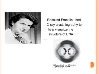
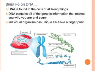
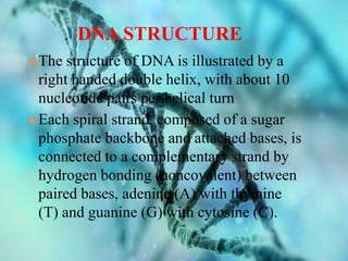
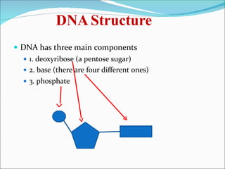
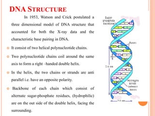
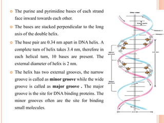
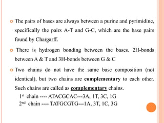
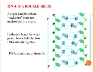
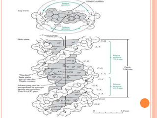
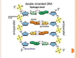
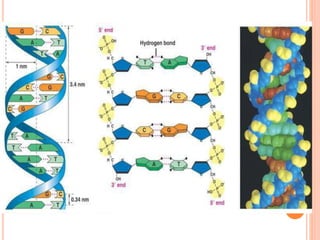
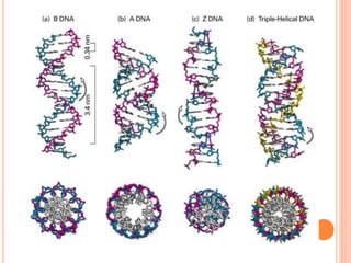
Assignment Presentation
On
DNA STRUCTURE AND FUNCTION
Submitted by :
Desai vruddhi k.
M.Sc. (Agri.) GPB
S.D.Agriculture University,
Sardarkrushinagar
PRINCIPLES OF BIOTECHNOLOGY (MBB 501)
HISTORY
 In 1869, Miescher discovered
"nuclein" (DNA) in the cells from pus
& later he separated it into a protein and
an acid molecule. It came to known as
nucleic acid after 1874.
 1926 , Levene proposed “Tetra
nucleotide theory” which states that
Nucleic acid consists of only 4
nitridesas it gives 4 different
nucleotides on hydrolysis.
 Rosalind Franklin used
X-ray crystallography to
help visualize the
structure of DNA
 James D. Watson and Francis Crick, co-
originators of the doublehelix model.
BRIEFING ON DNA…
 DNA is found in the cells of all living things.
 DNA contains all of the genetic information that makes
you who you are and every
 individual organism has unique DNA like a finger print.
 It contained phosphorus in the form of phosphate
 Deoxyribo nucleic acid
 It is a molecule that encodes the genetic instructions.
 Most DNA molecules are double-stranded helices.
 Each molecule consists of two long biopolymers
made of simpler units called nucleotides—each
nucleotide is composed of a nucleobase recorded
using the letters G, A, T, and C.
 DNA is well-suited for biological information storage.
 DNA Stands for “DeoxyriboNcleic Acid”.
 Term DNA was given by Zaccharis
 DNA is biopolymer consist of nucleotide as monomeric
unit.
 DNA is double helical structure in eukaryote and
prokaryote, but in virus it may be double stranded or
single stranded and presented as monopartite or
multipartite.
 In eukaryotes, DNA is presented in nucleus surrounded
by nuclear membrane
 In prokaryotes, DNA is presented in nucleoid region of
Cytoplasm without nuclear membrane
 In virus, DNA is presented in the core of virus
surrounded by Protein layer (called as capsid).
WHAT IS DNA?
 DNA, or deoxyribonucleic acid, is the hereditary
material in humans and almost all other
organisms. Nearly every cell in a person’s body
has the same DNA.
 Where is it located?
 Most DNA is located in the cell nucleus (where
it is called nuclear DNA), but a small amount of
DNA can also be found in the mitochondria
(where it is called mitochondrial DNA or mtDNA).
DNA STRUCTURE
The structure of DNA is illustrated by a
right handed double helix, with about 10
nucleotide pairs per helical turn
Each spiral strand, composed of a sugar
phosphate backbone and attached bases, is
connected to a complementary strand by
hydrogen bonding (noncovalent) between
paired bases, adenine (A) with thymine
(T) and guanine (G) with cytosine (C).
DNA Structure
 DNA has three main components
 1. deoxyribose (a pentose sugar)
 2. base (there are four different ones)
 3. phosphate
THE SUGARS
The Bases
 They are divided into two groups
 Pyrimidines and purines
 Pyrimidines (made of one 6 member ring)
 Thymine
 Cytosine
 Purines (made of a 6 member ring, fused to a 5
member ring)
 Adenine
 Guanine
 The rings are not only made of carbon (specific
formulas and structures are not required for
IB)
CHEMICAL STRUCTURE OF DNAAND RNA
RNA DNA
Nucleotide
Nucleoside
1’
2’
4’
The C is
named 1’-5’
THE BASES
PURINES
PYRIMIDINES
NITROGENOUS BASES
TAUTOMERISM
Tautomers are isomers of a compound
which differ only in the position of the
protons and electrons. ... A reaction which
involves simple proton transfer in an
intramolecular fashion is called
a tautomerism. Keto-enol tautomerism is
a very common process, and is acid or
base catalysed.
FUNCTIONS
 Nucleotides are precursors of the
nucleic acids, deoxyribonucleic acid
(DNA) and ribonucleic acid (RNA).
 The nucleic acids are concerned with
the storage and transfer of genetic
information.
 The universal currency of energy, namely ATP, is a nucleotide
derivative Nucleotides are also components of important co-
enzymes like - NAD+ and FAD, and - metabolic regulators such as
cAMP and cGMP.
 Dna must be stable
 There are coding and noncoding regions found on DNA
 Coding region code for genes(proteins).
 Non-coding regions can be either DNA junk or help regulate protein
synthesis.
DNA STRUCTURE
In 1953, Watson and Crick postulated a
three dimensional model of DNA structure that
accounted for both the X-ray data and the
characteristic base pairing in DNA.
 It consist of two helical polynucleotide chains.
 Two polynucleotide chains coil around the same
axis to form a right –handed double helix.
 In the helix, the two chains or strands are anti
parallel i.e. have an opposite polarity.
 Backbone of each chain which consist of
alternate sugar-phosphate residues, (hydrophilic)
are on the out side of the double helix, facing the
surrounding.
 The purine and pyrimidine bases of each strand
face inward towards each other.
 The bases are stacked perpendicular to the long
axis of the double helix.
 The base pair are 0.34 nm apart in DNA helix. A
complete turn of helix takes 3.4 nm, therefore in
each helical turn, 10 bases are present. The
external diameter of helix is 2 nm.
 The helix has two external grooves, the narrow
groove is called as minor groove while the wide
groove is called as major groove . The major
groove is the site for DNA binding proteins. The
minor grooves often are the site for binding
small molecules.
 The pairs of bases are always between a purine and pyrimidine,
specifically the pairs A-T and G-C, which are the base pairs
found by Chargarff.
 There is hydrogen bonding between the bases. 2H-bonds
between A & T and 3H-bonds between G & C
 Two chains do not have the same base composition (not
identical), but two chains are complementary to each other.
Such chains are called as complementary chains.
1st chain ---- ATACGCAC---3A, 1T, 3C, 1G
2nd chain ---- TATGCGTG---1A, 3T, 1C, 3G
DNA IS A DOUBLE HELIX
P
A
P C
P
G
P T
P C
P
G
P
A
PC
PT
G
P
PC P
A sugar and phosphate
“backbone” connects
nucleotides in a chain.
P
G
P
DNA strands are antiparallel.
5’
3’
3’
5’
Hydrogen bonds between
paired bases hold the two
DNA strands together.
A-DNA
 A-DNA is one of the many possible
double helical structures of DNA.
 It is most active along with other forms.
 Helix has left-handed sense, shorter
more compact helical structure.
 It occurs only in dehydrated samples of
DNA, such as those used in crystallographic
experiments.
 Structure
 A-DNA is fairly similar to B-DNA.
 Slight increase in the number of bp/ rotation
(resulting in a tighter rotation angle), and smaller
rise/turn.
 deep major groove and a shallow minor groove.
 Favoured conformation at low water
concentrations.
 In a solution with higher salt concentrations or with
alcohol
added, the DNA structure may change to an A form,
which is still right-handed, but every 2.3 nm makes
a turn and there are 11 base pairs per turn.
B-DNA
 Most common DNA conformation
in vivo.
 Favoured conformation at high
water concentrations.
 Also known as Watson & Crick
model of DNA.
 First identified in fibre at 92%
relative humidity.
 Structure
 Narrower, more elongated helix than A.
 Wide major groove easily accessible to proteins
& Narrow minor groove.
 Base pairs nearly perpendicular to helix axis
One spiral is 3.4nm or 34Ǻ.
 Distance between two H-bonds is 0.34nm or
3.4Ǻ.
Z-DNA
 Z-DNA is one of the many possible
double helical structures of DNA.
 Helix has left-handed sense.
 It is most active double helical
structure.
 Can be formed in vivo, given proper
sequence and super helical tension, but
function remains obscure.
Side view of A-, B-, and Z-DNA. The helix axis of A-, B-, and Z-DNA.
.“DNA DENATURATION”
 DNA Denaturation is the separation of a double strand
into two single strands, which occurs when the hydrogen
bonds between the strands are broken.
HYPERCHROMIC EFFECT
 is the increas of absorbance (optical
density) of a material. The most
famous example is the
hyperchromicity of DNAthat occurs
when the DNA duplex is denatured.
The UV absorption is increased when
the two single DNA strands are being
separated, either by heat or by
addition of denaturant or by
increasing the pH level. The opposite,
a decrease of absorbance is
called hypochromicity.
CONCLUSION
 The secondary structure of DNA is important in
many events in cellular life. Replication,
transcription and regulation of expression of many
genes depends on local differences or changes in
DNA structure. Recombination which leads to
rearrangement of genes takes advantage of the
ability to form an unusual structure called a
Holliday’s structure. Also different kinds of
mutations occur as a result of specific DNA
structure.
DNA strcture and function

DNA strcture and function

  • 2.
    Assignment Presentation On DNA STRUCTUREAND FUNCTION Submitted by : Desai vruddhi k. M.Sc. (Agri.) GPB S.D.Agriculture University, Sardarkrushinagar PRINCIPLES OF BIOTECHNOLOGY (MBB 501)
  • 5.
    HISTORY  In 1869,Miescher discovered "nuclein" (DNA) in the cells from pus & later he separated it into a protein and an acid molecule. It came to known as nucleic acid after 1874.  1926 , Levene proposed “Tetra nucleotide theory” which states that Nucleic acid consists of only 4 nitridesas it gives 4 different nucleotides on hydrolysis.
  • 6.
     Rosalind Franklinused X-ray crystallography to help visualize the structure of DNA
  • 7.
     James D.Watson and Francis Crick, co- originators of the doublehelix model.
  • 8.
    BRIEFING ON DNA… DNA is found in the cells of all living things.  DNA contains all of the genetic information that makes you who you are and every  individual organism has unique DNA like a finger print.
  • 9.
     It containedphosphorus in the form of phosphate  Deoxyribo nucleic acid  It is a molecule that encodes the genetic instructions.  Most DNA molecules are double-stranded helices.  Each molecule consists of two long biopolymers made of simpler units called nucleotides—each nucleotide is composed of a nucleobase recorded using the letters G, A, T, and C.  DNA is well-suited for biological information storage.
  • 10.
     DNA Standsfor “DeoxyriboNcleic Acid”.  Term DNA was given by Zaccharis  DNA is biopolymer consist of nucleotide as monomeric unit.  DNA is double helical structure in eukaryote and prokaryote, but in virus it may be double stranded or single stranded and presented as monopartite or multipartite.  In eukaryotes, DNA is presented in nucleus surrounded by nuclear membrane  In prokaryotes, DNA is presented in nucleoid region of Cytoplasm without nuclear membrane  In virus, DNA is presented in the core of virus surrounded by Protein layer (called as capsid).
  • 11.
    WHAT IS DNA? DNA, or deoxyribonucleic acid, is the hereditary material in humans and almost all other organisms. Nearly every cell in a person’s body has the same DNA.  Where is it located?  Most DNA is located in the cell nucleus (where it is called nuclear DNA), but a small amount of DNA can also be found in the mitochondria (where it is called mitochondrial DNA or mtDNA).
  • 12.
    DNA STRUCTURE The structureof DNA is illustrated by a right handed double helix, with about 10 nucleotide pairs per helical turn Each spiral strand, composed of a sugar phosphate backbone and attached bases, is connected to a complementary strand by hydrogen bonding (noncovalent) between paired bases, adenine (A) with thymine (T) and guanine (G) with cytosine (C).
  • 13.
    DNA Structure  DNAhas three main components  1. deoxyribose (a pentose sugar)  2. base (there are four different ones)  3. phosphate
  • 14.
  • 15.
    The Bases  Theyare divided into two groups  Pyrimidines and purines  Pyrimidines (made of one 6 member ring)  Thymine  Cytosine  Purines (made of a 6 member ring, fused to a 5 member ring)  Adenine  Guanine  The rings are not only made of carbon (specific formulas and structures are not required for IB)
  • 16.
    CHEMICAL STRUCTURE OFDNAAND RNA RNA DNA Nucleotide Nucleoside 1’ 2’ 4’ The C is named 1’-5’
  • 17.
  • 18.
  • 19.
    TAUTOMERISM Tautomers are isomersof a compound which differ only in the position of the protons and electrons. ... A reaction which involves simple proton transfer in an intramolecular fashion is called a tautomerism. Keto-enol tautomerism is a very common process, and is acid or base catalysed.
  • 24.
    FUNCTIONS  Nucleotides areprecursors of the nucleic acids, deoxyribonucleic acid (DNA) and ribonucleic acid (RNA).  The nucleic acids are concerned with the storage and transfer of genetic information.  The universal currency of energy, namely ATP, is a nucleotide derivative Nucleotides are also components of important co- enzymes like - NAD+ and FAD, and - metabolic regulators such as cAMP and cGMP.  Dna must be stable  There are coding and noncoding regions found on DNA  Coding region code for genes(proteins).  Non-coding regions can be either DNA junk or help regulate protein synthesis.
  • 25.
    DNA STRUCTURE In 1953,Watson and Crick postulated a three dimensional model of DNA structure that accounted for both the X-ray data and the characteristic base pairing in DNA.  It consist of two helical polynucleotide chains.  Two polynucleotide chains coil around the same axis to form a right –handed double helix.  In the helix, the two chains or strands are anti parallel i.e. have an opposite polarity.  Backbone of each chain which consist of alternate sugar-phosphate residues, (hydrophilic) are on the out side of the double helix, facing the surrounding.
  • 26.
     The purineand pyrimidine bases of each strand face inward towards each other.  The bases are stacked perpendicular to the long axis of the double helix.  The base pair are 0.34 nm apart in DNA helix. A complete turn of helix takes 3.4 nm, therefore in each helical turn, 10 bases are present. The external diameter of helix is 2 nm.  The helix has two external grooves, the narrow groove is called as minor groove while the wide groove is called as major groove . The major groove is the site for DNA binding proteins. The minor grooves often are the site for binding small molecules.
  • 27.
     The pairsof bases are always between a purine and pyrimidine, specifically the pairs A-T and G-C, which are the base pairs found by Chargarff.  There is hydrogen bonding between the bases. 2H-bonds between A & T and 3H-bonds between G & C  Two chains do not have the same base composition (not identical), but two chains are complementary to each other. Such chains are called as complementary chains. 1st chain ---- ATACGCAC---3A, 1T, 3C, 1G 2nd chain ---- TATGCGTG---1A, 3T, 1C, 3G
  • 28.
    DNA IS ADOUBLE HELIX P A P C P G P T P C P G P A PC PT G P PC P A sugar and phosphate “backbone” connects nucleotides in a chain. P G P DNA strands are antiparallel. 5’ 3’ 3’ 5’ Hydrogen bonds between paired bases hold the two DNA strands together.
  • 32.
    A-DNA  A-DNA isone of the many possible double helical structures of DNA.  It is most active along with other forms.  Helix has left-handed sense, shorter more compact helical structure.  It occurs only in dehydrated samples of DNA, such as those used in crystallographic experiments.
  • 33.
     Structure  A-DNAis fairly similar to B-DNA.  Slight increase in the number of bp/ rotation (resulting in a tighter rotation angle), and smaller rise/turn.  deep major groove and a shallow minor groove.  Favoured conformation at low water concentrations.  In a solution with higher salt concentrations or with alcohol added, the DNA structure may change to an A form, which is still right-handed, but every 2.3 nm makes a turn and there are 11 base pairs per turn.
  • 35.
    B-DNA  Most commonDNA conformation in vivo.  Favoured conformation at high water concentrations.  Also known as Watson & Crick model of DNA.  First identified in fibre at 92% relative humidity.
  • 36.
     Structure  Narrower,more elongated helix than A.  Wide major groove easily accessible to proteins & Narrow minor groove.  Base pairs nearly perpendicular to helix axis One spiral is 3.4nm or 34Ǻ.  Distance between two H-bonds is 0.34nm or 3.4Ǻ.
  • 37.
    Z-DNA  Z-DNA isone of the many possible double helical structures of DNA.  Helix has left-handed sense.  It is most active double helical structure.  Can be formed in vivo, given proper sequence and super helical tension, but function remains obscure.
  • 39.
    Side view ofA-, B-, and Z-DNA. The helix axis of A-, B-, and Z-DNA.
  • 41.
    .“DNA DENATURATION”  DNADenaturation is the separation of a double strand into two single strands, which occurs when the hydrogen bonds between the strands are broken.
  • 42.
    HYPERCHROMIC EFFECT  isthe increas of absorbance (optical density) of a material. The most famous example is the hyperchromicity of DNAthat occurs when the DNA duplex is denatured. The UV absorption is increased when the two single DNA strands are being separated, either by heat or by addition of denaturant or by increasing the pH level. The opposite, a decrease of absorbance is called hypochromicity.
  • 43.
    CONCLUSION  The secondarystructure of DNA is important in many events in cellular life. Replication, transcription and regulation of expression of many genes depends on local differences or changes in DNA structure. Recombination which leads to rearrangement of genes takes advantage of the ability to form an unusual structure called a Holliday’s structure. Also different kinds of mutations occur as a result of specific DNA structure.