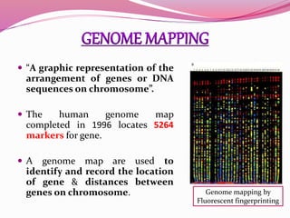
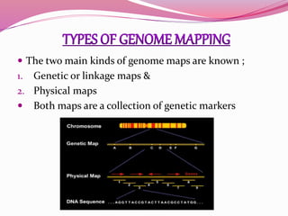
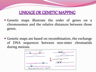
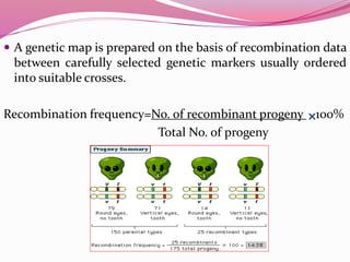
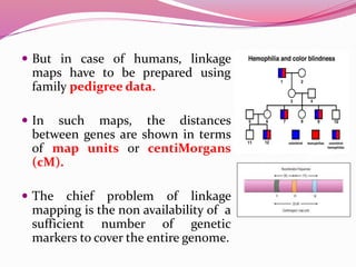
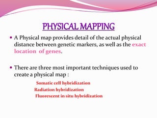
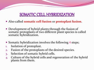
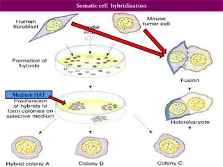
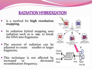
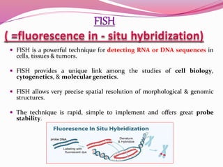
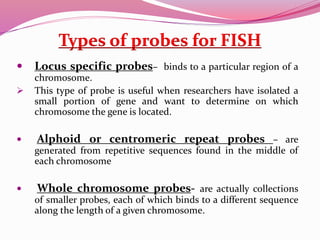
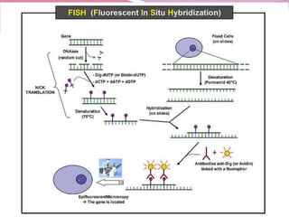
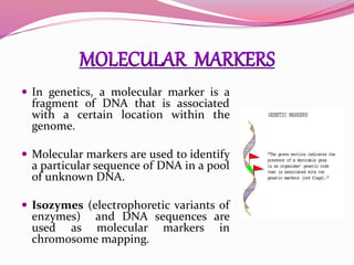
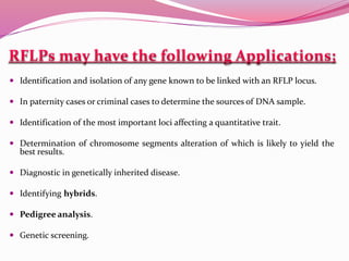
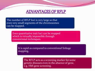
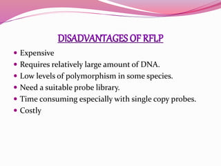
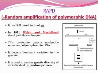
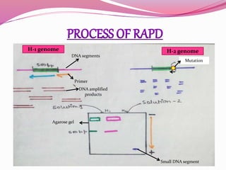
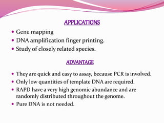
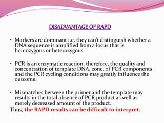
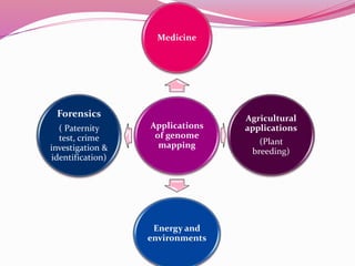
 The haploid set of chromosomes
in a gamete or microorganisms, or
in each cell of multicellular
organisms.
 It is encoded either in DNA or for
many types of viruses, in RNA.
 Each genome contains all of the
information needed to build and
maintain that organisms.
 “A graphic representation of the
arrangement of genes or DNA
sequences on chromosome”.
 The human genome map
completed in 1996 locates 5264
markers for gene.
 A genome map are used to
identify and record the location
of gene & distances between
genes on chromosome. Genome mapping by
Fluorescent fingerprinting
 The two main kinds of genome maps are known ;
1. Genetic or linkage maps &
2. Physical maps
 Both maps are a collection of genetic markers
 Genetic maps illustrate the order of genes on a
chromosomes and the relative distances between those
genes.
 Genetic maps are based on recombination, the exchange
of DNA sequences between non-sister chromatids
during meiosis.
 A genetic map is prepared on the basis of recombination data
between carefully selected genetic markers usually ordered
into suitable crosses.
Recombination frequency=No. of recombinant progeny 100%
Total No. of progeny
 But in case of humans, linkage
maps have to be prepared using
family pedigree data.
 In such maps, the distances
between genes are shown in terms
of map units or centiMorgans
(cM).
 The chief problem of linkage
mapping is the non availability of a
sufficient number of genetic
markers to cover the entire genome.
 A Physical map provides detail of the actual physical
distance between genetic markers, as well as the exact
location of genes.
 There are three most important techniques used to
create a physical map :
Somatic cell hybridization
Radiation hybridization
Fluorescent in situ hybridization
 Also called somatic cell fusion or protoplast fusion.
 Development of hybrid plants through the fusion of
somatic protoplasts of two different plant species is called
somatic hybridization.
 Somatic hybridization involves the following 4 steps;
1. Isolation of protoplast.
2. Fusion of the protoplasts of the desired species.
3. Selection of somatic hybrid cells.
4. Culture of the hybrid cells and regeneration of the hybrid
plants from them.
Somatic cell hybridization
Medium HAT
 is a method for high resolution
mapping.
 In radiation hybrid mapping uses
radiation such as x- ray, to break
the DNA into fragments.
 The amount of radiation can be
adjusted to create smaller or larger
fragments.
 This technique is not affected by
increased or decreased
recombination frequency.
 FISH is a powerful technique for detecting RNA or DNA sequences in
cells, tissues & tumors.
 FISH provides a unique link among the studies of cell biology,
cytogenetics, & molecular genetics.
 FISH allows very precise spatial resolution of morphological & genomic
structures.
 The technique is rapid, simple to implement and offers great probe
stability.
Types of probes for FISH
 Locus specific probes– binds to a particular region of a
chromosome.
 This type of probe is useful when researchers have isolated a
small portion of gene and want to determine on which
chromosome the gene is located.
 Alphoid or centromeric repeat probes – are
generated from repetitive sequences found in the middle of
each chromosome
 Whole chromosome probes- are actually collections
of smaller probes, each of which binds to a different sequence
along the length of a given chromosome.
 FISH has a large number of applications in molecular
biology and medical science including;
• Gene mapping
• Diagnosis of chromosomal abnormalities
• Studies of cellular structure and function
 In clinical research;
• Prenatal diagnosis, Cancer diagnosis
• Molecular cytogenetic of birth defects & mental retardation
• Diagnosis of infectious diseases &
• Detection of aberrant gene expression
 In laboratory research FISH can be used for;
• To study the evolution of genomes
• Analyzing nuclear organization
• Visualization of chromosomal territories and chromatin in
interphase cells etc.
 Chromosome painting –
 In genetics, a molecular marker is a
fragment of DNA that is associated
with a certain location within the
genome.
 Molecular markers are used to identify
a particular sequence of DNA in a pool
of unknown DNA.
 Isozymes (electrophoretic variants of
enzymes) and DNA sequences are
used as molecular markers in
chromosome mapping.
 A molecular genetic marker may can be divided into
two classes ;
a) An Biochemical marker which detect variation at
the gene product level such as changes in proteins
and amino acids.
b) Molecular markers which detect variation at the
DNA level such as Nucleotide changes, Deletion,
Duplication.
 A RFLP is a genetic marker that can be
examined by cleaving the DNA into
fragments with a restriction enzymes.
 A restriction enzyme cuts the DNA
molecule at every occurrence of a
particular sequences called
restriction site.
 For example, HindII enzyme cuts at
GTGCAC or GTTAAC.
 If we apply a restriction enzyme on
DNA, it is cut at every occurrence of
the restriction site into a million
restriction fragments each a few
thousands nucleotide long.
 Identification and isolation of any gene known to be linked with an RFLP locus.
 In paternity cases or criminal cases to determine the sources of DNA sample.
 Identification of the most important loci affecting a quantitative trait.
 Determination of chromosome segments alteration of which is likely to yield the
best results.
 Diagnostic in genetically inherited disease.
 Identifying hybrids.
 Pedigree analysis.
 Genetic screening.
The number of RFLP loci is very large so that
even very small segments of the chromosomes
can be mapped.
Even quantitative trait loci can be mapped
which is virtually impossible through
conventional techniques.
It is rapid as compared to conventional linkage
mapping.
The RFLP acts as a screening marker for some
genetic diseases even in the absence of gene,
e.g. HbS gene screening.
 Expensive
 Requires relatively large amount of DNA.
 Low levels of polymorphism in some species.
 Need a suitable probe library.
 Time consuming especially with single copy probes.
 Costly
 It is a PCR based technology.
 In 1991 Welsh and Maclelland
developed this technique.
 This procedure detects nucleotide
sequence polymorphism in DNA.
 It detects dominant variation in the
genome.
 It is used to analyse genetic diversity of
an individual by random primers.
Primer
Agarose gel
Mutation
DNA amplified
products
DNA segments
Small DNA segment
H-1 genome H-2 genome
 Gene mapping
 DNA amplification finger printing.
 Study of closely related species.
 They are quick and easy to assay, because PCR is involved.
 Only low quantities of template DNA are required.
 RAPD have a very high genomic abundance and are
randomly distributed throughout the genome.
 Pure DNA is not needed.
• Markers are dominant i.e. they can’t distinguish whether a
DNA sequence is amplified from a locus that is
homozygous or heterozygous.
• PCR is an enzymatic reaction, therefore, the quality and
concentration of template DNA, conc. of PCR components
and the PCR cycling conditions may greatly influence the
outcome.
• Mismatches between the primer and the template may
results in the total absence of PCR product as well as
merely decreased amount of the product.
Thus, the RAPD results can be difficult to interpret.
Applications
of genome
mapping
Medicine
Agricultural
applications
(Plant
breeding)
Energy and
environments
Forensics
( Paternity
test, crime
investigation &
identification)
RFLP RAPD
1. Large quantity of purified DNA
required.
1. Quantity of DNA required for analysis
is small.
2. Different species specific probes are
required.
2. Same primers with arbitrary sequence
can be used for different species
3.Comparatively slower processing due to
more steps involved.
3. Fewer steps in procedure therefore it is
rapid (Five times quicker than RFLP)
4. Technique comparatively more reliable 4. Technique comparatively less reliable
5. Can detect allelic variants 5. Cannot detect allelic variants
6. 1-3 loci detected 6. 1-10 loci detected
7. Method involves:
a) digestion of extracted DNA by
restriction enzymes,
b) gel electrophoresis of fragments,
c) southern blot by specific probes
and detection of specific sequences
7. Method involves:
a) extraction of DNA,
b) amplification by PCR using
random primers,
c) gel electrophoresis of amplified
DNA and visualization of markers
RFLP RAPD
 Genome mapping – Wikipedia the free encyclopedia
 Biotechnology – B. D. Singh
 Genetics principle and analysis – Daniel L. Hartl
- Elizabeth W. Jones
 Biotechnology – S. S. Purohit
 Biotechnology (Applying the genetic revolution)-
David P. Clark & Nanette J. Pazdernik
Genome mapping

Genome mapping

  • 2.
     The haploidset of chromosomes in a gamete or microorganisms, or in each cell of multicellular organisms.  It is encoded either in DNA or for many types of viruses, in RNA.  Each genome contains all of the information needed to build and maintain that organisms.
  • 3.
     “A graphicrepresentation of the arrangement of genes or DNA sequences on chromosome”.  The human genome map completed in 1996 locates 5264 markers for gene.  A genome map are used to identify and record the location of gene & distances between genes on chromosome. Genome mapping by Fluorescent fingerprinting
  • 4.
     The twomain kinds of genome maps are known ; 1. Genetic or linkage maps & 2. Physical maps  Both maps are a collection of genetic markers
  • 5.
     Genetic mapsillustrate the order of genes on a chromosomes and the relative distances between those genes.  Genetic maps are based on recombination, the exchange of DNA sequences between non-sister chromatids during meiosis.
  • 6.
     A geneticmap is prepared on the basis of recombination data between carefully selected genetic markers usually ordered into suitable crosses. Recombination frequency=No. of recombinant progeny 100% Total No. of progeny
  • 7.
     But incase of humans, linkage maps have to be prepared using family pedigree data.  In such maps, the distances between genes are shown in terms of map units or centiMorgans (cM).  The chief problem of linkage mapping is the non availability of a sufficient number of genetic markers to cover the entire genome.
  • 8.
     A Physicalmap provides detail of the actual physical distance between genetic markers, as well as the exact location of genes.  There are three most important techniques used to create a physical map : Somatic cell hybridization Radiation hybridization Fluorescent in situ hybridization
  • 9.
     Also calledsomatic cell fusion or protoplast fusion.  Development of hybrid plants through the fusion of somatic protoplasts of two different plant species is called somatic hybridization.  Somatic hybridization involves the following 4 steps; 1. Isolation of protoplast. 2. Fusion of the protoplasts of the desired species. 3. Selection of somatic hybrid cells. 4. Culture of the hybrid cells and regeneration of the hybrid plants from them.
  • 10.
  • 11.
     is amethod for high resolution mapping.  In radiation hybrid mapping uses radiation such as x- ray, to break the DNA into fragments.  The amount of radiation can be adjusted to create smaller or larger fragments.  This technique is not affected by increased or decreased recombination frequency.
  • 12.
     FISH isa powerful technique for detecting RNA or DNA sequences in cells, tissues & tumors.  FISH provides a unique link among the studies of cell biology, cytogenetics, & molecular genetics.  FISH allows very precise spatial resolution of morphological & genomic structures.  The technique is rapid, simple to implement and offers great probe stability.
  • 13.
    Types of probesfor FISH  Locus specific probes– binds to a particular region of a chromosome.  This type of probe is useful when researchers have isolated a small portion of gene and want to determine on which chromosome the gene is located.  Alphoid or centromeric repeat probes – are generated from repetitive sequences found in the middle of each chromosome  Whole chromosome probes- are actually collections of smaller probes, each of which binds to a different sequence along the length of a given chromosome.
  • 15.
     FISH hasa large number of applications in molecular biology and medical science including; • Gene mapping • Diagnosis of chromosomal abnormalities • Studies of cellular structure and function  In clinical research; • Prenatal diagnosis, Cancer diagnosis • Molecular cytogenetic of birth defects & mental retardation • Diagnosis of infectious diseases & • Detection of aberrant gene expression
  • 16.
     In laboratoryresearch FISH can be used for; • To study the evolution of genomes • Analyzing nuclear organization • Visualization of chromosomal territories and chromatin in interphase cells etc.  Chromosome painting –
  • 17.
     In genetics,a molecular marker is a fragment of DNA that is associated with a certain location within the genome.  Molecular markers are used to identify a particular sequence of DNA in a pool of unknown DNA.  Isozymes (electrophoretic variants of enzymes) and DNA sequences are used as molecular markers in chromosome mapping.
  • 18.
     A moleculargenetic marker may can be divided into two classes ; a) An Biochemical marker which detect variation at the gene product level such as changes in proteins and amino acids. b) Molecular markers which detect variation at the DNA level such as Nucleotide changes, Deletion, Duplication.
  • 19.
     A RFLPis a genetic marker that can be examined by cleaving the DNA into fragments with a restriction enzymes.  A restriction enzyme cuts the DNA molecule at every occurrence of a particular sequences called restriction site.  For example, HindII enzyme cuts at GTGCAC or GTTAAC.  If we apply a restriction enzyme on DNA, it is cut at every occurrence of the restriction site into a million restriction fragments each a few thousands nucleotide long.
  • 21.
     Identification andisolation of any gene known to be linked with an RFLP locus.  In paternity cases or criminal cases to determine the sources of DNA sample.  Identification of the most important loci affecting a quantitative trait.  Determination of chromosome segments alteration of which is likely to yield the best results.  Diagnostic in genetically inherited disease.  Identifying hybrids.  Pedigree analysis.  Genetic screening.
  • 22.
    The number ofRFLP loci is very large so that even very small segments of the chromosomes can be mapped. Even quantitative trait loci can be mapped which is virtually impossible through conventional techniques. It is rapid as compared to conventional linkage mapping. The RFLP acts as a screening marker for some genetic diseases even in the absence of gene, e.g. HbS gene screening.
  • 23.
     Expensive  Requiresrelatively large amount of DNA.  Low levels of polymorphism in some species.  Need a suitable probe library.  Time consuming especially with single copy probes.  Costly
  • 24.
     It isa PCR based technology.  In 1991 Welsh and Maclelland developed this technique.  This procedure detects nucleotide sequence polymorphism in DNA.  It detects dominant variation in the genome.  It is used to analyse genetic diversity of an individual by random primers.
  • 25.
    Primer Agarose gel Mutation DNA amplified products DNAsegments Small DNA segment H-1 genome H-2 genome
  • 26.
     Gene mapping DNA amplification finger printing.  Study of closely related species.  They are quick and easy to assay, because PCR is involved.  Only low quantities of template DNA are required.  RAPD have a very high genomic abundance and are randomly distributed throughout the genome.  Pure DNA is not needed.
  • 27.
    • Markers aredominant i.e. they can’t distinguish whether a DNA sequence is amplified from a locus that is homozygous or heterozygous. • PCR is an enzymatic reaction, therefore, the quality and concentration of template DNA, conc. of PCR components and the PCR cycling conditions may greatly influence the outcome. • Mismatches between the primer and the template may results in the total absence of PCR product as well as merely decreased amount of the product. Thus, the RAPD results can be difficult to interpret.
  • 28.
  • 29.
    RFLP RAPD 1. Largequantity of purified DNA required. 1. Quantity of DNA required for analysis is small. 2. Different species specific probes are required. 2. Same primers with arbitrary sequence can be used for different species 3.Comparatively slower processing due to more steps involved. 3. Fewer steps in procedure therefore it is rapid (Five times quicker than RFLP) 4. Technique comparatively more reliable 4. Technique comparatively less reliable
  • 30.
    5. Can detectallelic variants 5. Cannot detect allelic variants 6. 1-3 loci detected 6. 1-10 loci detected 7. Method involves: a) digestion of extracted DNA by restriction enzymes, b) gel electrophoresis of fragments, c) southern blot by specific probes and detection of specific sequences 7. Method involves: a) extraction of DNA, b) amplification by PCR using random primers, c) gel electrophoresis of amplified DNA and visualization of markers RFLP RAPD
  • 31.
     Genome mapping– Wikipedia the free encyclopedia  Biotechnology – B. D. Singh  Genetics principle and analysis – Daniel L. Hartl - Elizabeth W. Jones  Biotechnology – S. S. Purohit  Biotechnology (Applying the genetic revolution)- David P. Clark & Nanette J. Pazdernik