Downloaded 2,462 times


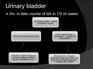


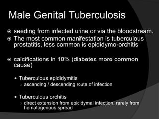


The document discusses renal tuberculosis (TB), its epidemiology, clinical features, pathogenesis, and imaging findings. It emphasizes the importance of early diagnosis due to the risk of renal failure and details how TB commonly affects the urinary tract, highlighting complications and diagnostic modalities like CT and intravenous urography. The document also describes various imaging characteristics and complications associated with renal and genitourinary TB.


