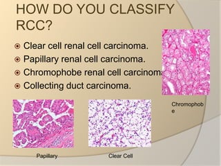
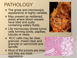
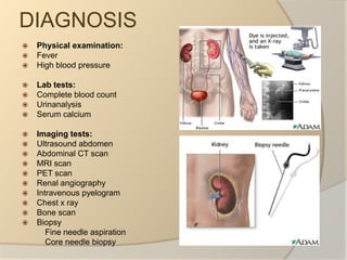
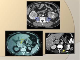
RENAL CELL CARCINOMA Designed By: Farooq Shah
WHAT IS RENAL CELL CARCIMONA (RCC)?Cancer arising from the lining of proximal convoluted tubule.The most common type of kidney cancer.Also known as Renal Adenocarcinoma or Grawitz's Tumor.Most lethal of all the genitourinary tumors.
HOW DO YOU CLASSIFY RCC?Clear cell renal cell carcinoma.Papillary renal cell carcinoma.Chromophobe renal cell carcinoma.Collecting duct carcinoma.ChromophobeClear Cell Papillary
EPIDIMOLOGY OF RCCThe incidence of renal cell carcinoma is rising steadily.More common in men than women, male to female ratio is 1.6:1.Blacks at an higher risk than whites.
WHAT LEADS TO RCC?Cigarette smoking.Obesity.Hypertension.Family history of the disease.Patients with inherited diseases like von HippelLindau disease.Hysterectomy is associated with doubled risk.Dialysis patients with acquired cystic disease of kidney show greater risk.
PATHOLOGYThe gross and microscopic appearance is highly variable.May present as reddened areas where blood vessels have bled and cysts containing watery fluids.Lite microscopy shows tumor cells forming cords, papillae, tubules or nests.  RCC cells may be clear, granular, mixed clear and granular or sarcomoid and spindle.Most of the tumors are mixed and they are most aggressive.
SIGNS AND SYMPTOMSClassic triad of :  Hematuria,,                                Flank pain                               Abdominal mass     Signs may include:Malaise,weight loss and anorexiaAbnormal urine colorPolycythemiaAnemiaFracture of hipVaricocele.enlargement of testicle on left sidePallor or plethoraHirsutismConstipationHypertensionHypercalcemiaLeg and ankle swelling
DIAGNOSISPhysical examination:FeverHigh blood pressureLab tests: Complete blood countUrinanalysisSerum calciumImaging tests:Ultrasound abdomen Abdominal CT scanMRI scanPET scanRenal angiographyIntravenous pyelogramChest x rayBone scanBiopsy          Fine needle aspiration          Core needle biopsy
STAGINGBased on examination,imaging and biopsyAJCC (TNM) staging system:T categories for kidney cancer:T0: No evidence of primary tumorT1: The tumor is only in the kidney and is 7cm or less across             T1a: The tumor is 4cm across or smaller             T1b: The tumor is larger than 4cm but not larger than 7cm T2: The tumor is larger than 7cm across but is still in the kidney             T2a: The tumor is more than 7cm but not more than 10             T2b: The tumor is more than 10cm acrossT3: The tumor is growing into a major vein or tissue around the kidney but not into adrenals or beyond Gerota,s fascia              T3a: The tumor is growing into the main vein or into fatty tissue              around the kidney              T3b: The tumor is growing into the venacava leading into the heart               T3c: The tumor has grown into the part of venacava that is within the chest or growing into the wall of that blood vesselT4: The tumor has spread beyond Gerota,s fascia. It may have grown into the adrenal gland
STAGING (CONTINUED)
STAGING (CONTINUED)N categories for kidney cancer:N0: No spread to nearby lymph nodesN1: tumor has spread to nearby lymph nodesM categories for kidney cancer:M0: There is no spread to distant lymph nodes or other organsM1: Distant metastasis is present ,distant lymph nodes and to organs like lungs, bone, brain and liver
HOW TO TREAT RCC?If only in kidneys, it can be cured 90% of the time with surgery.If it has spread outside the kidneys into the nodes or the main vein, it must be treated with cytoreductive surgery.RRC is resistant to chemo and radiotherapy in most casesMay respond to immunotherapyPARTIAL NEPHRECTOMY:For treating small renal tumors(< 4cm)Bilateral renal cell carcinomaIt can be done via laproscopic techniquesRADICAL NEPHRECTOMY:Surgical removal of kidney along with adrenal gland, retroperitoneal lymphnodes, perinephric fat and Gerota's fasciaIn cases where the tumor has spread into the renal vein, IVC and right atrium, this portion of tumor can be surgically removed as well.Medications like tyrosine kinase inhibitors including nexavar and rapamycin have shown to improve the prognosis for advanced RCC.
PROGNOSISFor tumors less than 4cm 5 year survival rate is 90-95%For larger tumors confined to kidneys without venous invasion survival is 80-85%For tumors that extend through the renal capsule n local fascia survivability reduces to near 60%For metastasis to lymph nodes survival rate is around 5-15%For spread to other organs 5 year survival rate  is less than 5%
THANK YOU

Renal Cell Carcinoma

  • 1.
    RENAL CELL CARCINOMADesigned By: Farooq Shah
  • 2.
    WHAT IS RENALCELL CARCIMONA (RCC)?Cancer arising from the lining of proximal convoluted tubule.The most common type of kidney cancer.Also known as Renal Adenocarcinoma or Grawitz's Tumor.Most lethal of all the genitourinary tumors.
  • 3.
    HOW DO YOUCLASSIFY RCC?Clear cell renal cell carcinoma.Papillary renal cell carcinoma.Chromophobe renal cell carcinoma.Collecting duct carcinoma.ChromophobeClear Cell Papillary
  • 4.
    EPIDIMOLOGY OF RCCTheincidence of renal cell carcinoma is rising steadily.More common in men than women, male to female ratio is 1.6:1.Blacks at an higher risk than whites.
  • 5.
    WHAT LEADS TORCC?Cigarette smoking.Obesity.Hypertension.Family history of the disease.Patients with inherited diseases like von HippelLindau disease.Hysterectomy is associated with doubled risk.Dialysis patients with acquired cystic disease of kidney show greater risk.
  • 6.
    PATHOLOGYThe gross andmicroscopic appearance is highly variable.May present as reddened areas where blood vessels have bled and cysts containing watery fluids.Lite microscopy shows tumor cells forming cords, papillae, tubules or nests. RCC cells may be clear, granular, mixed clear and granular or sarcomoid and spindle.Most of the tumors are mixed and they are most aggressive.
  • 7.
    SIGNS AND SYMPTOMSClassictriad of :  Hematuria,,                              Flank pain Abdominal mass Signs may include:Malaise,weight loss and anorexiaAbnormal urine colorPolycythemiaAnemiaFracture of hipVaricocele.enlargement of testicle on left sidePallor or plethoraHirsutismConstipationHypertensionHypercalcemiaLeg and ankle swelling
  • 8.
    DIAGNOSISPhysical examination:FeverHigh bloodpressureLab tests: Complete blood countUrinanalysisSerum calciumImaging tests:Ultrasound abdomen Abdominal CT scanMRI scanPET scanRenal angiographyIntravenous pyelogramChest x rayBone scanBiopsy Fine needle aspiration Core needle biopsy
  • 9.
    STAGINGBased on examination,imagingand biopsyAJCC (TNM) staging system:T categories for kidney cancer:T0: No evidence of primary tumorT1: The tumor is only in the kidney and is 7cm or less across             T1a: The tumor is 4cm across or smaller             T1b: The tumor is larger than 4cm but not larger than 7cm T2: The tumor is larger than 7cm across but is still in the kidney             T2a: The tumor is more than 7cm but not more than 10             T2b: The tumor is more than 10cm acrossT3: The tumor is growing into a major vein or tissue around the kidney but not into adrenals or beyond Gerota,s fascia              T3a: The tumor is growing into the main vein or into fatty tissue around the kidney              T3b: The tumor is growing into the venacava leading into the heart               T3c: The tumor has grown into the part of venacava that is within the chest or growing into the wall of that blood vesselT4: The tumor has spread beyond Gerota,s fascia. It may have grown into the adrenal gland
  • 10.
  • 11.
    STAGING (CONTINUED)N categoriesfor kidney cancer:N0: No spread to nearby lymph nodesN1: tumor has spread to nearby lymph nodesM categories for kidney cancer:M0: There is no spread to distant lymph nodes or other organsM1: Distant metastasis is present ,distant lymph nodes and to organs like lungs, bone, brain and liver
  • 12.
    HOW TO TREATRCC?If only in kidneys, it can be cured 90% of the time with surgery.If it has spread outside the kidneys into the nodes or the main vein, it must be treated with cytoreductive surgery.RRC is resistant to chemo and radiotherapy in most casesMay respond to immunotherapyPARTIAL NEPHRECTOMY:For treating small renal tumors(< 4cm)Bilateral renal cell carcinomaIt can be done via laproscopic techniquesRADICAL NEPHRECTOMY:Surgical removal of kidney along with adrenal gland, retroperitoneal lymphnodes, perinephric fat and Gerota's fasciaIn cases where the tumor has spread into the renal vein, IVC and right atrium, this portion of tumor can be surgically removed as well.Medications like tyrosine kinase inhibitors including nexavar and rapamycin have shown to improve the prognosis for advanced RCC.
  • 13.
    PROGNOSISFor tumors lessthan 4cm 5 year survival rate is 90-95%For larger tumors confined to kidneys without venous invasion survival is 80-85%For tumors that extend through the renal capsule n local fascia survivability reduces to near 60%For metastasis to lymph nodes survival rate is around 5-15%For spread to other organs 5 year survival rate  is less than 5%
  • 15.