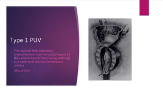
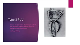
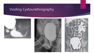
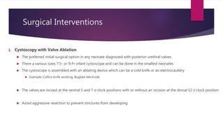
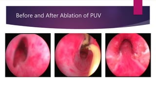
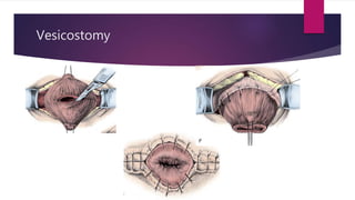
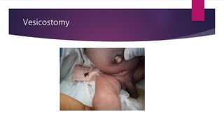
Posterior Urethral Valve
DR. M. YIGAH
Introduction
 It is a congenital condition characterized by the presence of abnormal obstructing urethral
membranes in the proximal urethra .
 Commonest cause of bladder outlet obstruction in infants
 Abnormal development of the wolffian and müllerian ducts based on an autopsy study
 The presentation of posterior urethral valves is often antenatal, and pathognomonic findings on
postnatal imaging confirm the diagnosis
 Surgical procedure may be short but the management may be life-long
 the embryonic insults to the bladder, kidneys and other organs can be life-long
Epidemiology
 In the UK 1 out of every 500 pregnancies has an anomaly of the urinary tract.
 The incidence of lower urinary tract obstruction (LUTO) in the UK is 2.2 per 10,000 births,
► Incidence of PUV in the UK is 1.4 per 10,000 births.
► In the US incidence ranges from 1 per 8000 to 1 per 25,000 live births.
• In Nigeria
► There were 3–8 reported cases per annum (Uba et al, 2007)
► A hospital in Port Harcourt observed that it accounted for 1 in 2,447 children (Jaja et al, 2012)
► 71% were already in renal failure at presentation (Odetunde et al, 2012)
 It affects male babies
Hugh Hampton Young
Historical Overview
 Valve-like leaflets was recognized in1769 by Morgagni
 Post-mortem findings were confirmed by Langenbeck in 1802.
 Hugh Hampton Young described the first endoscopic diagnosis
of a urethral obstruction that was termed posterior urethral
valves.
 Also described the various types of PUV
 Randall carried out the first endoscopic resection of valves in
1920
Gross Anatomy of the Urethra
Pathology
“ The prostatic urethra is markedly dilated. The vesical neck raised and bladder orifice relaxed. Deep
pittings penetrate down into either lateral wall of the prostatic urethra and at the extremity the
verumontanum is seen a fine frenulum which extends distally for about 1 cm, and in dividing, forms
what is apparently a definite valve on either side of the urethra, rising from the floor to each side
wall. Randall carried out the first endoscopic resection of valves in 1920”
 Randall 1921
Pathology
Crescentic duplication of the urethral mucosa forming paper-thin valves arise from
the verumontanum and extend downward and laterally to the side walls of the
urethra at apex of the prostate.
 There may be no hinderance to retrograde instrumentation but the valves come
together to obstruct the flow of urine
 Prostatic urethra becomes dilated
 Bladder hypertrophy with trabeculations, sacculations and diverticula
 Hydroureter and hydronephrosis
Type 1 PUV
• The mucosal folds extending
anteroinferiorly from the caudal aspect of
the verumontanum often fusing anteriorly
at a lower level like the membranous
urethra.
• 95% of PUV
Type 2 PUV
Bicuspid valves as leaflets radiating
from the verumontanum proximally to
the bladder neck
Type 3 PUV
• Valves as concentric diaphragms within
the prostatic urethra, either above or
below the verumontanum
• (~5% of PUVs)
Pathophysiology
 A vicious cycle of obstructive uropathy and renal dysplasia.
 Dam of urine in the bladder and upper segments of the urinary tree.
 Thickened bladder wall with reduced bladder compliance (remodelling of the bladder)
 Vesicoureteric reflux
 Hydroureters and Hydronephrosis
 Progressive renal calyceal dilatation and renal damage
 Impairment of concentration of urine by the renal tubules
 Polyuria
 Worsening bladder dysfunction - end-stage bladder, or Bladder valve syndrome.
 Worsening renal function leading to ESKD.
Diagnosis
 Clinical Features
 Maternal signs of oligohydramnios
 Physical stigmata of oligohydramnios
 Potter facies
 Clubfeet and deformed hands, and
 poor abdominal muscle tone.
 Failure pass urine after birth
 Infant may pass urine with weak stream with intermittency
 The bladder is distended and the kidneys may be palpable
 An infant affected by PUV may be affected by comorbidities such as
 Pulmonary hypoplasia – tachypnoea, poor respiratory effort, cyanosis
 Urinomas – neonatal ascites, respiratory distress
Diagnosis
 Clinical Features (Delayed Presentation)
 Recurrent Urinary tract infection (UTI)
 Diurnal enuresis in boys older than 5 years
 Secondary diurnal enuresis
 Voiding pain or dysfunction
 Abnormal urinary stream
 Poor growth, hypertension, and lethargy
 PUVs are sometimes discovered during evaluation of abdominal mass or renal failure
Diagnosis
1. Ultrasonography
Some of the cardinal features of a LUTO are
a. thickened, dilated bladder along with
b. bilateral hydroureter and pelvocaliectasis
c. Oligohydramnios
d. Dilated posterior urethra displaying the “keyhole sign”
 Increased renal echogenicity in PUV is an indicator of renal damage.
 Very difficult to differentiate PUV from other causes of LUTO like urethral atresia or prune-belly syndrome, bilateral
primary obstructing megaureters
Diagnosis
2. Voiding Cystourethrography
Definitive radiologic study in confirming the diagnosis of posterior urethral valves.
 Visualization of bladder leaflets
 The bladder often appears thickened and trabeculated with multiple diverticuli.
 Hypertrophied bladder neck
 High-grade vesicoureteral reflux may be seen
2. Radionuclide Renal Scan
 The radionuclide renal scan offers quantification of differential renal function
 Cortical deficits seen on the study may imply renal dysplasia
Voiding Cystourethrography
Diagnosis
 Supportive Investigation
 BUE and Cr
 Chest Xray
 FBC
 Urine RE and CS
Chest Xrays
 Bronchopulmonary dysplasia
 Reduced lung volume
 left upper and lower lobe atelectasis
Differential diagnosis
 Anterior urethral valves
 Urethral stricture disease
 Detrusor sphincter dyssynergy
 Diurnal urinary incontinence
 Pediatric renal insufficiency
 Urinary tract infection
Treatment
 The treatment goals are
 To restore flow of urine through the urethra and enable normal cyclic filling and
emptying of the bladder, which is superior to urinary diversion and passive urine
drainage.
 Halt the deterioration of renal function
 Prevent complications of PUV
 Improve the quality life
Surgical Interventions
1. Cystoscopy with Valve Ablation
 The preferred initial surgical option in any neonate diagnosed with posterior urethral valves
 There a various sizes 7.5- or 9-Fr infant cystoscope and can be done in the smallest neonates
 The cystoscope is assembled with an ablating device which can be a cold knife or an electrocaultery
 Example; Collins knife working, Bugbee electrode
 The valves are incised at the ventral 5 and 7 o’clock positions with or without an incision at the dorsal 12 o’clock position
 Avoid aggressive resection to prevent strictures from developing
Before and After Ablation of PUV
Cystoscopy with Valve Ablation
1. Cystoscopy with Valve Ablation
 A urethral catheter is typically placed for at least 24 hours after the procedure.
 Baby is monitored post-op for improvements in renal parameters or respiratory issues
 VCUG must be repeated after valve ablation within 1 month to ensure that the valves are no longer visible.
Surgical Interventions
2. Vesicostomy
 very low-birth-weight infant whose urethra cannot accommodate an endoscope
 Continued impaired renal function, high bladder urine volumes, and upper tract deterioration after valve ablation or
urethral catheterization
Vesicostomy
Vesicostomy
Surgical Interventions
3. Upper Tract Diversion
 Cutaneous ureterostomy or Pyelostomy
 It was believed direct decompression of the kidneys will produce direct, low-pressure urinary drainage,
allowing optimization of renal function.
 It is complex and not better than the lower urinary diversion.
 It not just about the diversion but also about the renal dysplasia
4. Circumcision
 vesicoureteral reflux, incomplete bladder emptying, and severe upper urinary tract dilation predispose to
Pyelonrphritis
 Circumcision reduces that risk of urinary tract infection by 83% to 92%, a reduction to a level of risk similar to
that for unaffected boys (Wiswell et al, 1988; Mukherjee et al, 2009)
Antenatal Surgical Interventions
 Antenatal interventions are considered when
 Oligohydramnios,
 Dilated bladder
 Severe hydroureteronephrosis without renal cortical cystic lesions
 Normal karyotype.
 Vesicoamniotic shunting
 Foetal cystoscopy with valve ablation
 Cutaneous ureterostomy
 vesicostomy
Prognostic Factors
a. Serum Creatinine at 1 year of life and (1 month post treatment)
 Cr < 0.8 mg/dL appears to indicate a minimal risk
 Cr > 1.2 mg/dL higher risk of developing end-stage renal failure
b. Age at diagnosis
c. Renal dysplasia
 Hyperechogenic kidneys, cystic cortical changes & loss of corticomedullary differentiation
d. Recurrent urinary tract infections
e. Bladder dysfunction.
Quality of Life
 Patients with posterior urethral valve have a lifetime of repercussion.
 Erectile dysfunction
 Infertility.
 Persistence of lower urinary tract symptoms – urgency, frequency, incontinence.
 Increased risk of UTI.
 Development of end stage kidney disease.
 Associated co-morbidities of renal transplantation.
References
1. Uba AF, Chirdan LB, Ihezue CH, Ramyil VL, Dakum NK. Posterior urethral valves in chilhood:
Experience in a centre with scarce facilities. Afr J Urol. 2007;13:124–31.
2. Jaja T, Anochie IC, Eke FU. Posterior urethral valve in childhood in Port Harcourt, Nigeria. Port
Harcourt Med J. 2012;6:10–6.
3. Odetunde OI, Odetunde OA, Ademuyiwa AO, Okafor HU, Ekwochi U, Azubuike JC, et al.
Outcome of late presentation of posterior urethral valves in a resource-limited economy:
Challenges in management. Int J Nephrol 2012. 2012 345298
4. Baja
5. Medscape

Posterior urethral valve

  • 1.
  • 2.
    Introduction  It isa congenital condition characterized by the presence of abnormal obstructing urethral membranes in the proximal urethra .  Commonest cause of bladder outlet obstruction in infants  Abnormal development of the wolffian and müllerian ducts based on an autopsy study  The presentation of posterior urethral valves is often antenatal, and pathognomonic findings on postnatal imaging confirm the diagnosis  Surgical procedure may be short but the management may be life-long  the embryonic insults to the bladder, kidneys and other organs can be life-long
  • 3.
    Epidemiology  In theUK 1 out of every 500 pregnancies has an anomaly of the urinary tract.  The incidence of lower urinary tract obstruction (LUTO) in the UK is 2.2 per 10,000 births, ► Incidence of PUV in the UK is 1.4 per 10,000 births. ► In the US incidence ranges from 1 per 8000 to 1 per 25,000 live births. • In Nigeria ► There were 3–8 reported cases per annum (Uba et al, 2007) ► A hospital in Port Harcourt observed that it accounted for 1 in 2,447 children (Jaja et al, 2012) ► 71% were already in renal failure at presentation (Odetunde et al, 2012)  It affects male babies
  • 4.
    Hugh Hampton Young HistoricalOverview  Valve-like leaflets was recognized in1769 by Morgagni  Post-mortem findings were confirmed by Langenbeck in 1802.  Hugh Hampton Young described the first endoscopic diagnosis of a urethral obstruction that was termed posterior urethral valves.  Also described the various types of PUV  Randall carried out the first endoscopic resection of valves in 1920
  • 5.
    Gross Anatomy ofthe Urethra
  • 6.
    Pathology “ The prostaticurethra is markedly dilated. The vesical neck raised and bladder orifice relaxed. Deep pittings penetrate down into either lateral wall of the prostatic urethra and at the extremity the verumontanum is seen a fine frenulum which extends distally for about 1 cm, and in dividing, forms what is apparently a definite valve on either side of the urethra, rising from the floor to each side wall. Randall carried out the first endoscopic resection of valves in 1920”  Randall 1921
  • 7.
    Pathology Crescentic duplication ofthe urethral mucosa forming paper-thin valves arise from the verumontanum and extend downward and laterally to the side walls of the urethra at apex of the prostate.  There may be no hinderance to retrograde instrumentation but the valves come together to obstruct the flow of urine  Prostatic urethra becomes dilated  Bladder hypertrophy with trabeculations, sacculations and diverticula  Hydroureter and hydronephrosis
  • 8.
    Type 1 PUV •The mucosal folds extending anteroinferiorly from the caudal aspect of the verumontanum often fusing anteriorly at a lower level like the membranous urethra. • 95% of PUV
  • 9.
    Type 2 PUV Bicuspidvalves as leaflets radiating from the verumontanum proximally to the bladder neck
  • 10.
    Type 3 PUV •Valves as concentric diaphragms within the prostatic urethra, either above or below the verumontanum • (~5% of PUVs)
  • 11.
    Pathophysiology  A viciouscycle of obstructive uropathy and renal dysplasia.  Dam of urine in the bladder and upper segments of the urinary tree.  Thickened bladder wall with reduced bladder compliance (remodelling of the bladder)  Vesicoureteric reflux  Hydroureters and Hydronephrosis  Progressive renal calyceal dilatation and renal damage  Impairment of concentration of urine by the renal tubules  Polyuria  Worsening bladder dysfunction - end-stage bladder, or Bladder valve syndrome.  Worsening renal function leading to ESKD.
  • 12.
    Diagnosis  Clinical Features Maternal signs of oligohydramnios  Physical stigmata of oligohydramnios  Potter facies  Clubfeet and deformed hands, and  poor abdominal muscle tone.  Failure pass urine after birth  Infant may pass urine with weak stream with intermittency  The bladder is distended and the kidneys may be palpable  An infant affected by PUV may be affected by comorbidities such as  Pulmonary hypoplasia – tachypnoea, poor respiratory effort, cyanosis  Urinomas – neonatal ascites, respiratory distress
  • 13.
    Diagnosis  Clinical Features(Delayed Presentation)  Recurrent Urinary tract infection (UTI)  Diurnal enuresis in boys older than 5 years  Secondary diurnal enuresis  Voiding pain or dysfunction  Abnormal urinary stream  Poor growth, hypertension, and lethargy  PUVs are sometimes discovered during evaluation of abdominal mass or renal failure
  • 14.
    Diagnosis 1. Ultrasonography Some ofthe cardinal features of a LUTO are a. thickened, dilated bladder along with b. bilateral hydroureter and pelvocaliectasis c. Oligohydramnios d. Dilated posterior urethra displaying the “keyhole sign”  Increased renal echogenicity in PUV is an indicator of renal damage.  Very difficult to differentiate PUV from other causes of LUTO like urethral atresia or prune-belly syndrome, bilateral primary obstructing megaureters
  • 15.
    Diagnosis 2. Voiding Cystourethrography Definitiveradiologic study in confirming the diagnosis of posterior urethral valves.  Visualization of bladder leaflets  The bladder often appears thickened and trabeculated with multiple diverticuli.  Hypertrophied bladder neck  High-grade vesicoureteral reflux may be seen 2. Radionuclide Renal Scan  The radionuclide renal scan offers quantification of differential renal function  Cortical deficits seen on the study may imply renal dysplasia
  • 16.
  • 17.
    Diagnosis  Supportive Investigation BUE and Cr  Chest Xray  FBC  Urine RE and CS
  • 18.
    Chest Xrays  Bronchopulmonarydysplasia  Reduced lung volume  left upper and lower lobe atelectasis
  • 19.
    Differential diagnosis  Anteriorurethral valves  Urethral stricture disease  Detrusor sphincter dyssynergy  Diurnal urinary incontinence  Pediatric renal insufficiency  Urinary tract infection
  • 20.
    Treatment  The treatmentgoals are  To restore flow of urine through the urethra and enable normal cyclic filling and emptying of the bladder, which is superior to urinary diversion and passive urine drainage.  Halt the deterioration of renal function  Prevent complications of PUV  Improve the quality life
  • 21.
    Surgical Interventions 1. Cystoscopywith Valve Ablation  The preferred initial surgical option in any neonate diagnosed with posterior urethral valves  There a various sizes 7.5- or 9-Fr infant cystoscope and can be done in the smallest neonates  The cystoscope is assembled with an ablating device which can be a cold knife or an electrocaultery  Example; Collins knife working, Bugbee electrode  The valves are incised at the ventral 5 and 7 o’clock positions with or without an incision at the dorsal 12 o’clock position  Avoid aggressive resection to prevent strictures from developing
  • 22.
    Before and AfterAblation of PUV
  • 23.
    Cystoscopy with ValveAblation 1. Cystoscopy with Valve Ablation  A urethral catheter is typically placed for at least 24 hours after the procedure.  Baby is monitored post-op for improvements in renal parameters or respiratory issues  VCUG must be repeated after valve ablation within 1 month to ensure that the valves are no longer visible.
  • 24.
    Surgical Interventions 2. Vesicostomy very low-birth-weight infant whose urethra cannot accommodate an endoscope  Continued impaired renal function, high bladder urine volumes, and upper tract deterioration after valve ablation or urethral catheterization
  • 25.
  • 26.
  • 27.
    Surgical Interventions 3. UpperTract Diversion  Cutaneous ureterostomy or Pyelostomy  It was believed direct decompression of the kidneys will produce direct, low-pressure urinary drainage, allowing optimization of renal function.  It is complex and not better than the lower urinary diversion.  It not just about the diversion but also about the renal dysplasia 4. Circumcision  vesicoureteral reflux, incomplete bladder emptying, and severe upper urinary tract dilation predispose to Pyelonrphritis  Circumcision reduces that risk of urinary tract infection by 83% to 92%, a reduction to a level of risk similar to that for unaffected boys (Wiswell et al, 1988; Mukherjee et al, 2009)
  • 28.
    Antenatal Surgical Interventions Antenatal interventions are considered when  Oligohydramnios,  Dilated bladder  Severe hydroureteronephrosis without renal cortical cystic lesions  Normal karyotype.  Vesicoamniotic shunting  Foetal cystoscopy with valve ablation  Cutaneous ureterostomy  vesicostomy
  • 29.
    Prognostic Factors a. SerumCreatinine at 1 year of life and (1 month post treatment)  Cr < 0.8 mg/dL appears to indicate a minimal risk  Cr > 1.2 mg/dL higher risk of developing end-stage renal failure b. Age at diagnosis c. Renal dysplasia  Hyperechogenic kidneys, cystic cortical changes & loss of corticomedullary differentiation d. Recurrent urinary tract infections e. Bladder dysfunction.
  • 30.
    Quality of Life Patients with posterior urethral valve have a lifetime of repercussion.  Erectile dysfunction  Infertility.  Persistence of lower urinary tract symptoms – urgency, frequency, incontinence.  Increased risk of UTI.  Development of end stage kidney disease.  Associated co-morbidities of renal transplantation.
  • 31.
    References 1. Uba AF,Chirdan LB, Ihezue CH, Ramyil VL, Dakum NK. Posterior urethral valves in chilhood: Experience in a centre with scarce facilities. Afr J Urol. 2007;13:124–31. 2. Jaja T, Anochie IC, Eke FU. Posterior urethral valve in childhood in Port Harcourt, Nigeria. Port Harcourt Med J. 2012;6:10–6. 3. Odetunde OI, Odetunde OA, Ademuyiwa AO, Okafor HU, Ekwochi U, Azubuike JC, et al. Outcome of late presentation of posterior urethral valves in a resource-limited economy: Challenges in management. Int J Nephrol 2012. 2012 345298 4. Baja 5. Medscape

Editor's Notes

  • #4 Obstructive uropathy accounts for most of these cases
  • #6 The seminal colliculus of the Veromontanum (Lt – mountain ridge) is a landmark on the floor of the prostatic urethra where the ejaculatory duct opens into the urethra. It also important in classifying several urethral developmental disorders.
  • #12 Bladder outlet obstruction leads to detrusor hypertrophy, which increases the voiding pressures initially as the bladder strives to complete emptying. As further remodeling of the bladder occurs, however, the postvoid residual begins to increase as the urine output increases. Ultimately, the bladder cannot meet the demand of emptying and the detrusor decompensates.
  • #13 A 5- or 7-Fr feeding tube, or similar caliber urinary catheter should be inserted per urethra in an infant presenting to the neonatal intensive care unit with a presumed diagnosis of LUTO. Catheter placement into the bladder may be impeded by the hypertrophied and often elevated bladder neck and curling of the catheter within the dilated posterior urethra. In such cases, a coudé-tipped catheter or, alternatively, using a stylet to curl the tip of the feeding tube dorsally will facilitate bladder drainage. The association between posterior urethral valves, oligohydramnios, and pulmonary hypoplasia is recognized but the etiology of hypoplasia is unclear and likely multifactorial Urinomas - A urinoma is the result of a breach of the integrity of the pelvis or calices of the kidney or of the ureter.[4] The urine collection in the perirenal fat causes an inflammatory response with lipolysis resulting in its fibrous encapsulation
  • #14 Daytime accidental wetting is more likely than bed-wetting to develop after a child has had bladder control for at least 6 months to 1 year (secondary diurnal enuresis).
  • #15 Improved the antenatal diagnosis of PUV and other LUTO
  • #16 This study should be completed in the early postnatal period after renal and bladder sonography, and as soon as an infant with suspected prenatal findings of valves is hemodynamically stabilized and able to undergo the contrast study
  • #17  2. cystogram, same patient. Free vesicoureteral reflux and vesical trabeculation with diverticula.
  • #18 Laboratory evaluation of a newborn with a diagnosis of posterior urethral valves will, as with any newborn, reflect maternal values and must be interpreted with caution. After 48 hours, the maternal blood mediated through the placenta should clear, and the infant’s baseline laboratory values may be monitored.
  • #22 In medicine, the removal or destruction of a body part or tissue or its function With the miniaturization of endoscopes in the age of fiberoptic and now digital technology, cystoscopy can be accomplished in even the smallest neonate and endoscopic valve ablation is the preferred approach at most centers today. Posterior urethral valves are thin and associated with minimal vascularity, and aggressive resection should be avoided. Use of a hot loop resectoscope for valve ablation, primarily in older children 4..
  • #24 It is not uncommon that it is left in for a file while the infant is monitored for improvements in renal parameters or respiratory issues, often in an intensive care environment. It is not uncommon to see immediate signs of diminished bladder pressures, including some improvement in renal dilation and in the volume of vesicoureteral reflux
  • #25 With miniaturization of endoscopic technology, vesicostomy is reserved primarily for the very low-birth-weight infant whose urethra cannot accommodate an endoscope, as well as a child with continued impaired renal function, high bladder urine volumes, and upper tract deterioration after valve ablation or urethral catheterization
  • #28 The overall risk of urinary tract infection in children with posterior urethral valves is 50% to 60%—several magnitudes greater than the 1% risk for unaffected boys It is known that there is bacterial colonization of the foreskin during the first 6 months of life that may be an important risk factor for the development of UTIs
  • #29 Advances in fiber-optics and endoscopic miniaturization popularized foetal interventions. Vesicoamniotic shunting - Biard and colleagues (2005) reported a mean 5.83 years of follow-up on 20 pregnancies with a singleton male fetus that underwent vesicoamniotic shunting for clear evidence of isolated LUTO and good or borderline urinary sampling parameters. This study found an overall 1-year survival of 91%, and health-related qualityof- life parameters were similar to those in the unaffected, healthy child population. Data has been difficult because of recruitment issues
  • #30 Despite numerous advances in antenatal diagnosis and intervention, and rapid postnatal evaluation and treatment, the lifetime prevalence of end-stage renal disease in boys with posterior urethral valves is between 20% and 50%. Age at diagnosis remains an unclear predictor of future renal outcomes. The assumption that antenatal diagnosis would lead to more rapid diagnosis of posterior urethral valves and therefore forestall renal injury has not been sustained. Indeed, Heikkilä and associates (2011) found that patients diagnosed in the presonography era (before 1982) had a risk of end-stage renal disease of 16.8%, as compared to 36.6% developing renal failure if diagnosed during the post-sonography era. Another review found a similar variation with 41% of those presenting before 1 year of age having poor long-term renal outcome compared to 15% of those presenting a fter 1 year of age Another common assumption, not corroborated, was that the later-presenting children likely had a milder variant of valve disease, allowing them to go undetected for some years before presenting with more vague symptoms of voiding dysfunction. However, some reports found much worse outcomes in children presenting outside of the neonatal period with posterior urethral valves, with delayed presentation associated with a significantly higher risk of azotemia, higher serum creatinine, and worse long-term renal outcomes
  • #31 Understanding these long-term risk factors and their impact on quality of life is necessary for counseling, preparing, and treating valve patients as they reach adulthood.