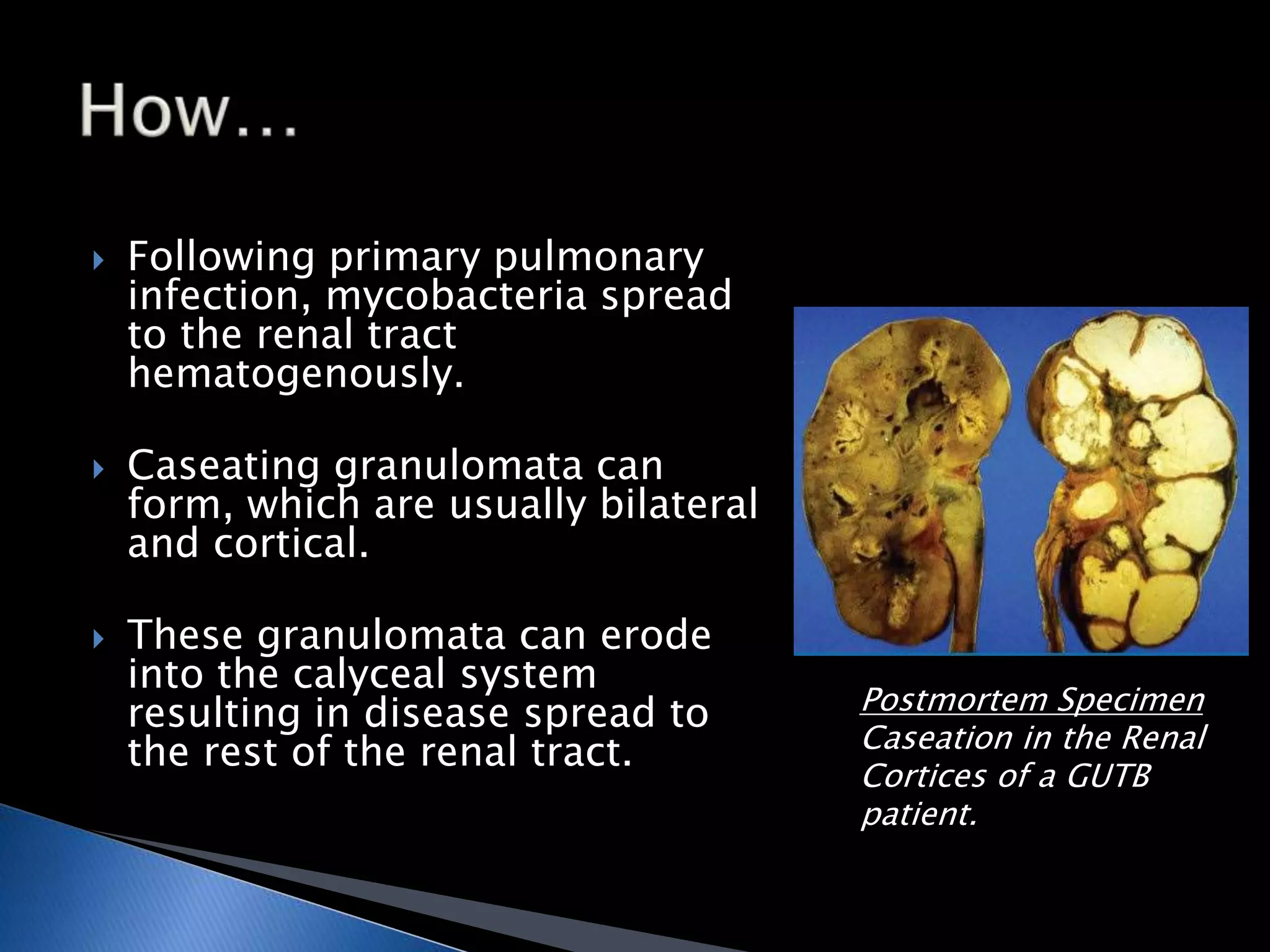
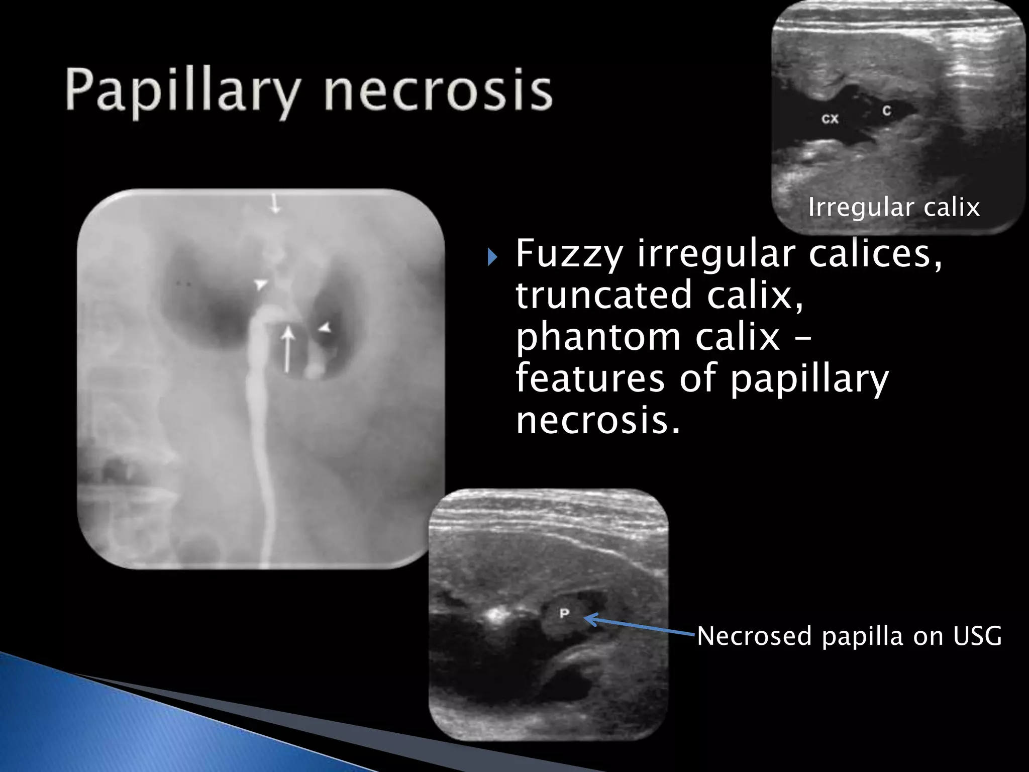
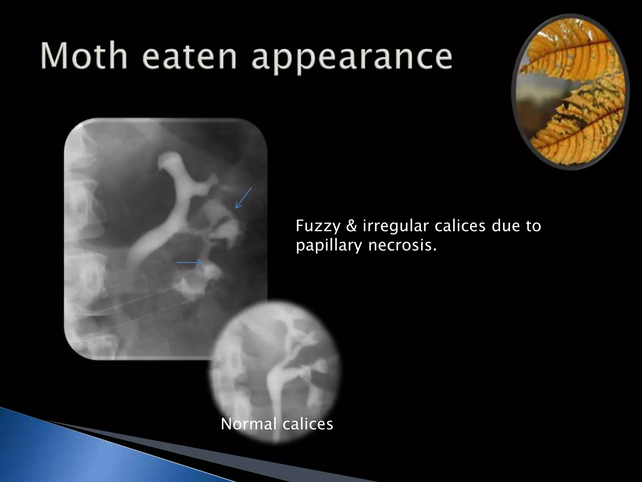
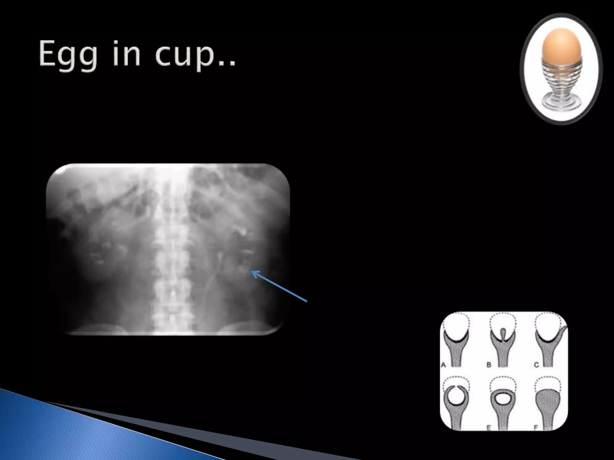
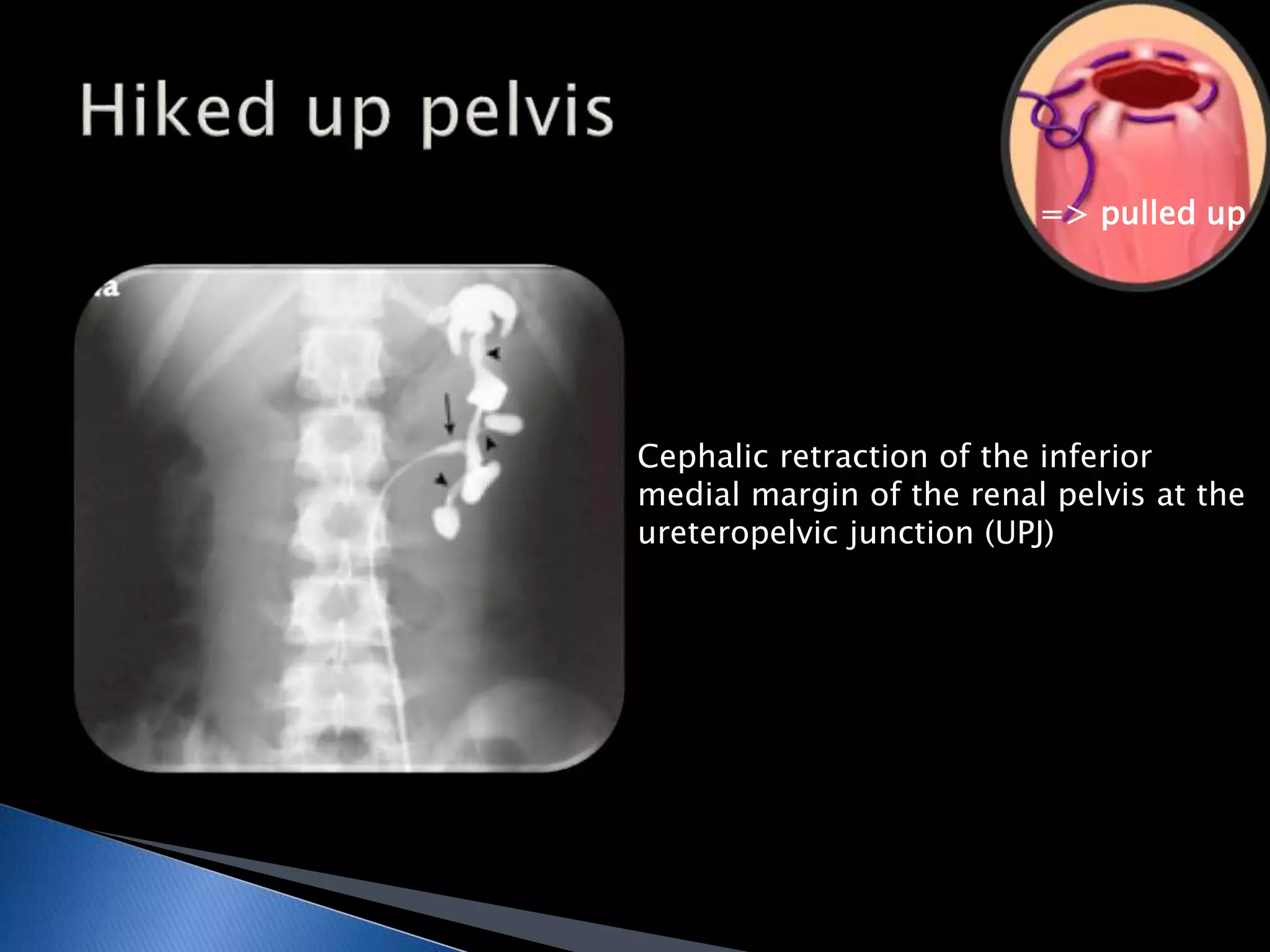
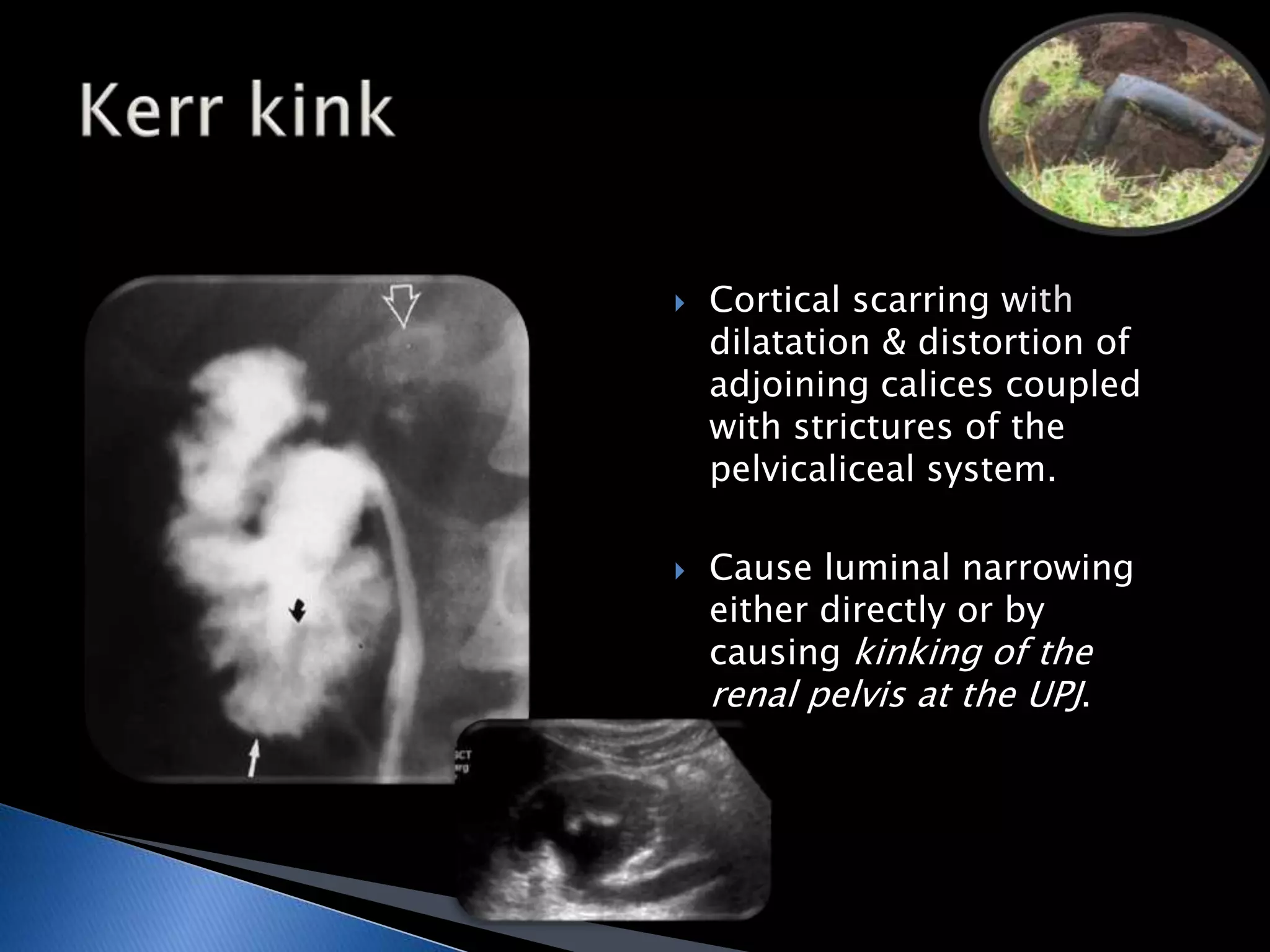
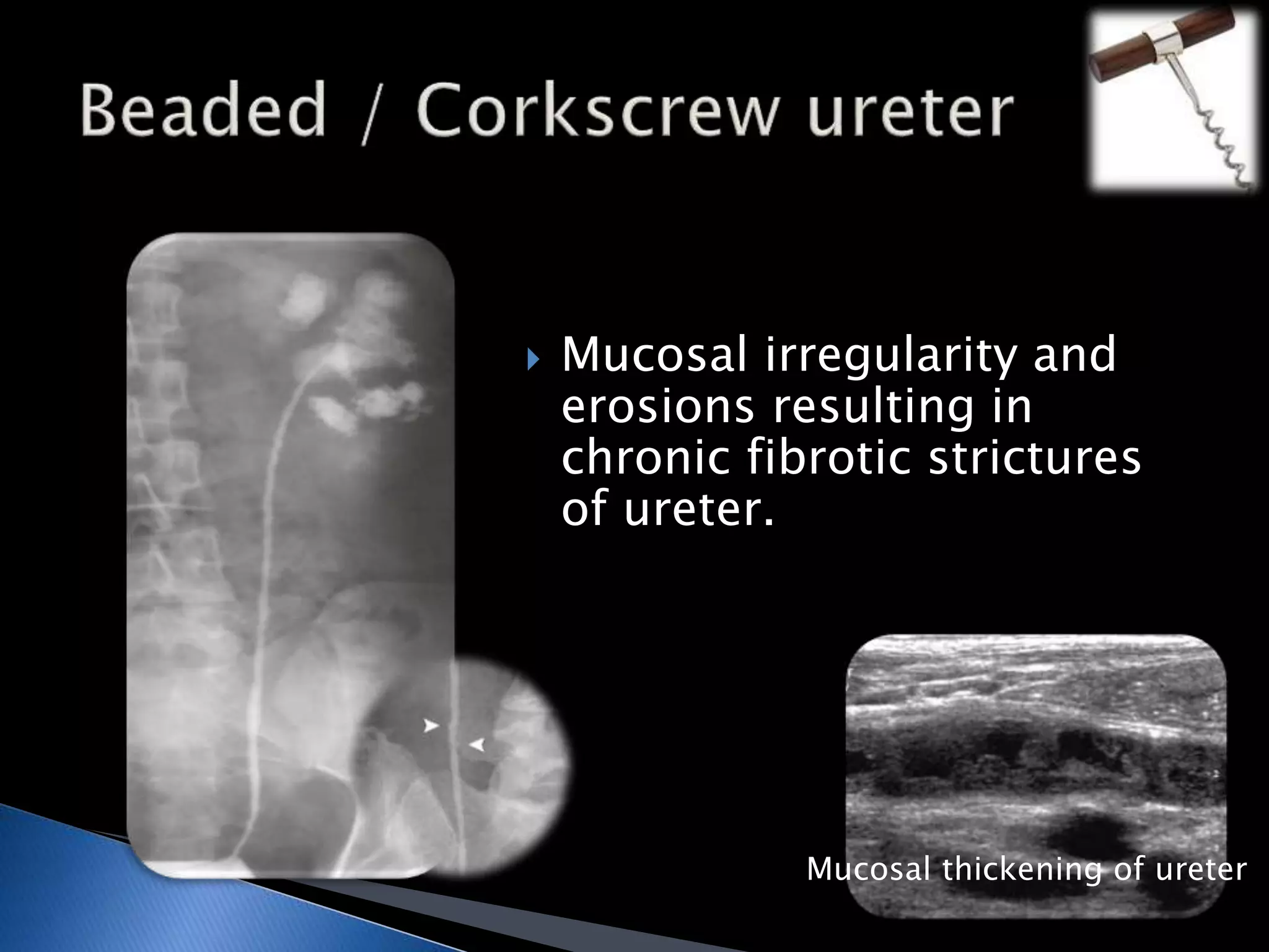
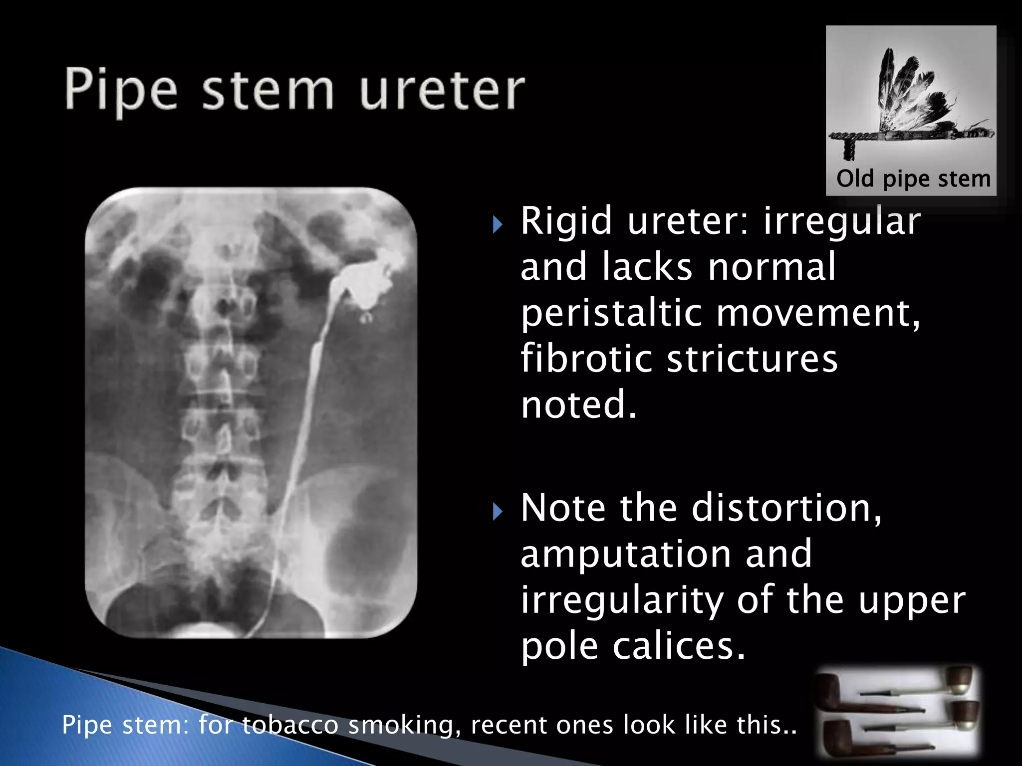
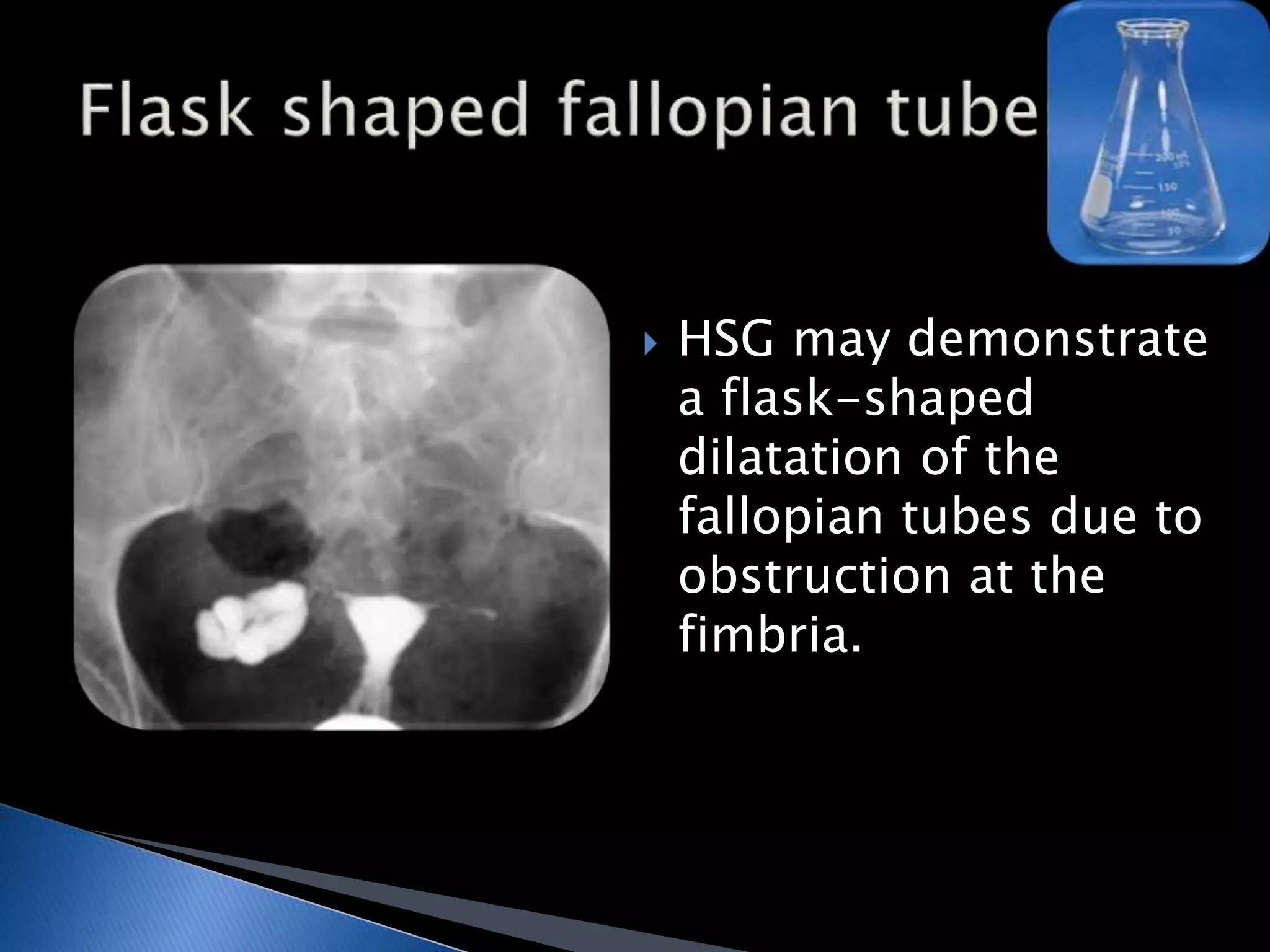
Dr. Sudha Kiran Das, 
Asst Professor, 
Radiology, JSSMC - Mysore
 Second most common form of extrapulmonary 
tuberculosis after lymph node tuberculosis in developing 
countries. 
 The kidneys are the most common site of GUTB and are 
infected through hematogenous spread; from the kidneys, 
the bacilli can spread to the renal tract, prostate and 
epididymis. 
 CT and intravenous urography can aid diagnosis— 
calcification, multiple strictures and fibrosis are suggestive 
features on imaging. 
 GUTB is strongly associated with infertility in women, as 
the Fallopian tubes are affected in most cases, and rates of 
successful pregnancy remain low even after treatment.
 Following primary pulmonary 
infection, mycobacteria spread 
to the renal tract 
hematogenously. 
 Caseating granulomata can 
form, which are usually bilateral 
and cortical. 
 These granulomata can erode 
into the calyceal system 
resulting in disease spread to 
the rest of the renal tract. 
Postmortem Specimen 
Caseation in the Renal 
Cortices of a GUTB 
patient.
Irregular calix 
 Fuzzy irregular calices, 
truncated calix, 
phantom calix – 
features of papillary 
necrosis. 
Necrosed papilla on USG
Fuzzy & irregular calices due to 
papillary necrosis. 
Normal calices
 Papillary (forniceal) 
excavation. 
 The necrotic papillary 
tip may remain within 
the excavated calix, 
producing a signet ring 
sign when the calix is 
filled with contrast 
material.
Phantom calix 
Infundibular stenosis
Ghost - like 
RGP 
Decreased nephrographic opacity and nonfilling of the collecting 
system elements at the lower pole of left kidney – phantom calices 
(ghost : exist, but not visualised, the same are visualized on RGP).
On IVP : 
Collecting system shows contrast 
material in a large papillary cavity, the 
“golf ball” (∗). 
Blunted calyx, the “tee,” is adjacent 
(arrow).
=> pulled up 
Cephalic retraction of the inferior 
medial margin of the renal pelvis at the 
ureteropelvic junction (UPJ)
 Cortical scarring with 
dilatation & distortion of 
adjoining calices coupled 
with strictures of the 
pelvicaliceal system. 
 Cause luminal narrowing 
either directly or by 
causing kinking of the 
renal pelvis at the UPJ.
 Ulcerations causing 
mucosal irregularity 
of ureter.
 Mucosal irregularity and 
erosions resulting in 
chronic fibrotic strictures 
of ureter. 
Mucosal thickening of ureter
 Rigid ureter: irregular 
and lacks normal 
peristaltic movement, 
fibrotic strictures 
noted. 
 Note the distortion, 
amputation and 
irregularity of the upper 
pole calices. 
Pipe stem: for tobacco smoking, recent ones look like this.. 
Old pipe stem
 IVP: cobra head sign, 
the lucent halo is 
however thick, 
irregular and less 
well defined. 
 DD’s: calculus / 
tumor. 
Rao A, Yvette K, Chacko N. Tuberculosis of urinary bladder presenting as 
pseudoureterocele. Indian J Med Sci 2005;59:272-3
 Diminutive and 
irregular urinary 
bladder – simulating a 
thimble.
 Autonephrectomy. 
 Diffuse, uniform, 
extensive parenchymal 
calcifications forming a 
cast of the kidney with 
autonephrectomy. 
 End stage of GuTB.
 HSG may demonstrate 
a flask-shaped 
dilatation of the 
fallopian tubes due to 
obstruction at the 
fimbria.
 Focal irregularity and 
areas of calcification 
occur within the lumen 
of the fallopian tubes.
 Caseous ulceration of the 
mucosa of the fallopian 
tube produces an irregular 
contour of the lumen of 
the tubes. 
 Diverticular cavities may 
surround the ampulla and 
give a “tuft” like 
appearance.
 Scarring fallopian 
tubes. 
 Irregular and 
rigid.
 Multiple constrictions 
along the course of 
fallopian tube on 
HSG due to fibrotic 
strictures.
 Scarring results in 
a “T” shaped 
uterine cavity.
 Prostatic abscess, 
T2-weighted MRI shows 
a peripheral enhancing 
cystic mass with 
radiating, streaky areas 
of low signal 
intensity.
Do mail us back at (jssmcrad@gmail.com) - if you come across more signs that 
can be added to this “sign soup”.

Genitourinary Tuberculosis

  • 1.
    Dr. Sudha KiranDas, Asst Professor, Radiology, JSSMC - Mysore
  • 2.
     Second mostcommon form of extrapulmonary tuberculosis after lymph node tuberculosis in developing countries.  The kidneys are the most common site of GUTB and are infected through hematogenous spread; from the kidneys, the bacilli can spread to the renal tract, prostate and epididymis.  CT and intravenous urography can aid diagnosis— calcification, multiple strictures and fibrosis are suggestive features on imaging.  GUTB is strongly associated with infertility in women, as the Fallopian tubes are affected in most cases, and rates of successful pregnancy remain low even after treatment.
  • 3.
     Following primarypulmonary infection, mycobacteria spread to the renal tract hematogenously.  Caseating granulomata can form, which are usually bilateral and cortical.  These granulomata can erode into the calyceal system resulting in disease spread to the rest of the renal tract. Postmortem Specimen Caseation in the Renal Cortices of a GUTB patient.
  • 5.
    Irregular calix Fuzzy irregular calices, truncated calix, phantom calix – features of papillary necrosis. Necrosed papilla on USG
  • 6.
    Fuzzy & irregularcalices due to papillary necrosis. Normal calices
  • 9.
     Papillary (forniceal) excavation.  The necrotic papillary tip may remain within the excavated calix, producing a signet ring sign when the calix is filled with contrast material.
  • 10.
  • 11.
    Ghost - like RGP Decreased nephrographic opacity and nonfilling of the collecting system elements at the lower pole of left kidney – phantom calices (ghost : exist, but not visualised, the same are visualized on RGP).
  • 12.
    On IVP : Collecting system shows contrast material in a large papillary cavity, the “golf ball” (∗). Blunted calyx, the “tee,” is adjacent (arrow).
  • 13.
    => pulled up Cephalic retraction of the inferior medial margin of the renal pelvis at the ureteropelvic junction (UPJ)
  • 14.
     Cortical scarringwith dilatation & distortion of adjoining calices coupled with strictures of the pelvicaliceal system.  Cause luminal narrowing either directly or by causing kinking of the renal pelvis at the UPJ.
  • 15.
     Ulcerations causing mucosal irregularity of ureter.
  • 16.
     Mucosal irregularityand erosions resulting in chronic fibrotic strictures of ureter. Mucosal thickening of ureter
  • 17.
     Rigid ureter:irregular and lacks normal peristaltic movement, fibrotic strictures noted.  Note the distortion, amputation and irregularity of the upper pole calices. Pipe stem: for tobacco smoking, recent ones look like this.. Old pipe stem
  • 18.
     IVP: cobrahead sign, the lucent halo is however thick, irregular and less well defined.  DD’s: calculus / tumor. Rao A, Yvette K, Chacko N. Tuberculosis of urinary bladder presenting as pseudoureterocele. Indian J Med Sci 2005;59:272-3
  • 19.
     Diminutive and irregular urinary bladder – simulating a thimble.
  • 20.
     Autonephrectomy. Diffuse, uniform, extensive parenchymal calcifications forming a cast of the kidney with autonephrectomy.  End stage of GuTB.
  • 22.
     HSG maydemonstrate a flask-shaped dilatation of the fallopian tubes due to obstruction at the fimbria.
  • 23.
     Focal irregularityand areas of calcification occur within the lumen of the fallopian tubes.
  • 24.
     Caseous ulcerationof the mucosa of the fallopian tube produces an irregular contour of the lumen of the tubes.  Diverticular cavities may surround the ampulla and give a “tuft” like appearance.
  • 25.
     Scarring fallopian tubes.  Irregular and rigid.
  • 26.
     Multiple constrictions along the course of fallopian tube on HSG due to fibrotic strictures.
  • 27.
     Scarring resultsin a “T” shaped uterine cavity.
  • 28.
     Prostatic abscess, T2-weighted MRI shows a peripheral enhancing cystic mass with radiating, streaky areas of low signal intensity.
  • 29.
    Do mail usback at (jssmcrad@gmail.com) - if you come across more signs that can be added to this “sign soup”.