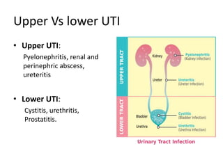
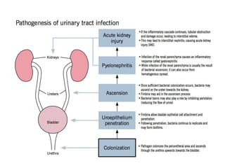
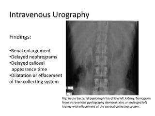
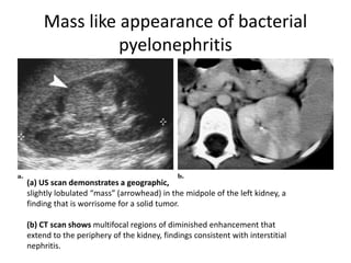
Radiological Approach to Urinary
Tract Infection
Dr Milan Silwal
MD Resident, Radiodiagnosis
NAMS ,Bir Hospital
Introduction
• UTI refers to a symptomatic bacterial infection within
the urinary tract.
• UTIs occur when there is colonisation of microbes in
the uroepithelium and a subsequent inflammatory
response.
• Urinary tract infections are the most common
urologic disease.
Except for the distal urethra, the urinary tract is normally sterile
due to host defenses against bacterial colonization.
Uncomplicated vs. complicated UTI
 uncomplicated: an otherwise healthy patient with a
structurally and functionally normal urinary tract
 complicated: factors are present that decrease the
likelihood of therapy being effective
– urinary tract is structurally or functionally abnormal
– immunocompromised status
– especially virulent pathogen
– factors that may make a patient complicated
• male
• childhood UTIs
• pregnancy
• elderly
• diabetes
Upper Vs lower UTI
• Upper UTI:
Pyelonephritis, renal and
perinephric abscess,
ureteritis
• Lower UTI:
Cystitis, urethritis,
Prostatitis.
Upper UTIs:
Associated with renal pelvic, renal calyceal and renal parenchymal inflammation
• Pyelonephritis
– Acute
– Chronic
– Emphysematous
– Xanthogranulomatous
– Fungal
• Abscess
– Renal
– Pyonephrosis
– Perinephric
• Tuberculosis
• Schistosomiasis
• Hydatid disease
• Reflux nephropathy
• Rare
– Pyeloureteritis cystica
– Leucoplakia and
Malacoplakia
Risk Factors For Upper UTI
• Immune suppression (AIDS, diabetes, corticosteroid therapy)
• Recent treatment with antibiotics for urothelial bacterial flora
imbalance,
• Urinary catheter.
• Pregnancy.
• Obstruction to the urinary collecting system:
 Urinary lithiasis,
 Deformity to the urinary system,
 Urothelial neoplasm,
 Bladder neuropathy
• In general, imaging is not necessary for diagnosis and
treatment of uncomplicated UTI in adult patients.
• However, diagnostic imaging demonstrates the
extent and nature of the urologic infections and
their potential complications.
• Guidelines for selection of pts with UTI for
radiologic evaluation:
- all neonates with 1st UTI
- all males with 1st UTI at any age
- all pts with recurrent UTI
- all pts with pyelonephritis
Acute Bacterial Pyelonephritis
• Epidemiology
Approximately five times more common in females with a sharp increase
following puberty .
• Clinical Features:
Rapid onset of High Fevers , Flank pain and Tenderness.
• Organsims :
 E. coli (most common)
 Klebsiella sp.
 Proteus sp.
 Enterobacter sp.
 Pseudomonas sp.
 Haemophilus influenzae
Plain Radiography
• Plain films have a limited role to play.
• They may demonstrate obstructing urinary
tract calculi and occasionally demonstrate gas
within the collecting system (emphysematous
pyelonephritis).
Intravenous Urography
Fig: Acute bacterial pyelonephritis of the left kidney. Tomogram
from intravenous pyelography demonstrates an enlarged left
kidney with effacement of the central collecting system.
Findings:
•Renal enlargement
•Delayed nephrograms
•Delayed caliceal
appearance time
•Dilatation or effacement
of the collecting system
Ultrasound
• Ultrasound is insensitive to the changes of acute
pyelonephritis, with most patients having 'normal' scan.
Possible features include:
– Particulate matter in the collecting system
– Reduced areas of cortical vascularity by using power Doppler
– Gas bubbles (emphysematous pyelonephritis)
– Abnormal echogenicity of the renal parenchyma
– Focal/segmental hypoechoic regions
Ultrasound is however useful in assessing for local
complications such as hydronephrosis, renal abscess
formation, renal infarction, perinephric collections.
USG
Fig: a) US scan shows a wedge-shaped hyperechoic focus (arrowhead)
in the upper pole of the right kidney related to acute bacterial pyelonephritis.
(b) Color flow US image demonstrates diminished flow through the involved
area.
CT
CT is the most sensitive modality for the renal tract.
Non-contrast CT
• Excellent for indentifying renal calculi, hemorrhage, renal enlargement,
inflammatory masses and obstruction.
Post-contrast CT
• one or more focal wedge like regions will appear swollen and demonstrate
reduced enhancement compared to the normal portions of the kidney.
• Delayed and persistent enhancement of the affected regions seen on 3-6
hours after contrast administration due to slow flow of contrast through
involved tubules.
CT
Fig:CT without contrast material shows focal hyper-attenuation areas in the upper
pole of the right kidney which does not present enhancement with contrast
administration,findings suggestive of hemorrhagic acute bacterial pyelonephritis.
CECT
Right pyelonephritis on a contrast-enhanced CT scan of the
abdomen and pelvis in the tubular venous phase: the renal
parenchyma has a ‘‘spoked wheel’’ appearance.
Mass like appearance of bacterial
pyelonephritis
(a) US scan demonstrates a geographic,
slightly lobulated “mass” (arrowhead) in the midpole of the left kidney, a
finding that is worrisome for a solid tumor.
(b) CT scan shows multifocal regions of diminished enhancement that
extend to the periphery of the kidney, findings consistent with interstitial
nephritis.
MRI
• MRI is usually reserved for patients who are pregnant,
and iodinated contrast agents are contraindicated.
• The kidney demonstrates wedge shaped regions of
altered signal:
• T1: affected region(s) appear hypointense compared
to normal kidney parenchyma
• T2: hyperintense compared to normal kidney
parenchyma
• T1 C+: reduced enhancement.
NUCLEAR MEDICINE
Scintiscan obtained with technetium 99m di-mercapto-succinic acid
:Demonstrates a photopenic, peripheral defect (arrow) in the upper lateral
margin of the right kidney that correlates with an area of acute bacterial
pyelonephritis.
Technetium-99m di-
mercapto-succinic acid
(DMSA) demonstrates a
similar reduction in
renal perfusion and
function, which one or
more wedge like defects
in the outline of the
kidneys .
Chronic Pyelonephritis
• A form of pyelonephritis where there are
longstanding sequelae of renal infection.
• When acute pyelonephritis occurs repeatedly,
usually in relation to occult VUR, this can lead to
the patient developing fibrosing interstitial
nephritis.
• Very often it progresses slowly into renal failure.
Radiographic Features
General features are often characterised by
• Renal scarring
• Renal atrophy
• Renal cortical thinning
• Compensatory hypertrophy of residual normal tissue
(which may mimic a mass lesion- pseudotumor)
• Calyceal clubbing: secondary to retraction of the
papilla from overlying scar
• Thickening and dilatation of the calyceal system
• Overall renal asymmetry
Chronic Pyelonephritis
Fig:(a) Unenhanced CT scan shows a small, deformed right kidney with multiple
deep scars and dystrophic calcifications.
(b) Photograph of the resected kidneys demonstrates extensive bilateral scar
formation.
Chronic pyelonephritis: a: axial view; b: coronal reconstruction. Pyelonephritis
scar tissue combining cortical retraction (whitearrows) and deformation of the
calyces with areas in between that are comparatively healthy seen on contrast-
enhanced CT scan
Tubercular Infection of Urinary Tract
• The genitourinary system is one of the most common sites of
involvement by extrapulmonary tuberculosis, accounting for
15 to 20 percent of infections outside the lungs.
• Approximately 4 to 8 percent of patients with pulmonary
tuberculosis will develop clinically significant genitourinary
infection.
• TB occurs in approximately 10 percent of patients with AIDS
and involves at least one extra-pulmonary site in nearly 50
percent of the patients, with kidneys being the most
commonly involved genitourinary site.
Pathogenesis
• Hematogeneous spread from the primary infection elsewhere.
• Latent Period: 5-20 years(so less common in case of Children)
• Initial phase :Involve both kidneys and lodged in the glomerular and
peritubular capillary bed.
• Most of them heal without sequelae.
• Bacilli erode the initial vascular location and spill into the tubules.
• This results in granuloma formation along the nephron.
• Granuloma, caseous necrosis, and cavitation represent
stages of progression of the infection.
• This may communicate with the pelvicalyceal system, and
the infection further spreads to the ureter and urinary
bladder.
• The pelvicalyceal and ureteral involvement appears as
mucosal ulcerations, focal or generalised dilatation or
cicatrisation.
• Fibrosis and calcium depositions represent healing.
Clinical Features
• Frequency of urination
• Dysuria
• Urgency
• Hematuria (50% of cases)
• Dull or chronic renal pain
• Constitutional symptoms like fever, weight loss, fatigue
are present in about 50 percent of the patients.
• Diagnosis of TB should be only considered in patients
with recurrent UTI that is not responding to
appropriate therapy.
Laboratory Investigations
• Sterile Pyuria
• Microscopic examinations: AFB
• Urine culture or growth of tubercle bacilli in inoculated guinea-pigs.
• Routine AFB culture (delay of 6-8 weeks) LJ media.
• PCR is highly sensitive (upto 94 percent), provides a quicker
diagnosis.
• FNAC is useful in those with a negative culture to define
granulomatous nature of the visible lesions.
Radiological Examinations
• Conventional radiological studies remain the
procedures of choice.
• Early findings are best detected on intravenous
urogaphy (IVU) while ultrasonography and CT are
useful for late or chronic changes.
• Whenever an imaging pattern of chronic renal
inflammatory disease is recognized, particularly in
the setting of periureteric or peripelvic fibrosis,
tuberculosis must be considered.
Plain Radiograph
• Important initial examination before IVU.
• Provide valuable information such as calcification or
evidence of skeletal involvement.
• Calcification is of two types:
(i) Amorphous granular: granulomatous masses
(ii) Dense punctate : healed tuberculoma.
Calcifications
Intravenous Urography
• very useful modality to detect early features
of urinary tract tuberculosis.
• However, the diagnosis can be made with
certainty on urography only if the lesion has
ulcerated into the calyx.
• may be normal in symptomatic renal
tuberculosis in the initial stages.
IVU findings on Kidney
The earliest urographic
abnormality is the loss of
definition of a minor calyx
producing an indistinct
feathery outline, irregularity
of the surface of one or
more papillae or calyces
with normal renal size and
contour.
Involvement of Renal Parenchyma
Fig: IVU shows a large cavity
communicating with a dilated,
irregular superior calyx
First : coalescence of granulomas
leading to unifocal or multifocal mass
lesions.
Caliectasis occurs with irregularity in
contour indicating erosion of the
pyramids and cortical necrosis.
Depending on the necrosis, a cavity
with irregular walls communicates
with a deformed calyx.
• The second form seen in advanced renal
tuberculosis is parenchymal surface scarring
over retracted papillae and dilated calyces.
• This may be focal or diffuse involving the
entire kidney and occurs due to tissue loss
leading to fibrosis.
• Associated calcification or calculi may be seen.
Figs 7.6A to D: (A) IVU reveals a large cavity communicating with the superior calyx of
right kidney, (B) US shows a hypoechoic focal lesion in the upper polar cortex with an
echogenic rim, (C) CT confirms the presence of the cavity seen on IVU and US, with
pooling of contrast on delayed images, (D) due to communication with the
pelvicalyceal system
Autonephrectomy
• Third form, represents end stage disease.
• Two types
A)Caseo-cavernous type:
enlarged sac filled with
caseous material,
with or without
calcification.
B) calcified shrunken
non-functioning kidney.
Fig.: Nephrostogram showing deformed and contracted renal pelvis with
proximal dilatation of the pelvicalyceal system. Superior calyx is
communicating with an irregular cavity and there is infundibular stenosis.
Ureter is also dilated due to lower end stricture
Subsequent stimulation of scirrhous reaction
causes stenosis and obstruction of parts of the
collecting system.
The common sites of strictures
being the neck of a calyx, PUJ and VUJ.
there may be dilatation of a single calyx
(hydrocalyx) or regional hydrocalycosis or
generalized dilatation of the pelvicalyceal
system.
• Phantom calyx: failure of contrast excretion by the
involved parenchyma due to complete obstruction of
the infundibulum or the calyx.
• Kerk’s kink : deformity of the pelvicalyceal system
may be caused by traction from a strictured
infundibulum or parenchymal fibrosis kinking the
pelvis.
URETER
• Involvement almost always secondary to renal TB.
• Initially Prolonged bacilluria and Atony leading to dilataion.
• Progress to mucosal ulceration- irregular segments.
• Finally necrosis and fibrosis – stricture formation.
• May produces a beaded or corkscrew appearance.
• Terminal segment - most commonly involved.
• Severe thickening of the wall produces a rigid shortened ureter with
narrow lumen termed as “pipe stem ureter”.
Fig.: IVU showing long irregular
stricture involving lower end of
left ureter with hydroureter,
Hydronephrosis and infundibular
stenosis of inferior calyx of left
kidney.
Also note changes of
tuberculosis involving L2-L3
vertebrae
Urinary Bladder
• Involved in upto one-third of patients of urinary tract TB
• Tubercular cystitis leads to edema of the bladder mucosa.
• Large tuberculomas in vesical wall can manifest radiologically as
filling defects simulating carcinoma.
• Advanced disease leads to irregular contracture with thick walls
and reduction of bladder capacity – the “thimble bladder”
• Fibrosis in the region of the trigone produces gaping of the
ureterovesical junction resulting in free vesicoureteric reflux.
Fig.: MCU study reveals a small
capacity bladder with reflux into
a dilated, tortuous right ureter
IVU reveals a small capacity
(thimble) urinary bladder, ureteral
stricture with proximal hydroureter
and hydronephrosis on left side.
Right kidney is non-functioning.
Urethral TB
• Tuberculosis of the male urethra is uncommon
and usually occurs secondary to renal infection.
Associations
• Prostatic abscess,
• Periurethral abscess
• Fistula formation.
It may result in a nonspecific stricture almost
always in the bulbo membranous urethra.
Ultrasonography
• Ultrasound is not used as the primary imaging
modality for diagnosis of urinary tract TB.
• The role of sonography is also to provide
guidance for the interventional procedures
of percutaneous nephrostomy (PCN),
antegrade dilatation of ureteral stricture and
drainage of the perinephric abscess.
USG findings of Renal TB
Fig:US reveals an anechoic cavity in the renal
cortex with a sonolucent track connecting the
cavity and the pelvicalyceal system
CT
• Indicated only in patients with strong clinical
suspicion but normal or equivocal findings on
urography and ultrasonography.
Useful in
• Demonstrating the extent of involvement,
• Length of ureteric stricture
• Adjoining retroperitoneal disease and associated
spinal or solid organ involvement.
Kidney
• Small poorly enhancing nodules, uneven caliectasis and
calcification.
• Isolated cortical involvement by focal lesions is well demonstrated
on CT.
• A cavity is well delineated with pooling of contrast in the delayed
images.
• Provide better visualisation of retroperitoneal structures.
• More detailed information on presence and pattern of calcification.
• Functional status of the kidney.
Figs: CECT of a 36 years old male
Showing multiple hypodense
lesions in the left kidney with
adjoining perinephric collection
and fascial thickening.
Fig: CECT abdomen: Renal pelvis is
contracted with a thickened wall
(arrow)
Ureter
Fig: CECT of a young girl reveals a poorly
functioning left kidney Shows ;
Thickenig of the left ureteric wall.(arrowhead)
Thickening of the
renal pelvis or ureter
is highly suggestive
of tuberculosis and
can be readily
identified on CT.
Differential Diagnosis
• Papillary necrosis.
• Focal lesions resemble other infections or
even neoplasms.
• Calcification and extensive inflammation can
mimic xanthogranulomatous pyelonephritis.
• Urinary bladder involvement may be confused
with neoplastic involvement.
Renal abscess
• Focal renal parenchymal inflammation progress to liquefaction –
abscess
– Sequel to untreated or resistant acute PN – Gm –ve bacilli or
anaerobic bacilli
– Hematogenous spread – Staph aureus
• Septic site elsewhere
• Impaired immunity, including DM
- superadded infection in a renal cyst
- direct involvement of perinephric space from pancreas, colon and
retroperitoneum.
• Multiple or b/l in hematogenous spread- also abscess elsewhere
• Better imaged with US or CT –
– Heterogenous lesion with
single or multiple central
cystic necrosis;
– irregular marginal
enhancement;
– echogenic debri
– Gas in severe cases
– Perinephric inflammatory
changes
• When substantial liquefied area – US or CT
guided percutaneous drain.
• In early stage – only tiny area of liquefaction;
thus better served by IV antibiotics and
subsequent scanning to monitor resolution
rather than premature intervention.
Perinephric abscess
• Renal infection of any severity – extension into the perinephric
space.
• Plain radiograph
– Loss of psoas shadow
– Poorly defined renal outline
• IVU
– Reduced or absent renal fxn – failure to excrete or concentrate
contrast
• USG or CT
– Fluid or semifluid collection, often containing debri and septation
– Sometimes gas
Pyonephrosis
 An infected and obstructed collecting system, which
frequently is enlarged.
 Causes- calculi, tumor, complications from
pyelonephritis (sloughed papilla), or strictures.
 Immediate intervention is required .
 If left untreated, a rapid, often permanent, decline in
renal function may result, and pt develop septic shock
 USG- dilatation of the PCS,
echogenic collecting system
debris, fluid-fluid levels within
the collecting system, and occ.
incomplete (dirty) echoes of
collecting system gas.
 Echogenic debris- sensitivity of
90%, specificity of 97%, and
accuracy of 96% in the
diagnosis of pyonephrosis
versus simple hydronephrosis
 CT
- thickening of the renal pelvic wall
(>2 mm),
- parenchymal or perinephric
inflammatory changes,
- dilatation and obstruction of the
collecting system,
- higher attenuation values of the
fluid within the renal
collecting system, and
- layering of contrast material above
and anterior to the purulent fluid on
excretory studies .
• If Untreated –
– danger of septicaemia
– Destruction of normal renal tissue
– Extension into surrounding tissues
• Rx – percuatenous drainage (only with adequate antibiotic cover
and minimal manipulation – gm –ve septicaemia and endotoxic
shock
Emphysematous Pyelonephritis
• A morbid infection of kidneys, with characteristic gas formation
within or around the kidneys.
• If not treated early, it may lead to fulminant sepsis and carries a
high mortality.
• More common in females.
Clinical presentation
• Flank pain, urinary tract obstruction with fever.
• Leukocytosis and hyperglycemia (in diabetics) are prominent lab
findings.
• Thrombocytopenia is particularly associated with poor prognosis
• Approximately 90% of patients have uncontrolled diabetes.
• Also seen in immunocompromised individuals or associated with
urolithiasis, neoplasms or sloughing of papilla.
• If diffuse – emergency nephrectomy
– Without surgery, mortality exceeds 50%
• If focal – conservative approach with IV antibiotics and CT guided drainage.
Causative organisms include
• E. Coli:
• Klebsiella pneumonia
• Proteus mirabilis
Radiographic features
Plain film and fluoroscopy (IVU)
May show mottled gas within
renal fossa or crescentic gas
collection within Gerota‘s fascia.
Linear gas shadows along
paraspinal region may also be
seen, representing
retroperitoneal air.
Fig:Bilateral emphysematous pyelonephritis.Abdominal radiograph
from intravenous pyelographic study demonstrates lucent air that
outlines both kidneys with gas along the left paraspinal region
USG
•May show an enlarged kidney
with coarse echoes within renal
parenchyma or collecting system.
•Dirty echogenic foci with
reverberation/ringdown
artifacts representing air ('dirty
shadowing') may also be seen
•Bowel gas over kidneys may lead
to false positive diagnosis.
CT
best diagnostic modality for
emphysematous pyelonephritis.
Ideal to assess the renal as well
as perirenal extent,
It may show following diagnostic
features:
•enlarged, destructed renal
parenchyma
•small bubbly or linear streaks of
gas, fluid collections, with gas-
fluid levels
• focal necrotic areas +/- abscess
Radiological Classification
• CT features of emphysematous pyelonephritis differentiates into two
types:
Type 1 (33%)
• Renal parenchymal destruction with streaky or mottled
appearance of gas
• Intra- or extrarenal fluid collections are characteristically
absent
• It is usually more aggressive and lead to death shortly, if not intervened
early. 69% mortality.
Type 2 (66%)
• Renal or extrarenal collections associated with bubbly or loculated gas, or
gas within pelvicalyceal system or ureter.
• Only 18% mortality.
Emphysematous pyelonephritis
classification
The Huang-Tseng CT classification system
• Class 1: gas in collecting system only
• Class 2: gas in renal parenchyma only (without extrarenal
extension)
• Class 3: gas in renal parenchyma with extrarenal extension
Class 3a: extension of gas or abscess to perinephric space
Class 3b: extension of gas or abscess to pararenal space
• Class 4: bilateral emphysematous pyelonephritis or solitary
kidney with emphysematous pyelonephritis
Xanthogranulomatous Pyelonephritis
• A rare form of chronic pyelonephritis and represents a
chronic granulomatous disease resulting in a nonfunctioning
kidney.
• Radiographic features are usually specific.
Epidemiology
• Most frequently presents in middle-aged to elderly patients.
• Female predilection (F:M 2:1)
• Presumably relating to an increased incidence of urinary
tract infections and thus struvite (staghorn) calculi.
Pathology
• Xanthogranulomatous pyelonephritis is, as the name
suggests, a chronic granulomatous process believed to
be the result of subacute/chronic infection inciting a
chronic but incomplete immune reaction .
• The kidney is eventually replaced by a mass of reactive
tissue, surrounding the usually present (90%) inciting
staghorn calculus with associated hydronephrosis of a
greater or lesser degree.
• Foamy (lipid laden) macrophages predominate .
Radiographic features
• Two forms of the disease are recognised both
macroscopically and on imaging :
1. Diffuse: 90% of cases
2. Focal: 10% of cases
Plain film
• Plain film findings are difficult to distinguish from
a routine staghorn calculus, although
fragmentation and enlargement of the
renal outline may be seen.
USG
• Ultrasound examination demonstrate an
enlarged and distorted renal outline, with loss
of the normal renal architecture and (usually)
a centrally located shadowing calculus.
• CT findings are most helpful in reaching the correct
diagnosis.
• The normal renal outline is lost and enlarged with
paradoxical contracted renal pelvis.
• The calyces, however, are dilated giving a multloculated
appearance that has been likened to the paw print of a
bear (bear's paw sign).
• Sometimes there is perinephric extension with thickening
of the Gerota's fascia. Calcification can be better delineated
on CT scan.
CT
NCCT showing staghorn calculus with dilated calices and
enlarged right kidney with few calcification
MRI
• MRI appearances mirror the heterogeneous
nature of the mass with solid and cystic
components surrounding a central staghorn
calculus.
• As such signal is heterogeneous on all
sequences.
Malacoplakia
• Malacoplakia of the urinary system is the commonest
manifestation of malacoplakia.
• The latter, meaning soft plaque, is a rare chronic
granulomatous condition that can affect any organ.
Epidemiology
• Malacoplakia has a peak incidence in middle age,
• Female-to-male ratio of 11:4.
• The disease is more common in patients who are
immuno compromised or those with diabetes mellitus.
• The histologic hallmark of the disorder is the
presence of basophilic inclusion called
Michaelis-Gutmann bodies in large
eosinophilic macrophage.
Location
• Within the urinary tract, the bladder is the
most frequently affected organ (40% of
patients with malacoplakia)
Radiographic Features
• Imaging characteristics of malacoplakia are varied, and most
commonly observed within the bladder, although plaques may also
occur in the ureters.
• Malacoplakia may present as multiple, polypoid, vascular, solid
masses or as circumferential wall thickening, associated with
vesicoureter reflux and dilatation of the upper urinary tract.
• These masses vary in size from a few millimeters to several
centimeters.
• Extremely aggressive – may involve adjacent structure; erodes
bone.
• Occ. Solitary, indistinguishable from renal mass on imaging features
alone.
Malakoplakia
(a, b) Unenhanced CT images obtained at different levels (a
higher than b) demonstrate symmetrically enlarged kidneys.
leukoplakia
• Multiple small plaques of chronic
inflammatory tissue arising from the
urothelium.
• Predilection to progress to premalignant
squamous metaplasia.
References
• Textbook of Radiology and Imaging , David
Sutton, 7th edition
• Diagnostic Radiology Genitourinary Imaging,
Berry, 3rd edition
• http://radiopedia.com
• Images: various internet sources
Thank You!

Urinary tract infections

  • 1.
    Radiological Approach toUrinary Tract Infection Dr Milan Silwal MD Resident, Radiodiagnosis NAMS ,Bir Hospital
  • 2.
    Introduction • UTI refersto a symptomatic bacterial infection within the urinary tract. • UTIs occur when there is colonisation of microbes in the uroepithelium and a subsequent inflammatory response. • Urinary tract infections are the most common urologic disease.
  • 3.
    Except for thedistal urethra, the urinary tract is normally sterile due to host defenses against bacterial colonization.
  • 4.
    Uncomplicated vs. complicatedUTI  uncomplicated: an otherwise healthy patient with a structurally and functionally normal urinary tract  complicated: factors are present that decrease the likelihood of therapy being effective – urinary tract is structurally or functionally abnormal – immunocompromised status – especially virulent pathogen – factors that may make a patient complicated • male • childhood UTIs • pregnancy • elderly • diabetes
  • 5.
    Upper Vs lowerUTI • Upper UTI: Pyelonephritis, renal and perinephric abscess, ureteritis • Lower UTI: Cystitis, urethritis, Prostatitis.
  • 6.
    Upper UTIs: Associated withrenal pelvic, renal calyceal and renal parenchymal inflammation • Pyelonephritis – Acute – Chronic – Emphysematous – Xanthogranulomatous – Fungal • Abscess – Renal – Pyonephrosis – Perinephric • Tuberculosis • Schistosomiasis • Hydatid disease • Reflux nephropathy • Rare – Pyeloureteritis cystica – Leucoplakia and Malacoplakia
  • 7.
    Risk Factors ForUpper UTI • Immune suppression (AIDS, diabetes, corticosteroid therapy) • Recent treatment with antibiotics for urothelial bacterial flora imbalance, • Urinary catheter. • Pregnancy. • Obstruction to the urinary collecting system:  Urinary lithiasis,  Deformity to the urinary system,  Urothelial neoplasm,  Bladder neuropathy
  • 8.
    • In general,imaging is not necessary for diagnosis and treatment of uncomplicated UTI in adult patients. • However, diagnostic imaging demonstrates the extent and nature of the urologic infections and their potential complications.
  • 9.
    • Guidelines forselection of pts with UTI for radiologic evaluation: - all neonates with 1st UTI - all males with 1st UTI at any age - all pts with recurrent UTI - all pts with pyelonephritis
  • 10.
    Acute Bacterial Pyelonephritis •Epidemiology Approximately five times more common in females with a sharp increase following puberty . • Clinical Features: Rapid onset of High Fevers , Flank pain and Tenderness. • Organsims :  E. coli (most common)  Klebsiella sp.  Proteus sp.  Enterobacter sp.  Pseudomonas sp.  Haemophilus influenzae
  • 12.
    Plain Radiography • Plainfilms have a limited role to play. • They may demonstrate obstructing urinary tract calculi and occasionally demonstrate gas within the collecting system (emphysematous pyelonephritis).
  • 13.
    Intravenous Urography Fig: Acutebacterial pyelonephritis of the left kidney. Tomogram from intravenous pyelography demonstrates an enlarged left kidney with effacement of the central collecting system. Findings: •Renal enlargement •Delayed nephrograms •Delayed caliceal appearance time •Dilatation or effacement of the collecting system
  • 14.
    Ultrasound • Ultrasound isinsensitive to the changes of acute pyelonephritis, with most patients having 'normal' scan. Possible features include: – Particulate matter in the collecting system – Reduced areas of cortical vascularity by using power Doppler – Gas bubbles (emphysematous pyelonephritis) – Abnormal echogenicity of the renal parenchyma – Focal/segmental hypoechoic regions Ultrasound is however useful in assessing for local complications such as hydronephrosis, renal abscess formation, renal infarction, perinephric collections.
  • 15.
    USG Fig: a) USscan shows a wedge-shaped hyperechoic focus (arrowhead) in the upper pole of the right kidney related to acute bacterial pyelonephritis. (b) Color flow US image demonstrates diminished flow through the involved area.
  • 16.
    CT CT is themost sensitive modality for the renal tract. Non-contrast CT • Excellent for indentifying renal calculi, hemorrhage, renal enlargement, inflammatory masses and obstruction. Post-contrast CT • one or more focal wedge like regions will appear swollen and demonstrate reduced enhancement compared to the normal portions of the kidney. • Delayed and persistent enhancement of the affected regions seen on 3-6 hours after contrast administration due to slow flow of contrast through involved tubules.
  • 17.
    CT Fig:CT without contrastmaterial shows focal hyper-attenuation areas in the upper pole of the right kidney which does not present enhancement with contrast administration,findings suggestive of hemorrhagic acute bacterial pyelonephritis.
  • 18.
    CECT Right pyelonephritis ona contrast-enhanced CT scan of the abdomen and pelvis in the tubular venous phase: the renal parenchyma has a ‘‘spoked wheel’’ appearance.
  • 19.
    Mass like appearanceof bacterial pyelonephritis (a) US scan demonstrates a geographic, slightly lobulated “mass” (arrowhead) in the midpole of the left kidney, a finding that is worrisome for a solid tumor. (b) CT scan shows multifocal regions of diminished enhancement that extend to the periphery of the kidney, findings consistent with interstitial nephritis.
  • 20.
    MRI • MRI isusually reserved for patients who are pregnant, and iodinated contrast agents are contraindicated. • The kidney demonstrates wedge shaped regions of altered signal: • T1: affected region(s) appear hypointense compared to normal kidney parenchyma • T2: hyperintense compared to normal kidney parenchyma • T1 C+: reduced enhancement.
  • 21.
    NUCLEAR MEDICINE Scintiscan obtainedwith technetium 99m di-mercapto-succinic acid :Demonstrates a photopenic, peripheral defect (arrow) in the upper lateral margin of the right kidney that correlates with an area of acute bacterial pyelonephritis. Technetium-99m di- mercapto-succinic acid (DMSA) demonstrates a similar reduction in renal perfusion and function, which one or more wedge like defects in the outline of the kidneys .
  • 22.
    Chronic Pyelonephritis • Aform of pyelonephritis where there are longstanding sequelae of renal infection. • When acute pyelonephritis occurs repeatedly, usually in relation to occult VUR, this can lead to the patient developing fibrosing interstitial nephritis. • Very often it progresses slowly into renal failure.
  • 23.
    Radiographic Features General featuresare often characterised by • Renal scarring • Renal atrophy • Renal cortical thinning • Compensatory hypertrophy of residual normal tissue (which may mimic a mass lesion- pseudotumor) • Calyceal clubbing: secondary to retraction of the papilla from overlying scar • Thickening and dilatation of the calyceal system • Overall renal asymmetry
  • 24.
    Chronic Pyelonephritis Fig:(a) UnenhancedCT scan shows a small, deformed right kidney with multiple deep scars and dystrophic calcifications. (b) Photograph of the resected kidneys demonstrates extensive bilateral scar formation.
  • 25.
    Chronic pyelonephritis: a:axial view; b: coronal reconstruction. Pyelonephritis scar tissue combining cortical retraction (whitearrows) and deformation of the calyces with areas in between that are comparatively healthy seen on contrast- enhanced CT scan
  • 26.
    Tubercular Infection ofUrinary Tract • The genitourinary system is one of the most common sites of involvement by extrapulmonary tuberculosis, accounting for 15 to 20 percent of infections outside the lungs. • Approximately 4 to 8 percent of patients with pulmonary tuberculosis will develop clinically significant genitourinary infection. • TB occurs in approximately 10 percent of patients with AIDS and involves at least one extra-pulmonary site in nearly 50 percent of the patients, with kidneys being the most commonly involved genitourinary site.
  • 27.
    Pathogenesis • Hematogeneous spreadfrom the primary infection elsewhere. • Latent Period: 5-20 years(so less common in case of Children) • Initial phase :Involve both kidneys and lodged in the glomerular and peritubular capillary bed. • Most of them heal without sequelae. • Bacilli erode the initial vascular location and spill into the tubules. • This results in granuloma formation along the nephron.
  • 28.
    • Granuloma, caseousnecrosis, and cavitation represent stages of progression of the infection. • This may communicate with the pelvicalyceal system, and the infection further spreads to the ureter and urinary bladder. • The pelvicalyceal and ureteral involvement appears as mucosal ulcerations, focal or generalised dilatation or cicatrisation. • Fibrosis and calcium depositions represent healing.
  • 29.
    Clinical Features • Frequencyof urination • Dysuria • Urgency • Hematuria (50% of cases) • Dull or chronic renal pain • Constitutional symptoms like fever, weight loss, fatigue are present in about 50 percent of the patients. • Diagnosis of TB should be only considered in patients with recurrent UTI that is not responding to appropriate therapy.
  • 30.
    Laboratory Investigations • SterilePyuria • Microscopic examinations: AFB • Urine culture or growth of tubercle bacilli in inoculated guinea-pigs. • Routine AFB culture (delay of 6-8 weeks) LJ media. • PCR is highly sensitive (upto 94 percent), provides a quicker diagnosis. • FNAC is useful in those with a negative culture to define granulomatous nature of the visible lesions.
  • 31.
    Radiological Examinations • Conventionalradiological studies remain the procedures of choice. • Early findings are best detected on intravenous urogaphy (IVU) while ultrasonography and CT are useful for late or chronic changes. • Whenever an imaging pattern of chronic renal inflammatory disease is recognized, particularly in the setting of periureteric or peripelvic fibrosis, tuberculosis must be considered.
  • 32.
    Plain Radiograph • Importantinitial examination before IVU. • Provide valuable information such as calcification or evidence of skeletal involvement. • Calcification is of two types: (i) Amorphous granular: granulomatous masses (ii) Dense punctate : healed tuberculoma.
  • 33.
  • 34.
    Intravenous Urography • veryuseful modality to detect early features of urinary tract tuberculosis. • However, the diagnosis can be made with certainty on urography only if the lesion has ulcerated into the calyx. • may be normal in symptomatic renal tuberculosis in the initial stages.
  • 35.
    IVU findings onKidney The earliest urographic abnormality is the loss of definition of a minor calyx producing an indistinct feathery outline, irregularity of the surface of one or more papillae or calyces with normal renal size and contour.
  • 36.
    Involvement of RenalParenchyma Fig: IVU shows a large cavity communicating with a dilated, irregular superior calyx First : coalescence of granulomas leading to unifocal or multifocal mass lesions. Caliectasis occurs with irregularity in contour indicating erosion of the pyramids and cortical necrosis. Depending on the necrosis, a cavity with irregular walls communicates with a deformed calyx.
  • 37.
    • The secondform seen in advanced renal tuberculosis is parenchymal surface scarring over retracted papillae and dilated calyces. • This may be focal or diffuse involving the entire kidney and occurs due to tissue loss leading to fibrosis. • Associated calcification or calculi may be seen.
  • 38.
    Figs 7.6A toD: (A) IVU reveals a large cavity communicating with the superior calyx of right kidney, (B) US shows a hypoechoic focal lesion in the upper polar cortex with an echogenic rim, (C) CT confirms the presence of the cavity seen on IVU and US, with pooling of contrast on delayed images, (D) due to communication with the pelvicalyceal system
  • 39.
    Autonephrectomy • Third form,represents end stage disease. • Two types A)Caseo-cavernous type: enlarged sac filled with caseous material, with or without calcification. B) calcified shrunken non-functioning kidney.
  • 40.
    Fig.: Nephrostogram showingdeformed and contracted renal pelvis with proximal dilatation of the pelvicalyceal system. Superior calyx is communicating with an irregular cavity and there is infundibular stenosis. Ureter is also dilated due to lower end stricture Subsequent stimulation of scirrhous reaction causes stenosis and obstruction of parts of the collecting system. The common sites of strictures being the neck of a calyx, PUJ and VUJ. there may be dilatation of a single calyx (hydrocalyx) or regional hydrocalycosis or generalized dilatation of the pelvicalyceal system.
  • 41.
    • Phantom calyx:failure of contrast excretion by the involved parenchyma due to complete obstruction of the infundibulum or the calyx. • Kerk’s kink : deformity of the pelvicalyceal system may be caused by traction from a strictured infundibulum or parenchymal fibrosis kinking the pelvis.
  • 42.
    URETER • Involvement almostalways secondary to renal TB. • Initially Prolonged bacilluria and Atony leading to dilataion. • Progress to mucosal ulceration- irregular segments. • Finally necrosis and fibrosis – stricture formation. • May produces a beaded or corkscrew appearance. • Terminal segment - most commonly involved. • Severe thickening of the wall produces a rigid shortened ureter with narrow lumen termed as “pipe stem ureter”.
  • 43.
    Fig.: IVU showinglong irregular stricture involving lower end of left ureter with hydroureter, Hydronephrosis and infundibular stenosis of inferior calyx of left kidney. Also note changes of tuberculosis involving L2-L3 vertebrae
  • 44.
    Urinary Bladder • Involvedin upto one-third of patients of urinary tract TB • Tubercular cystitis leads to edema of the bladder mucosa. • Large tuberculomas in vesical wall can manifest radiologically as filling defects simulating carcinoma. • Advanced disease leads to irregular contracture with thick walls and reduction of bladder capacity – the “thimble bladder” • Fibrosis in the region of the trigone produces gaping of the ureterovesical junction resulting in free vesicoureteric reflux.
  • 45.
    Fig.: MCU studyreveals a small capacity bladder with reflux into a dilated, tortuous right ureter IVU reveals a small capacity (thimble) urinary bladder, ureteral stricture with proximal hydroureter and hydronephrosis on left side. Right kidney is non-functioning.
  • 46.
    Urethral TB • Tuberculosisof the male urethra is uncommon and usually occurs secondary to renal infection. Associations • Prostatic abscess, • Periurethral abscess • Fistula formation. It may result in a nonspecific stricture almost always in the bulbo membranous urethra.
  • 47.
    Ultrasonography • Ultrasound isnot used as the primary imaging modality for diagnosis of urinary tract TB. • The role of sonography is also to provide guidance for the interventional procedures of percutaneous nephrostomy (PCN), antegrade dilatation of ureteral stricture and drainage of the perinephric abscess.
  • 48.
    USG findings ofRenal TB Fig:US reveals an anechoic cavity in the renal cortex with a sonolucent track connecting the cavity and the pelvicalyceal system
  • 49.
    CT • Indicated onlyin patients with strong clinical suspicion but normal or equivocal findings on urography and ultrasonography. Useful in • Demonstrating the extent of involvement, • Length of ureteric stricture • Adjoining retroperitoneal disease and associated spinal or solid organ involvement.
  • 50.
    Kidney • Small poorlyenhancing nodules, uneven caliectasis and calcification. • Isolated cortical involvement by focal lesions is well demonstrated on CT. • A cavity is well delineated with pooling of contrast in the delayed images. • Provide better visualisation of retroperitoneal structures. • More detailed information on presence and pattern of calcification. • Functional status of the kidney.
  • 51.
    Figs: CECT ofa 36 years old male Showing multiple hypodense lesions in the left kidney with adjoining perinephric collection and fascial thickening. Fig: CECT abdomen: Renal pelvis is contracted with a thickened wall (arrow)
  • 52.
    Ureter Fig: CECT ofa young girl reveals a poorly functioning left kidney Shows ; Thickenig of the left ureteric wall.(arrowhead) Thickening of the renal pelvis or ureter is highly suggestive of tuberculosis and can be readily identified on CT.
  • 53.
    Differential Diagnosis • Papillarynecrosis. • Focal lesions resemble other infections or even neoplasms. • Calcification and extensive inflammation can mimic xanthogranulomatous pyelonephritis. • Urinary bladder involvement may be confused with neoplastic involvement.
  • 54.
    Renal abscess • Focalrenal parenchymal inflammation progress to liquefaction – abscess – Sequel to untreated or resistant acute PN – Gm –ve bacilli or anaerobic bacilli – Hematogenous spread – Staph aureus • Septic site elsewhere • Impaired immunity, including DM - superadded infection in a renal cyst - direct involvement of perinephric space from pancreas, colon and retroperitoneum. • Multiple or b/l in hematogenous spread- also abscess elsewhere
  • 55.
    • Better imagedwith US or CT – – Heterogenous lesion with single or multiple central cystic necrosis; – irregular marginal enhancement; – echogenic debri – Gas in severe cases – Perinephric inflammatory changes
  • 56.
    • When substantialliquefied area – US or CT guided percutaneous drain. • In early stage – only tiny area of liquefaction; thus better served by IV antibiotics and subsequent scanning to monitor resolution rather than premature intervention.
  • 57.
    Perinephric abscess • Renalinfection of any severity – extension into the perinephric space. • Plain radiograph – Loss of psoas shadow – Poorly defined renal outline • IVU – Reduced or absent renal fxn – failure to excrete or concentrate contrast • USG or CT – Fluid or semifluid collection, often containing debri and septation – Sometimes gas
  • 59.
    Pyonephrosis  An infectedand obstructed collecting system, which frequently is enlarged.  Causes- calculi, tumor, complications from pyelonephritis (sloughed papilla), or strictures.  Immediate intervention is required .  If left untreated, a rapid, often permanent, decline in renal function may result, and pt develop septic shock
  • 60.
     USG- dilatationof the PCS, echogenic collecting system debris, fluid-fluid levels within the collecting system, and occ. incomplete (dirty) echoes of collecting system gas.  Echogenic debris- sensitivity of 90%, specificity of 97%, and accuracy of 96% in the diagnosis of pyonephrosis versus simple hydronephrosis
  • 61.
     CT - thickeningof the renal pelvic wall (>2 mm), - parenchymal or perinephric inflammatory changes, - dilatation and obstruction of the collecting system, - higher attenuation values of the fluid within the renal collecting system, and - layering of contrast material above and anterior to the purulent fluid on excretory studies .
  • 62.
    • If Untreated– – danger of septicaemia – Destruction of normal renal tissue – Extension into surrounding tissues • Rx – percuatenous drainage (only with adequate antibiotic cover and minimal manipulation – gm –ve septicaemia and endotoxic shock
  • 63.
    Emphysematous Pyelonephritis • Amorbid infection of kidneys, with characteristic gas formation within or around the kidneys. • If not treated early, it may lead to fulminant sepsis and carries a high mortality. • More common in females. Clinical presentation • Flank pain, urinary tract obstruction with fever. • Leukocytosis and hyperglycemia (in diabetics) are prominent lab findings. • Thrombocytopenia is particularly associated with poor prognosis
  • 64.
    • Approximately 90%of patients have uncontrolled diabetes. • Also seen in immunocompromised individuals or associated with urolithiasis, neoplasms or sloughing of papilla. • If diffuse – emergency nephrectomy – Without surgery, mortality exceeds 50% • If focal – conservative approach with IV antibiotics and CT guided drainage. Causative organisms include • E. Coli: • Klebsiella pneumonia • Proteus mirabilis
  • 65.
    Radiographic features Plain filmand fluoroscopy (IVU) May show mottled gas within renal fossa or crescentic gas collection within Gerota‘s fascia. Linear gas shadows along paraspinal region may also be seen, representing retroperitoneal air. Fig:Bilateral emphysematous pyelonephritis.Abdominal radiograph from intravenous pyelographic study demonstrates lucent air that outlines both kidneys with gas along the left paraspinal region
  • 66.
    USG •May show anenlarged kidney with coarse echoes within renal parenchyma or collecting system. •Dirty echogenic foci with reverberation/ringdown artifacts representing air ('dirty shadowing') may also be seen •Bowel gas over kidneys may lead to false positive diagnosis.
  • 67.
    CT best diagnostic modalityfor emphysematous pyelonephritis. Ideal to assess the renal as well as perirenal extent, It may show following diagnostic features: •enlarged, destructed renal parenchyma •small bubbly or linear streaks of gas, fluid collections, with gas- fluid levels • focal necrotic areas +/- abscess
  • 68.
    Radiological Classification • CTfeatures of emphysematous pyelonephritis differentiates into two types: Type 1 (33%) • Renal parenchymal destruction with streaky or mottled appearance of gas • Intra- or extrarenal fluid collections are characteristically absent • It is usually more aggressive and lead to death shortly, if not intervened early. 69% mortality. Type 2 (66%) • Renal or extrarenal collections associated with bubbly or loculated gas, or gas within pelvicalyceal system or ureter. • Only 18% mortality.
  • 69.
    Emphysematous pyelonephritis classification The Huang-TsengCT classification system • Class 1: gas in collecting system only • Class 2: gas in renal parenchyma only (without extrarenal extension) • Class 3: gas in renal parenchyma with extrarenal extension Class 3a: extension of gas or abscess to perinephric space Class 3b: extension of gas or abscess to pararenal space • Class 4: bilateral emphysematous pyelonephritis or solitary kidney with emphysematous pyelonephritis
  • 70.
    Xanthogranulomatous Pyelonephritis • Arare form of chronic pyelonephritis and represents a chronic granulomatous disease resulting in a nonfunctioning kidney. • Radiographic features are usually specific. Epidemiology • Most frequently presents in middle-aged to elderly patients. • Female predilection (F:M 2:1) • Presumably relating to an increased incidence of urinary tract infections and thus struvite (staghorn) calculi.
  • 71.
    Pathology • Xanthogranulomatous pyelonephritisis, as the name suggests, a chronic granulomatous process believed to be the result of subacute/chronic infection inciting a chronic but incomplete immune reaction . • The kidney is eventually replaced by a mass of reactive tissue, surrounding the usually present (90%) inciting staghorn calculus with associated hydronephrosis of a greater or lesser degree. • Foamy (lipid laden) macrophages predominate .
  • 72.
    Radiographic features • Twoforms of the disease are recognised both macroscopically and on imaging : 1. Diffuse: 90% of cases 2. Focal: 10% of cases Plain film • Plain film findings are difficult to distinguish from a routine staghorn calculus, although fragmentation and enlargement of the renal outline may be seen.
  • 73.
    USG • Ultrasound examinationdemonstrate an enlarged and distorted renal outline, with loss of the normal renal architecture and (usually) a centrally located shadowing calculus.
  • 74.
    • CT findingsare most helpful in reaching the correct diagnosis. • The normal renal outline is lost and enlarged with paradoxical contracted renal pelvis. • The calyces, however, are dilated giving a multloculated appearance that has been likened to the paw print of a bear (bear's paw sign). • Sometimes there is perinephric extension with thickening of the Gerota's fascia. Calcification can be better delineated on CT scan.
  • 75.
    CT NCCT showing staghorncalculus with dilated calices and enlarged right kidney with few calcification
  • 76.
    MRI • MRI appearancesmirror the heterogeneous nature of the mass with solid and cystic components surrounding a central staghorn calculus. • As such signal is heterogeneous on all sequences.
  • 77.
    Malacoplakia • Malacoplakia ofthe urinary system is the commonest manifestation of malacoplakia. • The latter, meaning soft plaque, is a rare chronic granulomatous condition that can affect any organ. Epidemiology • Malacoplakia has a peak incidence in middle age, • Female-to-male ratio of 11:4. • The disease is more common in patients who are immuno compromised or those with diabetes mellitus.
  • 78.
    • The histologichallmark of the disorder is the presence of basophilic inclusion called Michaelis-Gutmann bodies in large eosinophilic macrophage. Location • Within the urinary tract, the bladder is the most frequently affected organ (40% of patients with malacoplakia)
  • 79.
    Radiographic Features • Imagingcharacteristics of malacoplakia are varied, and most commonly observed within the bladder, although plaques may also occur in the ureters. • Malacoplakia may present as multiple, polypoid, vascular, solid masses or as circumferential wall thickening, associated with vesicoureter reflux and dilatation of the upper urinary tract. • These masses vary in size from a few millimeters to several centimeters. • Extremely aggressive – may involve adjacent structure; erodes bone. • Occ. Solitary, indistinguishable from renal mass on imaging features alone.
  • 80.
    Malakoplakia (a, b) UnenhancedCT images obtained at different levels (a higher than b) demonstrate symmetrically enlarged kidneys.
  • 81.
    leukoplakia • Multiple smallplaques of chronic inflammatory tissue arising from the urothelium. • Predilection to progress to premalignant squamous metaplasia.
  • 82.
    References • Textbook ofRadiology and Imaging , David Sutton, 7th edition • Diagnostic Radiology Genitourinary Imaging, Berry, 3rd edition • http://radiopedia.com • Images: various internet sources
  • 83.

Editor's Notes

  • #45 Caseo cavernous type simulate xantho granulomatous pyelonephritis
  • #62 When substantial liquefied area – US or CT guided percutaneous drain In early stage – only tiny area of liquefaction; thus better served by IV antibiotics and subsequent scanning to monitor resolution rather than premature intervention.
  • #70 A caveat to CT evaluation is that it is often difficult to distinguish simple hydronephrosis from pyonephrosis on the basis of fluid attenuation measurements