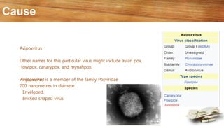
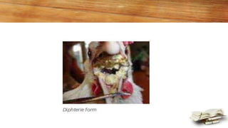
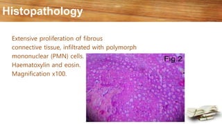
Presented by:
14-ARID-2011
14-ARID-2022
14-ARID-2033
14-ARID-2043
14-ARID-2053
Evening 9th semester
Fowl pox
Avian Pox, Avian Diphtheria
Fowl pox is a viral disease characterized by skin lesions
Another form of the disease, in which changes appear in the mouth and upper respiratory tract
Fowl pox
Cause
Avipoxvirus
Other names for this particular virus might include avian pox,
fowlpox, canarypox, and mynahpox.
Avipoxvirus is a member of the family Poxviridae
200 nanometres in diamete
Enveloped.
Bricked shaped virus
Species affected
Infects birds of both sexes
All ages and breed
Chickens, turkeys, pheasants and pigeons can be affected by different
Fowl Poxvirus strains
The incubation period of fowl pox is 4-10 days.
Nodules appear 5-8 days after infection
Scabs clear in 3-4 weeks in simple cases
Spread
I. Spread of the virus from one bird to another by direct contact is the main method of spread.
Most of the spread is the result of birds picking, fighting, or scratching one another.
Some break in the skin is equired for the virus to enter the cells, grow, and cause disease.
2. Infection also occurs by mechanical spread of virus to the injured skin. Individuals handling
birds at the time of vaccination may carry the virus on their hands and clothes, and may
unknowngly deposit the virus in the eyes of susceptible birds.
3. Mosquitoes are known to transmit the disease and produce eye infection. Mosquitoes can
infect a number of birds after a single feeding on a bird infected with fowl pox virus.
4. In a contaminated environment, presence of virus in the air from feathers and dried scabs
containing fowl pox virus, may cause skin and respiratory tract infection. The virus can survive in
dried scabs for months or even years.
5. Bad sanitary and hygienic conditions help in the spread of the disease.
Symptoms
Fowl pox can occur at any age
There are two forms of the disease
I) skin or cutaneous form (dry pox)
2) diphtheritic form (wet form)
Skin form
In the skin form, lesions (changes) appear on the unfeathered skin of the head, neck, comb,
wattles, eyelids, legs, and feet.
The lesions on the head, combs, and wattles are usually wart-like (nodular) in appearance, and
yellow to dark brown in colour
Diphtheritic form
In the diphtheritic form (wet form), small white nodules are observed in upper respiratory and
digestive tracts. These nodules merge together to form raised-yellow white cheesy patches. Most
lesions are found in the mouth, but may also be present in the larynx, trachea, and oesophagus.
These lesions cause difficulty in breathing.
Recently it has been found that most cases of diphtheritic fowl pox are characterized by the
formation of massive yellow cheesy necrotic masses in the larynx and adjacent trachea
The bird in such cases dies suddenly from asphyxiation (lack of oxygen). Lesions in the nares
(nose) give rise to nasal discharge, while those on the conjunctiva to eye discharge.
Fowl pox usually causes weakness and poor weight gain.
In layers, egg production is temporarily stopped.
Mortality is low (I to 2%) when skin lesions are present
But may be as high as 50% with the diphtheritic form , but is usually low.
Diagnosis
Wart-like lesions of the head particularly of the comb and around the eyes, or yellow cheesy
lesions in the larynx and adjacent trachea on postmortem, are diagnostic of fowl pox.
A definitive diagnosis can be made in a laboratory by histological examination (inclusion bodies)
or virus isolation in embryonated chicken eggs.
Differential diagnose
• Necrotic dermatitis.
• Leg mites.
Treatment
There is no satisfactory treatment.
But
Oxytetracyclin 300mg per gallon water for three days.
• Treat scabs with dilute iodine solution.
• Apply ointment to soften the scab. Mix 2 tablespoon of
sulfur powder with ½ cup Vaseline. Apply daily until
lesion heal.
• Mix diluted iodine solute in flock drinking water. 1
teaspoon of 1% iodine solution per gallon of drinking
water.
• Sanitize drinking water.
Control
Fowl pox can be prevented by vaccination
Precautions should be taken to minimize the spread of the vaccine virus, both on
the birds and in the environment
Carcasses of dead or affected birds should be buried or burnt.
Control mosquito. (anti mosquito spray)
Biosecurity
Zoonotic Risk:
There is no zoonotic risk associated with fowlpox virus.
VIDEO
Symptoms
Histopathology
Extensive proliferation of fibrous
connective tissue, infiltrated with polymorph
mononuclear (PMN) cells.
Haematoxylin and eosin.
Magnification x100.
Rounded and swollen keratinocytes with eosinophilic intracytoplasmic inclusion bodie
s (Bollinger bodies).
Haematoxylin and eosin.
Magnification x100.
Swollen keratinocytes with eosinophilic intracytoplasmic inclusion
bodies (Bollinger bodies). Haematoxylin and eosin.
Magnification x400.
Necropsy findings
A silent message for all class
Referance
Color Atlas of Poultry diseass
Booklet-Poultry-Diseases
Fowl pox

Fowl pox

  • 1.
  • 2.
    Avian Pox, AvianDiphtheria Fowl pox is a viral disease characterized by skin lesions Another form of the disease, in which changes appear in the mouth and upper respiratory tract Fowl pox
  • 3.
    Cause Avipoxvirus Other names forthis particular virus might include avian pox, fowlpox, canarypox, and mynahpox. Avipoxvirus is a member of the family Poxviridae 200 nanometres in diamete Enveloped. Bricked shaped virus
  • 4.
    Species affected Infects birdsof both sexes All ages and breed Chickens, turkeys, pheasants and pigeons can be affected by different Fowl Poxvirus strains The incubation period of fowl pox is 4-10 days. Nodules appear 5-8 days after infection Scabs clear in 3-4 weeks in simple cases
  • 5.
    Spread I. Spread ofthe virus from one bird to another by direct contact is the main method of spread. Most of the spread is the result of birds picking, fighting, or scratching one another. Some break in the skin is equired for the virus to enter the cells, grow, and cause disease. 2. Infection also occurs by mechanical spread of virus to the injured skin. Individuals handling birds at the time of vaccination may carry the virus on their hands and clothes, and may unknowngly deposit the virus in the eyes of susceptible birds. 3. Mosquitoes are known to transmit the disease and produce eye infection. Mosquitoes can infect a number of birds after a single feeding on a bird infected with fowl pox virus.
  • 6.
    4. In acontaminated environment, presence of virus in the air from feathers and dried scabs containing fowl pox virus, may cause skin and respiratory tract infection. The virus can survive in dried scabs for months or even years. 5. Bad sanitary and hygienic conditions help in the spread of the disease.
  • 7.
    Symptoms Fowl pox canoccur at any age There are two forms of the disease I) skin or cutaneous form (dry pox) 2) diphtheritic form (wet form)
  • 8.
    Skin form In theskin form, lesions (changes) appear on the unfeathered skin of the head, neck, comb, wattles, eyelids, legs, and feet. The lesions on the head, combs, and wattles are usually wart-like (nodular) in appearance, and yellow to dark brown in colour
  • 9.
    Diphtheritic form In thediphtheritic form (wet form), small white nodules are observed in upper respiratory and digestive tracts. These nodules merge together to form raised-yellow white cheesy patches. Most lesions are found in the mouth, but may also be present in the larynx, trachea, and oesophagus. These lesions cause difficulty in breathing. Recently it has been found that most cases of diphtheritic fowl pox are characterized by the formation of massive yellow cheesy necrotic masses in the larynx and adjacent trachea The bird in such cases dies suddenly from asphyxiation (lack of oxygen). Lesions in the nares (nose) give rise to nasal discharge, while those on the conjunctiva to eye discharge. Fowl pox usually causes weakness and poor weight gain. In layers, egg production is temporarily stopped.
  • 10.
    Mortality is low(I to 2%) when skin lesions are present But may be as high as 50% with the diphtheritic form , but is usually low.
  • 11.
    Diagnosis Wart-like lesions ofthe head particularly of the comb and around the eyes, or yellow cheesy lesions in the larynx and adjacent trachea on postmortem, are diagnostic of fowl pox. A definitive diagnosis can be made in a laboratory by histological examination (inclusion bodies) or virus isolation in embryonated chicken eggs.
  • 12.
    Differential diagnose • Necroticdermatitis. • Leg mites.
  • 13.
    Treatment There is nosatisfactory treatment. But Oxytetracyclin 300mg per gallon water for three days. • Treat scabs with dilute iodine solution. • Apply ointment to soften the scab. Mix 2 tablespoon of sulfur powder with ½ cup Vaseline. Apply daily until lesion heal. • Mix diluted iodine solute in flock drinking water. 1 teaspoon of 1% iodine solution per gallon of drinking water. • Sanitize drinking water.
  • 14.
    Control Fowl pox canbe prevented by vaccination Precautions should be taken to minimize the spread of the vaccine virus, both on the birds and in the environment
  • 15.
    Carcasses of deador affected birds should be buried or burnt. Control mosquito. (anti mosquito spray) Biosecurity
  • 16.
    Zoonotic Risk: There isno zoonotic risk associated with fowlpox virus.
  • 17.
  • 18.
  • 26.
    Histopathology Extensive proliferation offibrous connective tissue, infiltrated with polymorph mononuclear (PMN) cells. Haematoxylin and eosin. Magnification x100.
  • 27.
    Rounded and swollenkeratinocytes with eosinophilic intracytoplasmic inclusion bodie s (Bollinger bodies). Haematoxylin and eosin. Magnification x100.
  • 28.
    Swollen keratinocytes witheosinophilic intracytoplasmic inclusion bodies (Bollinger bodies). Haematoxylin and eosin. Magnification x400.
  • 29.
  • 30.
    A silent messagefor all class
  • 31.
    Referance Color Atlas ofPoultry diseass Booklet-Poultry-Diseases