The Heart: A hollow, muscular, cone-shaped organ that acts as a central pump for the circulatory system.
Location: Situated in the mediastinum (thoracic cavity) between the lungs, slightly to the left.
Dimensions / Size : Roughly the size of a closed fist; approx. 12 cm long, 9 cm wide, and weighs 250–350g.
Apex & Base: The broad upper part is the base; the pointed lower end is the apex (directed toward the left hip).
Layers of the Heart Wall
Epicardium (Outer): A thin, serous membrane that protects the heart and provides lubrication.
Myocardium (Middle): The thickest layer made of cardiac muscle fibers. Responsible for the pumping action (contraction).
Endocardium (Inner): A smooth endothelial lining that covers the chambers and valves, preventing blood from sticking to the walls.
Chambers
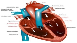
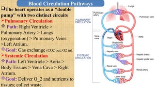
Right Atrium (RA): Receives deoxygenated blood from the body via Superior and Inferior Vena Cava.
Right Ventricle (RV): Pumps deoxygenated blood to the lungs via pulmonary artery.
Left Atrium (LA): Receives oxygenated blood from the lungs via Pulmonary Veins.
Left Ventricle (LV): Pumps oxygenated blood to the whole body via the Aorta
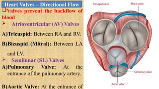
Atrioventricular (AV) Valves
Tricuspid: Between RA and RV.
Bicuspid (Mitral): Between LA and LV.
Semilunar (SL) Valves
Pulmonary Valve: At the entrance of the pulmonary artery.
Aortic Valve: At the entrance of the aorta.
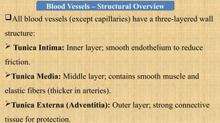
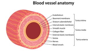
All blood vessels (except capillaries) have a three-layered wall structure:
Tunica Intima: Inner layer; smooth endothelium to reduce friction.
Tunica Media: Middle layer; contains smooth muscle and elastic fibers (thicker in arteries).
Tunica Externa (Adventitia): Outer layer; strong connective tissue for protection.
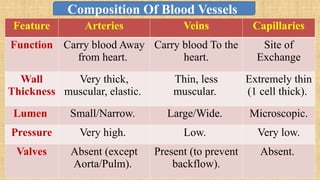
Arteries: Carry blood Away from heart.
Veins : Carry blood To the heart.
Capillaries: Site of Exchange
Structure: Composed only of a single layer of endothelial cells (Tunica Intima).
Function: This thinness allows for the diffusion of:
Gases: Oxygen and Carbon dioxide.
Nutrients: Glucose, amino acids.
Waste: Urea and metabolic byproducts.