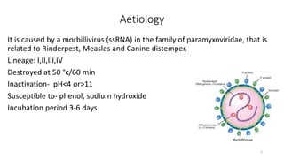
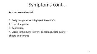
Peste des Petits Ruminants (PPR)
Presented By:
Bhuwan Raj Bhatta
Roll.No: 03(Three)
IAAS, Paklihawa Campus
1
Introduction
Peste des petits ruminants(PPR) is also known as ‘ovine
rinderpest’,‘Goat plague’.
It is an acute, highly contagious viral disease of goat and sheep.
It is characterized by the fever, sores in mouth, diarrhoea,
pneumonia and sometimes death.
2
Aetiology
It is caused by a morbillivirus (ssRNA) in the family of paramyxoviridae, that is
related to Rinderpest, Measles and Canine distemper.
Lineage: I,II,III,IV
Destroyed at 50 °c/60 min
Inactivation- pH<4 or>11
Susceptible to- phenol, sodium hydroxide
Incubation period 3-6 days.
3
Epidemiology
Origin and spread:
The first description of the disease was published on 1940 an
outbreak in Ivory cost, West Africa in goats and sheep on 1940 and
confirmed on 1972.
Pakistan and India on early 1990s (Calcutta goat markets), 1994 on
Tamil Nadu finally reaching Tibet on 2007.
4
Epidemiology cont.…
Geographical Repartition
As of 2017 the disease is present in West africa, part of Central Africa
(Gabon, Central African Republic), East Africa (north of the Equator),
the Middle East and the Indian subcontinent including Nepal and
Myanmar.
Morbidity- 50-90%
Mortality- higher in goat (55-85%) than compared to sheep (45-75%)
5
Outbreak in Nepal
On 2016 Feb at 18 Bajura, PPR killed almost 3170
goats and on 2016 Feb 20 in Humla also killed
almost 1800 goats.
PPR kills over 3000 sheep and goat in Jajarkot- 2015
6
Transmission
The virus is secreted in tears, nasal discharge, secretions from coughing,
and in the faeces of infected animals.
Water, feed, and bedding can also be contaminated with secretions.
Since animals excrete the virus before showing signs of the disease, it can
spread by movement of infected animals.
Virus also present in semen and embryo
Offspring can also affected from the infected mother through milk
7
Symptoms
They are similar to those of Rinderpest in cattle.
Hyper acute cases:
Hyper acute cases are found dead without previous symptoms.
They die with a serous, foamy or haemorrhagic discharge coming
out of the nose.
8
Symptoms cont...
Acute cases at onset
1. Body temperature is high (40.5 to 41 °C)
2. Loss of appetite
3. Depression
4. Ulcers in the gums (lower), dental pad, hard palate,
cheeks and tongue
9
Nn
Fig- Self auscultation Fig- Arched back (painful defecation)
10
Fig- ocular and mucopurulent nasal discharge Fig- sticky diarrhoea
11
Fig- inflammation and erosion
in mouth
12
Fig- Periodontitis Fig- In recovery case
13
Symptoms contd…
Evolution of acute cases:
• Nasal discharge become mucopurulent and may obstruct the nose
• Dry coughing develop
• pneumonia
• Death occur from 5-10 days after the onset of fever
• Some animals may recover but the dry, stertorous coughing often persist for some days
• Pregnant may abort
14
Pathogenesis
Entry- invade retropharyngeal lymph nodes and mucosa
from there respiratory and alimentary tract affected
proliferation and damage them
inflammation of alimentary inflammation of
respiratory
lead to diarrhoea, dehydration lead to pneumonia
and
and death respiratory distress
15
Post Mortem Lesion
Carcase will be dehydrated with faecal soiling
Congestion of the ileo-ceacal valve
Engorgement and blackening of the folds in the caecum, colon and
rectum(‘zebra striping’)
Enlarged spleen
Oedematous lymph nodes
Bronchopneumonia
16
Diagnosis
1. Based on the clinical signs.
2. Histo-pathological changes in the lungs.
3. Bronchopneumonia with antero-ventral consolidation.
4. Virus can be isolated from lymphoid tissue at necropsy,
including lymph nodes, Peyer’s patches and spleen.
5. Blood examination
6. Serological test:
• AGI
• PCR
• ELISA
• cDNA probe
17
Differential Diagnosis
• Bluetongue
• Rinderpest
• FMD
• Sheep pox
• Coccidiosis
• Salmonellosis
• E.coli
• C.C.P.P
18
Treatment
 There is no specific treatment, supportive therapy.
 Symptomatic treatment for diarrhoea and respiratory problem
 Hyper immune rinderpest serum
 OTC- 10ml mg/kg b.wt with levamisole 2.5 mg/kg b.wt for 3 days followed by
repetition at 3 days interval recommended for secondary bacterial invaders
 Lemon juice effective against scab formation
19
Prevention and Control
• Disease is prevented in many countries by strict quarantine
controls.
• Introduced disease has been eradicated from countries by
compulsory slaughter of affected flocks/herds.
• Sanitation and hygienic measures
• Do not introduce of new animal especially from areas where the
disease is prevalent
• Affected isolated and treated
20
Vaccine
• Tissue culture rinderpest vaccine, given at 3-4 months
• PPR culture vaccine (I.V.R.I)- 1ml s/c annually
• Raksha PPR- 1 ml s/c
• Live attenuated RP vaccine protection for one year only
• PPRV attenuated vaccine commercially available
• Genetically engineered recombinant vaccine is under trial (OIE, 2002)
• French attenuated PPR vaccine- immunity for at least 3 years
21
THANK YOU
22

PPR (Peste des Petits Ruminants)

  • 1.
    Peste des PetitsRuminants (PPR) Presented By: Bhuwan Raj Bhatta Roll.No: 03(Three) IAAS, Paklihawa Campus 1
  • 2.
    Introduction Peste des petitsruminants(PPR) is also known as ‘ovine rinderpest’,‘Goat plague’. It is an acute, highly contagious viral disease of goat and sheep. It is characterized by the fever, sores in mouth, diarrhoea, pneumonia and sometimes death. 2
  • 3.
    Aetiology It is causedby a morbillivirus (ssRNA) in the family of paramyxoviridae, that is related to Rinderpest, Measles and Canine distemper. Lineage: I,II,III,IV Destroyed at 50 °c/60 min Inactivation- pH<4 or>11 Susceptible to- phenol, sodium hydroxide Incubation period 3-6 days. 3
  • 4.
    Epidemiology Origin and spread: Thefirst description of the disease was published on 1940 an outbreak in Ivory cost, West Africa in goats and sheep on 1940 and confirmed on 1972. Pakistan and India on early 1990s (Calcutta goat markets), 1994 on Tamil Nadu finally reaching Tibet on 2007. 4
  • 5.
    Epidemiology cont.… Geographical Repartition Asof 2017 the disease is present in West africa, part of Central Africa (Gabon, Central African Republic), East Africa (north of the Equator), the Middle East and the Indian subcontinent including Nepal and Myanmar. Morbidity- 50-90% Mortality- higher in goat (55-85%) than compared to sheep (45-75%) 5
  • 6.
    Outbreak in Nepal On2016 Feb at 18 Bajura, PPR killed almost 3170 goats and on 2016 Feb 20 in Humla also killed almost 1800 goats. PPR kills over 3000 sheep and goat in Jajarkot- 2015 6
  • 7.
    Transmission The virus issecreted in tears, nasal discharge, secretions from coughing, and in the faeces of infected animals. Water, feed, and bedding can also be contaminated with secretions. Since animals excrete the virus before showing signs of the disease, it can spread by movement of infected animals. Virus also present in semen and embryo Offspring can also affected from the infected mother through milk 7
  • 8.
    Symptoms They are similarto those of Rinderpest in cattle. Hyper acute cases: Hyper acute cases are found dead without previous symptoms. They die with a serous, foamy or haemorrhagic discharge coming out of the nose. 8
  • 9.
    Symptoms cont... Acute casesat onset 1. Body temperature is high (40.5 to 41 °C) 2. Loss of appetite 3. Depression 4. Ulcers in the gums (lower), dental pad, hard palate, cheeks and tongue 9
  • 10.
    Nn Fig- Self auscultationFig- Arched back (painful defecation) 10
  • 11.
    Fig- ocular andmucopurulent nasal discharge Fig- sticky diarrhoea 11
  • 12.
    Fig- inflammation anderosion in mouth 12
  • 13.
    Fig- Periodontitis Fig-In recovery case 13
  • 14.
    Symptoms contd… Evolution ofacute cases: • Nasal discharge become mucopurulent and may obstruct the nose • Dry coughing develop • pneumonia • Death occur from 5-10 days after the onset of fever • Some animals may recover but the dry, stertorous coughing often persist for some days • Pregnant may abort 14
  • 15.
    Pathogenesis Entry- invade retropharyngeallymph nodes and mucosa from there respiratory and alimentary tract affected proliferation and damage them inflammation of alimentary inflammation of respiratory lead to diarrhoea, dehydration lead to pneumonia and and death respiratory distress 15
  • 16.
    Post Mortem Lesion Carcasewill be dehydrated with faecal soiling Congestion of the ileo-ceacal valve Engorgement and blackening of the folds in the caecum, colon and rectum(‘zebra striping’) Enlarged spleen Oedematous lymph nodes Bronchopneumonia 16
  • 17.
    Diagnosis 1. Based onthe clinical signs. 2. Histo-pathological changes in the lungs. 3. Bronchopneumonia with antero-ventral consolidation. 4. Virus can be isolated from lymphoid tissue at necropsy, including lymph nodes, Peyer’s patches and spleen. 5. Blood examination 6. Serological test: • AGI • PCR • ELISA • cDNA probe 17
  • 18.
    Differential Diagnosis • Bluetongue •Rinderpest • FMD • Sheep pox • Coccidiosis • Salmonellosis • E.coli • C.C.P.P 18
  • 19.
    Treatment  There isno specific treatment, supportive therapy.  Symptomatic treatment for diarrhoea and respiratory problem  Hyper immune rinderpest serum  OTC- 10ml mg/kg b.wt with levamisole 2.5 mg/kg b.wt for 3 days followed by repetition at 3 days interval recommended for secondary bacterial invaders  Lemon juice effective against scab formation 19
  • 20.
    Prevention and Control •Disease is prevented in many countries by strict quarantine controls. • Introduced disease has been eradicated from countries by compulsory slaughter of affected flocks/herds. • Sanitation and hygienic measures • Do not introduce of new animal especially from areas where the disease is prevalent • Affected isolated and treated 20
  • 21.
    Vaccine • Tissue culturerinderpest vaccine, given at 3-4 months • PPR culture vaccine (I.V.R.I)- 1ml s/c annually • Raksha PPR- 1 ml s/c • Live attenuated RP vaccine protection for one year only • PPRV attenuated vaccine commercially available • Genetically engineered recombinant vaccine is under trial (OIE, 2002) • French attenuated PPR vaccine- immunity for at least 3 years 21
  • 22.