Downloaded 532 times


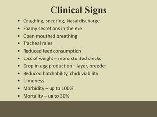


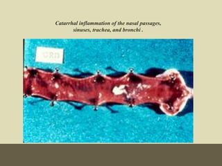


This document discusses Mycoplasma gallisepticum infection in poultry farms. Some key points: - M. gallisepticum is a highly successful pathogen that causes chronic respiratory disease in chickens and turkeys. Once infected, the infection remains for life. - It is transmitted both horizontally between flocks through contact/aerosols and vertically from parent to offspring through eggs. - Clinical signs include coughing, sneezing, and reduced egg production. Post-mortem lesions include sinusitis, tracheitis, and airsacculitis. - Diagnosis involves isolation of the bacteria or serological tests like ELISA. Treatment includes antibiotics like tetracyclines and tylos


