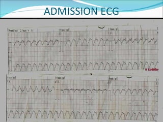
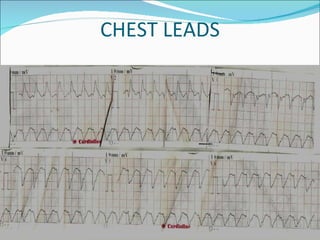
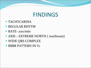
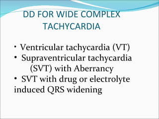
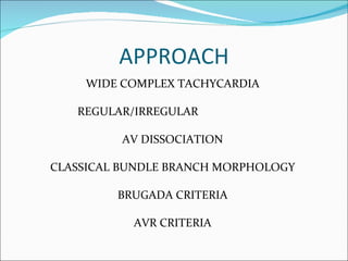
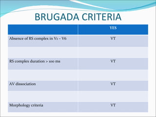
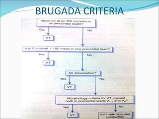
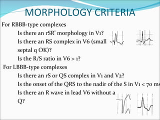
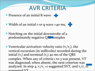
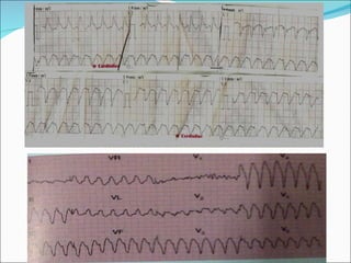
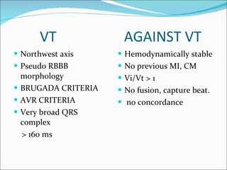
K.M.JEYABALAJI Dr.P. VIJAYARAGAVAN’S UNIT
HISTORY A 22 yr male patient came with complaints of Acute onset breathlessness Palpitation Profuse sweating Vague chest discomfort For past  1 hour
EXAMINATION Dyspnoeic, tachypnoeic,  Pulse- 180/ min REGULAR BP- 90/ 60 mmhg JVP- --- CVS- s1,s2 heard RS – NVBS P/A- soft CNS- NFND
ADMISSION ECG
CHEST LEADS
FINDINGS TACHYCARDIA REGULAR RHYTM RATE- 200/min AXIS – EXTREME NORTH ( northwest) WIDE QRS COMPLEX  RBBB PATTERN IN V1
DD FOR WIDE COMPLEX TACHYCARDIA Ventricular tachycardia (VT) Supraventricular tachycardia  (SVT) with Aberrancy SVT with drug or electrolyte  induced QRS widening
APPROACH WIDE COMPLEX TACHYCARDIA REGULAR/IRREGULAR  AV DISSOCIATION CLASSICAL BUNDLE BRANCH MORPHOLOGY BRUGADA CRITERIA AVR CRITERIA
BRUGADA CRITERIA YES Absence of RS complex in V1 – V6 VT RS complex duration > 100 ms VT AV dissociation  VT Morphology criteria VT
BRUGADA CRITERIA
MORPHOLOGY CRITERIA For RBBB-type complexes Is there an rSR’ morphology in V1? Is there an RS complex in V6 (small septal q OK)? Is the R/S ratio in V6 > 1? For LBBB-type complexes Is there an rS or QS complex in V1 and V2? Is the onset of the QRS to the nadir of the S in V1 < 70 ms? Is there an R wave in lead V6 without a Q?
AVR CRITERIA Presence of an initial R wave Width of an initial r or q wave >40 ms, Notching on the initial downstroke of a predominantly negative QRS complex Ventricular activation–velocity ratio (v i /v t ), the vertical excursion (in millivolts) recorded during the initial (v i ) and terminal (v t ) 40 ms of the QRS complex. When any of criteria 1 to 3 was present, VT was diagnosed; when absent, the next criterion was analyzed. In step 4, v i /v t  >1 suggested SVT, and v i /v t  ≤1 suggested VT.
 
VENTRICULAR TACHYCARDIA Absence of typical RBBB or LBBB morphology Extreme axis deviation (“northwest axis”) Very broad complexes (>160ms) AV dissociation (P and QRS complexes at different rates) Capture beats — occur when the sinoatrial node transiently ‘captures’ the ventricles, in the midst of AV dissociation, to produce a QRS complex of normal duration. Fusion beats — occur when a sinus and ventricular beat coincides to produce a hybrid complex.
Positive or negative concordance throughout the chest leads, i.e. leads V1-6 show entirely positive (R) or entirely negative (QS) complexes, with no RS complexes seen. Brugada’s sign –  The distance from the onset of the QRS complex to the nadir of the S-wave is > 100ms Josephson’s sign  – Notching near the nadir of the S-wave
VT CAPTURE BEAT  FUSION BEAT  BRUGADA SIGN , JOSEPHSON SIGN
NEGATIVE CONCORDANCE  POSITIVE CONCORDANCE
SVT WITH ABBERANCY •  Any SVT can be conducted with aberrancy: –  Sinus Tachycardia –  Atrial tachycardia –  Atrial flutter –  Atrioventricular nodal reentrant tachycardia  (AVNRT) –  Junctional Tachycardia –  Orthodromic Atrioventricular Reentrant Tachycardia   (AVRT)
VT  AGAINST VT Northwest axis Pseudo RBBB morphology BRUGADA CRITERIA AVR CRITERIA Very broad QRS complex > 160 ms Hemodynamically stable  No previous MI, CM Vi/Vt > 1 No fusion, capture beat. no concordance
FASCICULAR VT SUPERIOR AXIS  PSEUDO RBBB MORPHOLOGY HEMODYNAMICALLY STABLE
TAKE HOME MESSAGE No criteria is 100% sensitive nor specific Never  go blindly by ECG Give equal imortance to history, clinical presentation,  Vitals If you are 100% sure that it is SVT, then proceed. Having even 1% doubt, then treat it as VT
THANK YOU

ECG: Wide Complex Tachycardia

  • 1.
  • 2.
    HISTORY A 22yr male patient came with complaints of Acute onset breathlessness Palpitation Profuse sweating Vague chest discomfort For past 1 hour
  • 3.
    EXAMINATION Dyspnoeic, tachypnoeic, Pulse- 180/ min REGULAR BP- 90/ 60 mmhg JVP- --- CVS- s1,s2 heard RS – NVBS P/A- soft CNS- NFND
  • 4.
  • 5.
  • 6.
    FINDINGS TACHYCARDIA REGULARRHYTM RATE- 200/min AXIS – EXTREME NORTH ( northwest) WIDE QRS COMPLEX RBBB PATTERN IN V1
  • 7.
    DD FOR WIDECOMPLEX TACHYCARDIA Ventricular tachycardia (VT) Supraventricular tachycardia (SVT) with Aberrancy SVT with drug or electrolyte induced QRS widening
  • 8.
    APPROACH WIDE COMPLEXTACHYCARDIA REGULAR/IRREGULAR AV DISSOCIATION CLASSICAL BUNDLE BRANCH MORPHOLOGY BRUGADA CRITERIA AVR CRITERIA
  • 9.
    BRUGADA CRITERIA YESAbsence of RS complex in V1 – V6 VT RS complex duration > 100 ms VT AV dissociation VT Morphology criteria VT
  • 10.
  • 11.
    MORPHOLOGY CRITERIA ForRBBB-type complexes Is there an rSR’ morphology in V1? Is there an RS complex in V6 (small septal q OK)? Is the R/S ratio in V6 > 1? For LBBB-type complexes Is there an rS or QS complex in V1 and V2? Is the onset of the QRS to the nadir of the S in V1 < 70 ms? Is there an R wave in lead V6 without a Q?
  • 12.
    AVR CRITERIA Presenceof an initial R wave Width of an initial r or q wave >40 ms, Notching on the initial downstroke of a predominantly negative QRS complex Ventricular activation–velocity ratio (v i /v t ), the vertical excursion (in millivolts) recorded during the initial (v i ) and terminal (v t ) 40 ms of the QRS complex. When any of criteria 1 to 3 was present, VT was diagnosed; when absent, the next criterion was analyzed. In step 4, v i /v t >1 suggested SVT, and v i /v t ≤1 suggested VT.
  • 13.
  • 14.
    VENTRICULAR TACHYCARDIA Absenceof typical RBBB or LBBB morphology Extreme axis deviation (“northwest axis”) Very broad complexes (>160ms) AV dissociation (P and QRS complexes at different rates) Capture beats — occur when the sinoatrial node transiently ‘captures’ the ventricles, in the midst of AV dissociation, to produce a QRS complex of normal duration. Fusion beats — occur when a sinus and ventricular beat coincides to produce a hybrid complex.
  • 15.
    Positive or negative concordance throughoutthe chest leads, i.e. leads V1-6 show entirely positive (R) or entirely negative (QS) complexes, with no RS complexes seen. Brugada’s sign –  The distance from the onset of the QRS complex to the nadir of the S-wave is > 100ms Josephson’s sign  – Notching near the nadir of the S-wave
  • 16.
    VT CAPTURE BEAT FUSION BEAT BRUGADA SIGN , JOSEPHSON SIGN
  • 17.
    NEGATIVE CONCORDANCE POSITIVE CONCORDANCE
  • 18.
    SVT WITH ABBERANCY• Any SVT can be conducted with aberrancy: – Sinus Tachycardia – Atrial tachycardia – Atrial flutter – Atrioventricular nodal reentrant tachycardia (AVNRT) – Junctional Tachycardia – Orthodromic Atrioventricular Reentrant Tachycardia (AVRT)
  • 19.
    VT AGAINSTVT Northwest axis Pseudo RBBB morphology BRUGADA CRITERIA AVR CRITERIA Very broad QRS complex > 160 ms Hemodynamically stable No previous MI, CM Vi/Vt > 1 No fusion, capture beat. no concordance
  • 20.
    FASCICULAR VT SUPERIORAXIS PSEUDO RBBB MORPHOLOGY HEMODYNAMICALLY STABLE
  • 21.
    TAKE HOME MESSAGENo criteria is 100% sensitive nor specific Never go blindly by ECG Give equal imortance to history, clinical presentation, Vitals If you are 100% sure that it is SVT, then proceed. Having even 1% doubt, then treat it as VT
  • 22.