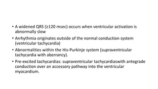
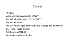
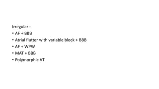
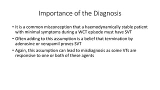
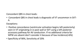
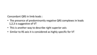
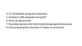
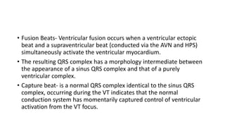
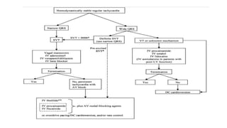
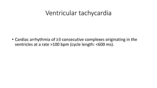
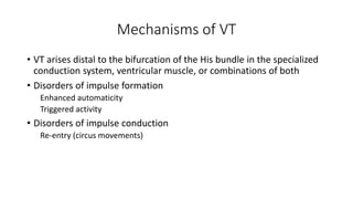
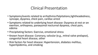
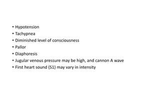
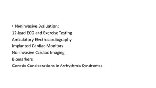
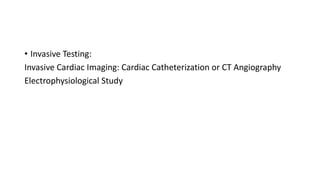
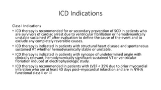
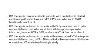
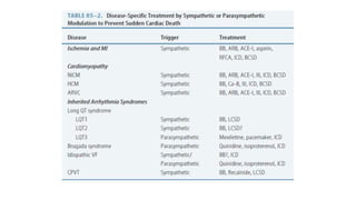
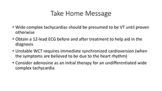
Wide complex tachycardia refers to a cardiac rhythm with a rate over 100 beats per minute and a QRS duration of 120 ms or more. It can be caused by ventricular tachycardia originating in the ventricles, or supraventricular tachycardia with aberrancy or pre-excitation. Differentiating the two is important as treatment differs. The electrocardiogram and various diagnostic criteria and algorithms using features like QRS duration, axis, and morphology can help determine the origin of the arrhythmia. Treatment involves terminating unstable rhythms with cardioversion or medications, while implanting defibrillators may help prevent recurrence in some patients.
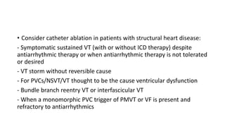
differentiating between supraventicular tachycardia and ventricular tachycardia in wide complex rhythm is always confusing and management is totally different. correct diagnosis will make dramatic difference in patient management.
Case scenario of a patient with idiopathic ventricular Tachycardia (VT), followed by a topic review including diagnosis and management guidelines. It is defined as Monomorphic VT in patients without any structural heart disease or coronary disease”. Classified on the basis of site of origin broadly into three different categories i.e Outflow Tract VT, Annular VT, Fascicular VT
Idiopathic VT refers to VT occurring in structurally normal hearts in the absence of myocardial scarring. Classification of monomorphic idiopathic VT includes outflow tract VT, fascicular VT, papillary muscle VT,annular VT, and miscellaneous (VT from the body of the RV and crux of
the heart). It is commonly seen in young patients and usually has a benign course. The 12-lead lectrocardiogram is critical in distinguishing the specific form and locations of idiopathic VT. Treatment options include medical therapy specific to the underlying mechanism of VT or catheter
ablation.
Its a medical presentation describing how to approach to various cardiac arrhythmias in systematic way. Illustrated with more ECG photographs from standard sources.
Ventricular tachycardia are difficult to understand. it is classified in to two types. 1. VT in structurally normal heart, 2. VT in heart with structural diseases. I have tried to simplify the VT in structurally normal heart, which may be helpful to many students and learners.
June 3, 2024 Anti-Semitism Letter Sent to MIT President Kornbluth and MIT Cor...Levi Shapiro
Letter from the Congress of the United States regarding Anti-Semitism sent June 3rd to MIT President Sally Kornbluth, MIT Corp Chair, Mark Gorenberg
Dear Dr. Kornbluth and Mr. Gorenberg,
The US House of Representatives is deeply concerned by ongoing and pervasive acts of antisemitic
harassment and intimidation at the Massachusetts Institute of Technology (MIT). Failing to act decisively to ensure a safe learning environment for all students would be a grave dereliction of your responsibilities as President of MIT and Chair of the MIT Corporation.
This Congress will not stand idly by and allow an environment hostile to Jewish students to persist. The House believes that your institution is in violation of Title VI of the Civil Rights Act, and the inability or
unwillingness to rectify this violation through action requires accountability.
Postsecondary education is a unique opportunity for students to learn and have their ideas and beliefs challenged. However, universities receiving hundreds of millions of federal funds annually have denied
students that opportunity and have been hijacked to become venues for the promotion of terrorism, antisemitic harassment and intimidation, unlawful encampments, and in some cases, assaults and riots.
The House of Representatives will not countenance the use of federal funds to indoctrinate students into hateful, antisemitic, anti-American supporters of terrorism. Investigations into campus antisemitism by the Committee on Education and the Workforce and the Committee on Ways and Means have been expanded into a Congress-wide probe across all relevant jurisdictions to address this national crisis. The undersigned Committees will conduct oversight into the use of federal funds at MIT and its learning environment under authorities granted to each Committee.
• The Committee on Education and the Workforce has been investigating your institution since December 7, 2023. The Committee has broad jurisdiction over postsecondary education, including its compliance with Title VI of the Civil Rights Act, campus safety concerns over disruptions to the learning environment, and the awarding of federal student aid under the Higher Education Act.
• The Committee on Oversight and Accountability is investigating the sources of funding and other support flowing to groups espousing pro-Hamas propaganda and engaged in antisemitic harassment and intimidation of students. The Committee on Oversight and Accountability is the principal oversight committee of the US House of Representatives and has broad authority to investigate “any matter” at “any time” under House Rule X.
• The Committee on Ways and Means has been investigating several universities since November 15, 2023, when the Committee held a hearing entitled From Ivory Towers to Dark Corners: Investigating the Nexus Between Antisemitism, Tax-Exempt Universities, and Terror Financing. The Committee followed the hearing with letters to those institutions on January 10, 202
Synthetic Fiber Construction in lab .pptxPavel ( NSTU)
Synthetic fiber production is a fascinating and complex field that blends chemistry, engineering, and environmental science. By understanding these aspects, students can gain a comprehensive view of synthetic fiber production, its impact on society and the environment, and the potential for future innovations. Synthetic fibers play a crucial role in modern society, impacting various aspects of daily life, industry, and the environment. ynthetic fibers are integral to modern life, offering a range of benefits from cost-effectiveness and versatility to innovative applications and performance characteristics. While they pose environmental challenges, ongoing research and development aim to create more sustainable and eco-friendly alternatives. Understanding the importance of synthetic fibers helps in appreciating their role in the economy, industry, and daily life, while also emphasizing the need for sustainable practices and innovation.
Palestine last event orientationfvgnh .pptxRaedMohamed3
An EFL lesson about the current events in Palestine. It is intended to be for intermediate students who wish to increase their listening skills through a short lesson in power point.
How to Make a Field invisible in Odoo 17Celine George
It is possible to hide or invisible some fields in odoo. Commonly using “invisible” attribute in the field definition to invisible the fields. This slide will show how to make a field invisible in odoo 17.
Biological screening of herbal drugs: Introduction and Need for
Phyto-Pharmacological Screening, New Strategies for evaluating
Natural Products, In vitro evaluation techniques for Antioxidants, Antimicrobial and Anticancer drugs. In vivo evaluation techniques
for Anti-inflammatory, Antiulcer, Anticancer, Wound healing, Antidiabetic, Hepatoprotective, Cardio protective, Diuretics and
Antifertility, Toxicity studies as per OECD guidelines
The French Revolution, which began in 1789, was a period of radical social and political upheaval in France. It marked the decline of absolute monarchies, the rise of secular and democratic republics, and the eventual rise of Napoleon Bonaparte. This revolutionary period is crucial in understanding the transition from feudalism to modernity in Europe.
For more information, visit-www.vavaclasses.com
Macroeconomics- Movie Location
This will be used as part of your Personal Professional Portfolio once graded.
Objective:
Prepare a presentation or a paper using research, basic comparative analysis, data organization and application of economic information. You will make an informed assessment of an economic climate outside of the United States to accomplish an entertainment industry objective.
Embracing GenAI - A Strategic ImperativePeter Windle
Artificial Intelligence (AI) technologies such as Generative AI, Image Generators and Large Language Models have had a dramatic impact on teaching, learning and assessment over the past 18 months. The most immediate threat AI posed was to Academic Integrity with Higher Education Institutes (HEIs) focusing their efforts on combating the use of GenAI in assessment. Guidelines were developed for staff and students, policies put in place too. Innovative educators have forged paths in the use of Generative AI for teaching, learning and assessments leading to pockets of transformation springing up across HEIs, often with little or no top-down guidance, support or direction.
This Gasta posits a strategic approach to integrating AI into HEIs to prepare staff, students and the curriculum for an evolving world and workplace. We will highlight the advantages of working with these technologies beyond the realm of teaching, learning and assessment by considering prompt engineering skills, industry impact, curriculum changes, and the need for staff upskilling. In contrast, not engaging strategically with Generative AI poses risks, including falling behind peers, missed opportunities and failing to ensure our graduates remain employable. The rapid evolution of AI technologies necessitates a proactive and strategic approach if we are to remain relevant.
Introduction to AI for Nonprofits with Tapp NetworkTechSoup
Dive into the world of AI! Experts Jon Hill and Tareq Monaur will guide you through AI's role in enhancing nonprofit websites and basic marketing strategies, making it easy to understand and apply.
Welcome to TechSoup New Member Orientation and Q&A (May 2024).pdfTechSoup
In this webinar you will learn how your organization can access TechSoup's wide variety of product discount and donation programs. From hardware to software, we'll give you a tour of the tools available to help your nonprofit with productivity, collaboration, financial management, donor tracking, security, and more.
2. • Wide complex tachycardia (WCT) refers to a cardiac rhythm where
rate is more than 100 beats per minute with a QRS duration of 120
ms or more on the surface electrocardiogram (ECG)
3. • Widening of the QRS complex is related to slower spread of ventricular
depolarization, either due to disease of the His-Purkinje network and/or reliance
on slower, muscle-to-muscle spread of depolarization.
1. propagation of a supraventricular impulse (atrial premature depolarizations
[APDs] or supraventricular tachycardia [SVT]) with block (preexisting or rate-
related) in one or more parts of the His-Purkinje network;
2. depolarizations originating in the ventricles themselves (ventricular premature
beats [VPDs] or ventricular tachycardia [VT]);
3. slowed propagation of a supraventricular impulse because of intra-myocardial
scar/fibrosis/hypertrophy; or
4. conduction of a supraventricular impulse from atrium to ventricle over an
accessory pathway (bypass tract) – so called “pre-excited” tachycardia.
4. • A widened QRS (≥120 msec) occurs when ventricular activation is
abnormally slow
• Arrhythmia originates outside of the normal conduction system
(ventricular tachycardia)
• Abnormalities within the His-Purkinje system (supraventricular
tachycardia with aberrancy).
• Pre-excited tachycardias: supraventricular tachycardiaswith antegrade
conduction over an accessory pathway into the ventricular
myocardium.
5. Causes :
• Regular :
Ventricular tachycardia(80% of WCT)
Any SVT with aberrancy (2nd MC WCT)
Any SVT with BBB
Any SVT with delayed conduction due to drugs and electrolytes
Class IA,IC ; hyperkalemia.
Antidromic AVRT(1-5%)
Pacemaker mediated rhythm
6. Irregular :
• AF + BBB
• Atrial flutter with variable block + BBB
• AF + WPW
• MAT + BBB
• Polymorphic VT
7. Importance of the Diagnosis
• It is a common misconception that a haemodynamically stable patient
with minimal symptoms during a WCT episode must have SVT
• Often adding to this assumption is a belief that termination by
adenosine or verapamil proves SVT
• Again, this assumption can lead to misdiagnosis as some VTs are
responsive to one or both of these agents
8. VT or SVT with aberration???
• History
• Physical Examination
• The Electrocardiogram
• Algorithms
• Electrophysiologic Testing
9. The likelihood of VT is also increased with:
• Age > 35 (positive predictive value of 85%)
• Structural heart disease
• Ischaemic heart disease
• Previous MI
• Congestive heart failure
• Cardiomyopathy
• Family history of sudden cardiac death (suggesting conditions such as
HOCM, congenital long QT syndrome, Brugada syndrome or
arrhythmogenic right ventricular dysplasia that are associated with
episodes of VT)
10. The likelihood of SVT with aberrancy is
increased if:
• Previous ECGs show a bundle branch block pattern with identical
morphology to the broad complex tachycardia.
• Previous ECGs show evidence of WPW (short PR < 120ms, broad QRS,
delta wave).
• The patient has a history of paroxysmal tachycardias that have been
successfully terminated with adenosine or vagal manoeuvres.
12. Features for differentiation by ECG
1. QRS duration
2. QRS axis
3. Concordant pattern
4. Precordial RS duration.
5. Morphological criteria - RBBB , LBBB , ambiguous chest lead pattern
6. Q wave presence
7. AV dissociation
8. Baseline QRS prolongation – QRS duration , QRS configuration.
9. aVR changes.
10.Lead II R-wave-peak-time (RWPT) criterion .
13.
14. QRS duration :
• > 160 ms with LBBB , >140 ms with RBBB - VT
• Wellens et al . Showed that 69% of VT had QRS duration of >140ms
and
Exceptions:
• Anti arrythmitic drugs non specifically prolong QRS duration.
• Pts with structurally normal heart may have VT with QRS duration of
120-140ms.(<140ms in12% , < 120 ms in 4%)
• QRS duration also depend site of origin of VT , septal VT none of SVT-
A showed QRS duration of >140ms
15. QRS axis :
• Frontal plane axis of -90 to +180 --- VT
• Shift in QRS axis of more than 40 from baseline --- VT(less specific)
• RBBB with LAD, LBBB with RAD --- VT.
• LAFB (-30 to -90) , LPFB (+110 to150) and RBBB (normal axis).
16. Concordant QRS in chest leads:
• Concordant QRS in chest leads is diagnostic of VT uncommon in SVT-
A.
Exceptions:
• Positive concordance (ventricular activation begins left posteriorly)
seen in VT originating in Lt post wall or SVT using a left posterior
accessory pathway for AV conduction. If no additional criteria for
WPW are absent don’t consider it because of low incidence(<6%)
• Specificity of 90%, Sensitivity of 20%
17. Concordant QRS in limb leads :
• The presence of predominantly negative QRS complexes in leads
1,2,3 is suggestive of VT
• This is another way to describe right superior axis
• Similar to RS axis it is considered as highly specific for VT
18. AV dissociation :
• The most specific ECG finding for VT
Clues for AV dissociation:
1. Clinically by cannon A waves , variable intensity of S1 , Variation in
SBP unrelated to respiration.
2. AV dissociation
3. AV ratio of less than 1
19. 4. 2:1 VA block(d/t retrograde conduction)
5. Variation in QRS amplitude during WCT
6. Fusion & capture beats
7. Recording separate atrial electro gram(oesophageal/transvenous)
8. Echo (evaluating RA contraction in relation to ventricular)
20. • Fusion Beats- Ventricular fusion occurs when a ventricular ectopic
beat and a supraventricular beat (conducted via the AVN and HPS)
simultaneously activate the ventricular myocardium.
• The resulting QRS complex has a morphology intermediate between
the appearance of a sinus QRS complex and that of a purely
ventricular complex.
• Capture beat- is a normal QRS complex identical to the sinus QRS
complex, occurring during the VT indicates that the normal
conduction system has momentarily captured control of ventricular
activation from the VT focus.
21.
22. Diagnostic approach/algorithms
• Sandler and Marriott Criteria (1965)
• Wellens(1978) ,
• Akhtar(1988) ,
• Brugada(1991)
• Griffith(1994)
• Bayesian(1995)
• aVR algorithms(2007)
• lead II R-wave-peak-time (RWPT) criterion(2010)
27. In RBBB pattern first “rabbit ear” is taller in VT while second “rabbit ear” is taller
in SVT.
In LBBB pattern the time from R-wave start to S-wave nadir is short in SVT and
long in VT.
29. Ultrasimple Brugada criterion: RW
to peak Time (RWPT)
• In 2010 Joseph Brugada et al. published a new criterion to
differentiate VT from SVT in wide complex tachycardias: the R wave
peak time in Lead II .
• They suggest measuring the duration of onset of the QRS to the first
change in polarity (either nadir Q or peak R) in lead II. If the RWPT is ≥
50ms the likelihood of a VT very high (positive likelihood ratio 34.8).
30. WELLEN’S CRITERIA
• AV DISSOCIATION
• LEFT AXIS DEVIATION
• CAPTURE OR FUSION BEATS
• QRS ≥ 140 msec
• PRECORDIAL QRS CONCORDANCE
• RSR’ IN V1, MONO OR BIPHASIC QRS IN V1,OR
• MONOPHASIC QS IN V6
31.
32. KINDWALL’S CRITERIA FOR VT IN
LBBB
• R wave in V1 or V2 >30 ms.
• Any Q wave in V6.
• Onset of QRS to nadir of S wave in V1 or V2 >60 ms.
• Notching on the downstroke of the S wave in V1 or V2.
34. Management
• If the patient is hemodynamically unstable, the first-choice therapy
for ventricular tachycardia (VT) is synchronized direct-current (DC)
cardioversion with 50 – 100 J
• If the patient is suffering from monomorphic VT and has a preserved
heart function, the first-line treatment is lidocaine. Alternatives
include either amiodarone or procainamide
• If the patient has polymorphic VT with a normal baseline QT interval,
AHA guidelines state that the first steps are to treat ischemia and
correct any electrolyte imbalance.
35. • If cardiac function is impaired, use amiodarone or lidocaine, followed
by synchronized DC cardioversion
• If the patient has polymorphic VT with a prolonged baseline QT
interval, ACLS guidelines state that any electrolyte imbalance should
be corrected. Following this, any one of these treatments can be
administered: magnesium sulfate, overdrive pacing, or lidocaine
• Long-term treatment of sustained ventricular arrhythmias includes
placement of an implantable cardioverter-defibrillator (ICD) and
possible adjunctive therapy with amiodarone or sotalol in certain
subsets of patients. Patients should be under the care of a
cardiologist or electrophysiologist
36.
37.
38. Ventricular tachycardia
• Cardiac arrhythmia of ≥3 consecutive complexes originating in the
ventricles at a rate >100 bpm (cycle length: <600 ms).
39. Types of VT
• Sustained: VT >30 s or requiring termination due to hemodynamic
compromise in <30 s.
• Nonsustained / unsustained: ≥3 beats, terminating spontaneously.
• Monomorphic: Stable single QRS morphology from beat to beat.
• Polymorphic: Changing or multiform QRS morphology from beat to
beat.
• Bidirectional: VT with a beat-to-beat alternation in the QRS frontal
plane axis, often seen in the setting of digitalis toxicity or
catecholaminergic polymorphic VT
40. • VT/VF storm: VT/VF storm (electrical storm or arrhythmic storm)
refers to a state of cardiac electrical instability that is defined by ≥3
episodes of sustained VT, VF, or appropriate shocks from an ICD
within 24h.
• Ventricular flutter: A regular VA ≈300 bpm (cycle length: 200 ms) with
a sinusoidal, monomorphic appearance; no isoelectric interval
between successive QRS complexes.
• Ventricular fibrillation:Rapid, grossly irregular electrical activity with
marked variability in electrocardiographic waveform, ventricular rate
usually >300 bpm (cycle length: <200 ms).
41. Mechanisms of VT
• VT arises distal to the bifurcation of the His bundle in the specialized
conduction system, ventricular muscle, or combinations of both
• Disorders of impulse formation
Enhanced automaticity
Triggered activity
• Disorders of impulse conduction
Re-entry (circus movements)
42. Clinical Presentation
• Symptoms/events related to arrhythmia:Palpitations,lightheadedness,
syncope, dyspnea, chest pain, cardiac arrest
• Symptoms related to underlying heart disease: Dyspnea at rest or on
exertion, orthopnea, paroxysmal nocturnal dyspnea, chest pain,
edema
• Precipitating factors: Exercise, emotional stress
• Known heart disease: Coronary, valvular (e.g., mitral valve prolapse),
congenital heart disease, other
• Risk factors for heart disease: Hypertension, diabetes mellitus,
hyperlipidemia, and smoking
43. • Hypotension
• Tachypnea
• Diminished level of consciousness
• Pallor
• Diaphoresis
• Jugular venous pressure may be high, and cannon A wave
• First heart sound (S1) may vary in intensity
47. Catheter Ablation
• The ablation strategy, risks and outcomes are related to the mechanism
and location of the VA.
• Most VA originate close to the subendocardium and are approached
through a transvenous (for the right ventricle) or transaortic/transeptal (for
the left ventricle) catheterization.
• Consider catheter ablation in patients without structural heart disease:
- Symptomatic sustained monomorphic VT in patient unresponsive to
antiarrhythmics or when antiarrhythmics are not tolerated or not desired by
the patient
- When a monomorphic PVC trigger of PMVT or VF is present and refractory
to antiarrhythmic
48. • Consider catheter ablation in patients with structural heart disease:
- Symptomatic sustained VT (with or without ICD therapy) despite
antiarrhythmic therapy or when antiarrhythmic therapy is not tolerated
or desired
- VT storm without reversible cause
- For PVCs/NSVT/VT thought to be the cause ventricular dysfunction
- Bundle branch reentry VT or interfascicular VT
- When a monomorphic PVC trigger of PMVT or VF is present and
refractory to antiarrhythmics
49. ICD Indications
Class I Indications
• ICD therapy is recommended for or secondary prevention of SCD in patients who
are survivors of cardiac arrest due to ventricular fibrillation or hemodynamlcally
unstable sustained VT after evaluation to define the cause of the event and to
exclude any completely reversible causes.
• ICD therapy is indicated in patients with structural heart disease and spontaneous
sustained VT whether hemodynamically stable or unstable.
• ICD therapy is indicated in patients with syncope of undetermined origin with
clinically relevant, hemodynamically significant sustained VT or ventricular
fibriation induced at electrophysiologic study.
• ICD therapy is recommended in patients with LVEF < 35% due to prior myocardial
infarction who are at least 40 days post–myocardial infarction and are in NYHA
functional class II or III
50. • ICD therapy is recommended in patients with nonischemic dilated
cardiomyopathy who have an LVEF ≤ 35% and who are in NYHA
functional class II or III.
• ICD therapy is indicated in patients with LV dysfunction due to prior
myocardial infarction who are at least 40 days post–myocardial
infarction, have an LVEF < 30%, and are in NYHA functional class I.
• ICD therapy is indicated in patients with nonsustained VT due to prior
myocardial infarction, LVEF < 40% and inducible ventricular fibrillation
or sustained VT at electrophysiologic study.
51.
52. Take Home Message
• Wide complex tachycardias should be presumed to be VT until proven
otherwise
• Obtain a 12-lead ECG before and after treatment to help aid in the
diagnosis
• Unstable WCT requires immediate synchronized cardioversion (when
the symptoms are believed to be due to the heart rhythm)
• Consider adenosine as an initial therapy for an undifferentiated wide
complex tachycardia
53. • Aberrancy, aberrant conduction and aberration are all terms used to
describe an abnormal conduction of electrical impulses through the heart.
• these waywardly conducted signals of SVT take longer to transmit through
the myocardium, and consequently produce a wide QRS complex in an
ECG.
• This aberrant conduction may be distinguished from a bundle branch block
(BBB), which it resembles, though the distinction is nearly impossible to
make in the field. BBBs are physiological blockages of the bundle branches
that cause part of the wave of depolarization to travel outside of the
normal cardiac conduction system, thus taking more time and widening the
QRS.
54. • Aberrancy's abnormal mechanism of conduction is caused by a
difference in refractory periods between the right and left bundle
branches, not a permanent physiological blockage
• Many SVTs with aberrancy are the result of increased atrial activity
(atrial fibrillation, multifocal atrial tachycardia, atrial flutter, etc.),
coupled with an increase in automaticity of the AV node.
• The premature signals caused by the atrial delinquency provide the
perfect environment for aberrancy to thrive.
55. • Which lead we should look for measuring the width of qrs ?
• Should we take the narrowest qrs or widest qrs or should we take the average ?
• Should we calculate how much the tachycardia has widened the qrs from the
baseline width of a given patient ? Is it not possible , what is wide for some may
be normal for another !
• If there is no isoelectric line and ST segment blends with qrs complex how to
mark end of qrs ?
• If limb leads show a narrow qrs and chest leads shows wide qrs what is the
significance ?
• In precordial leads if one lead alone shows a narrow qrs ,what is the significance
• Can a narrow qrs VT conduct with aberrancy and making it really wide ?