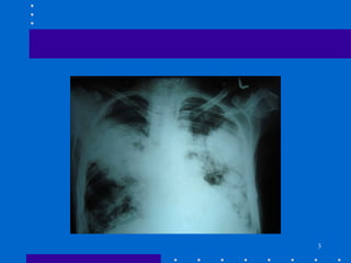
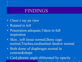
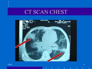
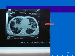
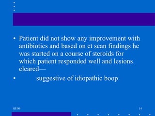
IMAGE OF THE WEEK PROF.DR.G.SUNDARAMURTY’S UNIT  S.DHANRAJ MD I YR
28yrs old male presented with following features +non-productive cough exertional dyspnea - two weeks fever, malaise, weight loss AUSCULTATION--- Bilateral coarse crackles and wheeze +
FINDINGS Chest x ray pa view Rotated to left Penetration adequate,Taken in full inspiration Skin , soft tissue normal,Bony cage normal,Trachea,mediastinal shadow normal Both dome of diaphragm normal in contour&shape Card.phrenic angle obliterated by opacity
Bilateral Heterogenous air space opacity occupying right midzone extending to lower zone and left mid& lower zone. 03/00
DIFFERENTIAL DIAGNOSIS USUAL INTERSTITIAL PNEUMONIA ACUTE INTERSTITIAL PNEUMONIA CHRONIC EOSINOPHILIC PNEUMONITIS ACUTE RESPIRATORY DISTRESS SYNDROME MYCOPLASMA, HIV, HSV, CMV, RUBEOLA, KLEBSIELLA, HAEMOPHILUS, LEGIONELLA, GRP. B- STREP, CRYPTOCOCCUS, NOCARDIA, PCP
INVESTIGATIONS ESR--- 10/22mm MANTOUX---Negative AFB---Negative HIV---Non reactive ANA---Negative
Patient was started on empirical antibiotics SPUTUM CULTURE– Negative FUNGAL CULTURE—Negative CT SCAN was taken 03/00
CT SCAN CHEST 03/00
03/00
ATOLL SIGN 03/00
03/00
FINDINGS Peribronchial & subpleural consolidation with irregular margins with air bronchogram Subpleural ground glass opacities ATOLL Sign—ring shaped opacity with central ground glass attenuation Interstitial thickening with ground glass opacities noted in midlobe/irregular/suprabasal segment of right lower lobe 03/00
Patient did not show any improvement with antibiotics and based on ct scan findings he was started on a course of steroids for which patient responded well and lesions cleared— suggestive of idiopathic boop 03/00
BOOP--INTRODUCTION Bronchiolitis  Obliterans Organizing Pneumonia - refers to a generic term of non-specific inflammatory reaction of small airways in response to exogenous/endogenous stimuli Comprises two types - based on histopathology Clinical features mimic pneumonia  without response to antibacterial therapy
BOOP- EPIDEMIOLOGY Smoking is  not  a risk factor
BOOP- Classification SECONDARY BOOP IDIOPATHIC BOOP
SECONDARY BOOP Connective tissue disorders - SLE, RA, Polymyositis - Dermatomyositis, Sjogren’s syndrome, MCTD, Ulcerative Colitis, Vasculitis Inhaled/Systemic Toxins - gases, nicotine, cocaine, CO, nitrogen, chlorine Drugs - Penicillamine, Amiodarone, Gold, Bleomycin, Mitomycin-c, Methotrexate,  Sulfasalazine
SECONDARY  BOOP Infections:  Mycoplasma, HIV, HSV, CMV, Rubeola, Klebsiella, Hemophilus, Legionella, Grp B- Strep, Cryptococcus, Nocardia, PCP Pediatric  RSV, Parainfluenza,  Adenovirus, Mycoplasma 03/00
SECONDARY BOOP Obstructive Pneumonitis Hypersensitivity Pneumonitis Aspiration Pneumonitis Chronic Eosinophilic Pneumonia Diffuse Alveolar Damage Myelodysplastic Syndrome Hematological malignancy 03/00
BOOP- IMAGING Chest Xray: Patchy peripheral bilateral migratory alveolar infiltrates 20-30% - reticular or nodular infiltrate Pleural effusions in  30% due to secondary BOOP CXR- can be normal in 4-10% Cavitation & lymphadenopathy are absent Focal consolidation is a marker for a good response to steroid therapy 03/00
BOOP-  IMAGING High Resolution CT scan of Chest:  patchy consolidation, ground glass opacity, nodularity with subpleural lower lobe predeliction. Bronchial wall thickening and dilatation denote severe disease Honey combing not seen in idiopathic BOOP
BOOP- Bronchoscopy Gold standard- Open lung or thoracoscopic lung biopsy for histopathology
BOOP--Treatment Spontaneous recovery occurs rarely Antibiotic therapy for underlying infections Withdrawal of offending toxin/ drug Supportive therapy Steroids for idiopathic BOOP  and  BOOP secondary to connective tissue disorders
BOOP-- STEROID Rx Idiopathic BOOP responds to steroids  better than BOOP due to connective tissue disorders Immunosuppressive agents - cyclophosphamide, azathioprine for those who fail to respond to steroid Rx
Usual interstitial pneumonia/idiopathic pulmonary fibrosis- -Massive fibrosis appearing as a honeycomb pattern on HRCT scans and traction bronchiectasis (lung architecture distortion) Irregular linear infiltrates generally in lower lung zones Acute interstitial pneumonia or Hammond rich syndrome- -Accelerated interstitial pneumonitis with fibrosis and ground-glass attenuation ,Interlobular septal thickening 00
Ch ronic eosinophilic pneumonia — Diffuse migratory, patchy alveolar infiltrates often along the pleural edges Ground-glass opacities Infective pneumonias (community-acquired, nosocomial, aspiration)-- Generally, either unilateral or bilateral infiltrates.  Aspiration pneumonia infiltrates common in gravity-dependent regions  Acute respiratory distress syndrome and diffuse alveolar damage- -   Focal infiltrates initially, with rapid progression to diffuse bilateral interstitial infiltrates.Alveolar concolidation often in dependent lung zones
THANK YOU 03/00

Imaging: BOOP

  • 1.
    IMAGE OF THEWEEK PROF.DR.G.SUNDARAMURTY’S UNIT S.DHANRAJ MD I YR
  • 2.
    28yrs old malepresented with following features +non-productive cough exertional dyspnea - two weeks fever, malaise, weight loss AUSCULTATION--- Bilateral coarse crackles and wheeze +
  • 4.
    FINDINGS Chest xray pa view Rotated to left Penetration adequate,Taken in full inspiration Skin , soft tissue normal,Bony cage normal,Trachea,mediastinal shadow normal Both dome of diaphragm normal in contour&shape Card.phrenic angle obliterated by opacity
  • 5.
    Bilateral Heterogenous airspace opacity occupying right midzone extending to lower zone and left mid& lower zone. 03/00
  • 6.
    DIFFERENTIAL DIAGNOSIS USUALINTERSTITIAL PNEUMONIA ACUTE INTERSTITIAL PNEUMONIA CHRONIC EOSINOPHILIC PNEUMONITIS ACUTE RESPIRATORY DISTRESS SYNDROME MYCOPLASMA, HIV, HSV, CMV, RUBEOLA, KLEBSIELLA, HAEMOPHILUS, LEGIONELLA, GRP. B- STREP, CRYPTOCOCCUS, NOCARDIA, PCP
  • 7.
    INVESTIGATIONS ESR--- 10/22mmMANTOUX---Negative AFB---Negative HIV---Non reactive ANA---Negative
  • 8.
    Patient was startedon empirical antibiotics SPUTUM CULTURE– Negative FUNGAL CULTURE—Negative CT SCAN was taken 03/00
  • 9.
  • 10.
  • 11.
  • 12.
  • 13.
    FINDINGS Peribronchial &subpleural consolidation with irregular margins with air bronchogram Subpleural ground glass opacities ATOLL Sign—ring shaped opacity with central ground glass attenuation Interstitial thickening with ground glass opacities noted in midlobe/irregular/suprabasal segment of right lower lobe 03/00
  • 14.
    Patient did notshow any improvement with antibiotics and based on ct scan findings he was started on a course of steroids for which patient responded well and lesions cleared— suggestive of idiopathic boop 03/00
  • 15.
    BOOP--INTRODUCTION Bronchiolitis Obliterans Organizing Pneumonia - refers to a generic term of non-specific inflammatory reaction of small airways in response to exogenous/endogenous stimuli Comprises two types - based on histopathology Clinical features mimic pneumonia without response to antibacterial therapy
  • 16.
    BOOP- EPIDEMIOLOGY Smokingis not a risk factor
  • 17.
    BOOP- Classification SECONDARYBOOP IDIOPATHIC BOOP
  • 18.
    SECONDARY BOOP Connectivetissue disorders - SLE, RA, Polymyositis - Dermatomyositis, Sjogren’s syndrome, MCTD, Ulcerative Colitis, Vasculitis Inhaled/Systemic Toxins - gases, nicotine, cocaine, CO, nitrogen, chlorine Drugs - Penicillamine, Amiodarone, Gold, Bleomycin, Mitomycin-c, Methotrexate, Sulfasalazine
  • 19.
    SECONDARY BOOPInfections: Mycoplasma, HIV, HSV, CMV, Rubeola, Klebsiella, Hemophilus, Legionella, Grp B- Strep, Cryptococcus, Nocardia, PCP Pediatric RSV, Parainfluenza, Adenovirus, Mycoplasma 03/00
  • 20.
    SECONDARY BOOP ObstructivePneumonitis Hypersensitivity Pneumonitis Aspiration Pneumonitis Chronic Eosinophilic Pneumonia Diffuse Alveolar Damage Myelodysplastic Syndrome Hematological malignancy 03/00
  • 21.
    BOOP- IMAGING ChestXray: Patchy peripheral bilateral migratory alveolar infiltrates 20-30% - reticular or nodular infiltrate Pleural effusions in 30% due to secondary BOOP CXR- can be normal in 4-10% Cavitation & lymphadenopathy are absent Focal consolidation is a marker for a good response to steroid therapy 03/00
  • 22.
    BOOP- IMAGINGHigh Resolution CT scan of Chest: patchy consolidation, ground glass opacity, nodularity with subpleural lower lobe predeliction. Bronchial wall thickening and dilatation denote severe disease Honey combing not seen in idiopathic BOOP
  • 23.
    BOOP- Bronchoscopy Goldstandard- Open lung or thoracoscopic lung biopsy for histopathology
  • 24.
    BOOP--Treatment Spontaneous recoveryoccurs rarely Antibiotic therapy for underlying infections Withdrawal of offending toxin/ drug Supportive therapy Steroids for idiopathic BOOP and BOOP secondary to connective tissue disorders
  • 25.
    BOOP-- STEROID RxIdiopathic BOOP responds to steroids better than BOOP due to connective tissue disorders Immunosuppressive agents - cyclophosphamide, azathioprine for those who fail to respond to steroid Rx
  • 26.
    Usual interstitial pneumonia/idiopathicpulmonary fibrosis- -Massive fibrosis appearing as a honeycomb pattern on HRCT scans and traction bronchiectasis (lung architecture distortion) Irregular linear infiltrates generally in lower lung zones Acute interstitial pneumonia or Hammond rich syndrome- -Accelerated interstitial pneumonitis with fibrosis and ground-glass attenuation ,Interlobular septal thickening 00
  • 27.
    Ch ronic eosinophilicpneumonia — Diffuse migratory, patchy alveolar infiltrates often along the pleural edges Ground-glass opacities Infective pneumonias (community-acquired, nosocomial, aspiration)-- Generally, either unilateral or bilateral infiltrates. Aspiration pneumonia infiltrates common in gravity-dependent regions Acute respiratory distress syndrome and diffuse alveolar damage- - Focal infiltrates initially, with rapid progression to diffuse bilateral interstitial infiltrates.Alveolar concolidation often in dependent lung zones
  • 28.