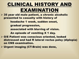
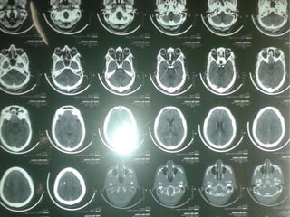
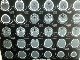
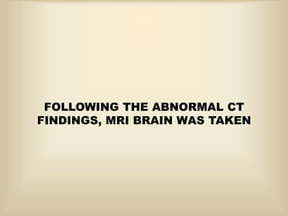
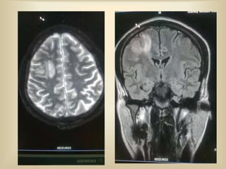
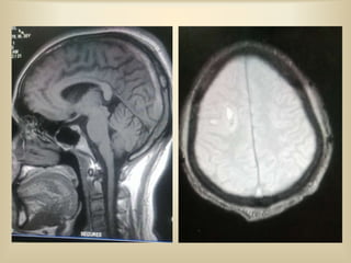
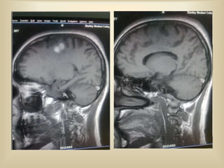
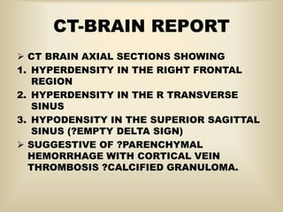
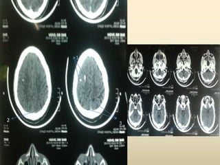
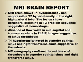
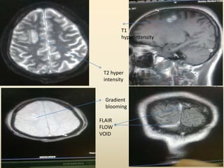
IMAGE OF THE WEEK
PROF.DR.G.SUNDARAMURTHY
M2 UNIT
P-VANJI NATHAN
CLINICAL HISTORY AND
EXAMINATION
 35 year old male patient, a chronic alcoholic
presented to casualty with history of
headache 1 week, sudden onset,
gradual progression,
associated with blurring of vision.
An episode of vomiting X 1 day.
 O/E Patient was conscious oriented, looked
distressed and had R lateral rectus palsy (diplopia)
on CNS examination.
 Urgent imaging (CT-Brain) was done,
FOLLOWING THE ABNORMAL CT
FINDINGS, MRI BRAIN WAS TAKEN
CT-BRAIN REPORT
 CT BRAIN AXIAL SECTIONS SHOWING
1. HYPERDENSITY IN THE RIGHT FRONTAL
REGION
2. HYPERDENSITY IN THE R TRANSVERSE
SINUS
3. HYPODENSITY IN THE SUPERIOR SAGITTAL
SINUS (?EMPTY DELTA SIGN)
 SUGGESTIVE OF ?PARENCHYMAL
HEMORRHAGE WITH CORTICAL VEIN
THROMBOSIS ?CALCIFIED GRANULOMA.
1
22
3
MRI BRAIN REPORT
 MRI brain shows T1 hyperintense / non
suppressible T2 hyperintensity in the right
high parietal lobe. The lesion shows
peripheral blooming in T2 gradient sequence
suggestive of hemorrhage.
 Absence of flow voids in mid sagittal and
transverse sinus in FLAIR images suggestive
of sinus thrombosis
 T1 hyperintensity noted in superior sagittal
sinus and right transverse sinus suggestive of
thrombosis.
 MR venography confirms the evidence of
thrombosis in superior sagittal sinus and right
transverse sinus.
T2 hyper
intensity
Gradient
blooming
T1
hyperintensity
FLAIR
FLOW
VOID
CEREBRAL VEINS-VALVELESS,THIN
EXTERNAL
SUPERIOR MIDDLE
SUPER
FICIAL
DEEP
CAVERNOUS OR
SPHENOPARIETAL
SINUSSUPERIOR
SAGITTAL
SINUS
BASAL
VEIN
INTERNAL-2 IN
NUMBER- JOIN TO
FORM GREAT
CEREBRAL VEIN
INFERIOR
SUP.CER.VN
OR CAV.SIN.
BASAL VN
RECEIVES
CT FINDINGS IN CVT
 Dense clot sign - Areas of thrombosis in sinuses
appearing as hyperdensities in unenhanced ct.
 Cord sign - Dense cortical veins-uncommon direct
sign of cvt in unenhanced ct.
 Venous infarction is the most non specific indirect
sign in unenhanced ct - presence of multiple
isolated lesions, subcortical involvement, more
than one arterial territory involved.
 Empty delta sign - Filling defect with enhancing
rim on enhanced ct.
 Contrast enhancement secondary to venous stasis
 Diffuse brain swelling with edema and decreased
ventricular size might be present.
 CT venogarhy allows direct visualisation of
thrombus.
MRI FINDINGS IN CVT
 Can confirmatively tell presence of CVT.
 Venous thrombus may be directly visualised.
 Patent dural sinuses are seen as flow void
especially when the image is orthogonal to blood
flow direction.
 Thrombus manifest as absence of flow void-best
seen in FLAIR and T2 weighted spin echo
sequences.
 T1 weighted - hyperintensity ; T2 weighted
gradient echo-exaggerated signal loss.
 MR venography - shows direct visualisation of
thrombus.
Imaging: Cortical Vein Thrombosis

Imaging: Cortical Vein Thrombosis

  • 1.
    IMAGE OF THEWEEK PROF.DR.G.SUNDARAMURTHY M2 UNIT P-VANJI NATHAN
  • 2.
    CLINICAL HISTORY AND EXAMINATION 35 year old male patient, a chronic alcoholic presented to casualty with history of headache 1 week, sudden onset, gradual progression, associated with blurring of vision. An episode of vomiting X 1 day.  O/E Patient was conscious oriented, looked distressed and had R lateral rectus palsy (diplopia) on CNS examination.  Urgent imaging (CT-Brain) was done,
  • 6.
    FOLLOWING THE ABNORMALCT FINDINGS, MRI BRAIN WAS TAKEN
  • 11.
    CT-BRAIN REPORT  CTBRAIN AXIAL SECTIONS SHOWING 1. HYPERDENSITY IN THE RIGHT FRONTAL REGION 2. HYPERDENSITY IN THE R TRANSVERSE SINUS 3. HYPODENSITY IN THE SUPERIOR SAGITTAL SINUS (?EMPTY DELTA SIGN)  SUGGESTIVE OF ?PARENCHYMAL HEMORRHAGE WITH CORTICAL VEIN THROMBOSIS ?CALCIFIED GRANULOMA.
  • 12.
  • 13.
    MRI BRAIN REPORT MRI brain shows T1 hyperintense / non suppressible T2 hyperintensity in the right high parietal lobe. The lesion shows peripheral blooming in T2 gradient sequence suggestive of hemorrhage.  Absence of flow voids in mid sagittal and transverse sinus in FLAIR images suggestive of sinus thrombosis  T1 hyperintensity noted in superior sagittal sinus and right transverse sinus suggestive of thrombosis.  MR venography confirms the evidence of thrombosis in superior sagittal sinus and right transverse sinus.
  • 14.
  • 16.
    CEREBRAL VEINS-VALVELESS,THIN EXTERNAL SUPERIOR MIDDLE SUPER FICIAL DEEP CAVERNOUSOR SPHENOPARIETAL SINUSSUPERIOR SAGITTAL SINUS BASAL VEIN INTERNAL-2 IN NUMBER- JOIN TO FORM GREAT CEREBRAL VEIN INFERIOR SUP.CER.VN OR CAV.SIN. BASAL VN RECEIVES
  • 19.
    CT FINDINGS INCVT  Dense clot sign - Areas of thrombosis in sinuses appearing as hyperdensities in unenhanced ct.  Cord sign - Dense cortical veins-uncommon direct sign of cvt in unenhanced ct.  Venous infarction is the most non specific indirect sign in unenhanced ct - presence of multiple isolated lesions, subcortical involvement, more than one arterial territory involved.  Empty delta sign - Filling defect with enhancing rim on enhanced ct.  Contrast enhancement secondary to venous stasis  Diffuse brain swelling with edema and decreased ventricular size might be present.  CT venogarhy allows direct visualisation of thrombus.
  • 21.
    MRI FINDINGS INCVT  Can confirmatively tell presence of CVT.  Venous thrombus may be directly visualised.  Patent dural sinuses are seen as flow void especially when the image is orthogonal to blood flow direction.  Thrombus manifest as absence of flow void-best seen in FLAIR and T2 weighted spin echo sequences.  T1 weighted - hyperintensity ; T2 weighted gradient echo-exaggerated signal loss.  MR venography - shows direct visualisation of thrombus.