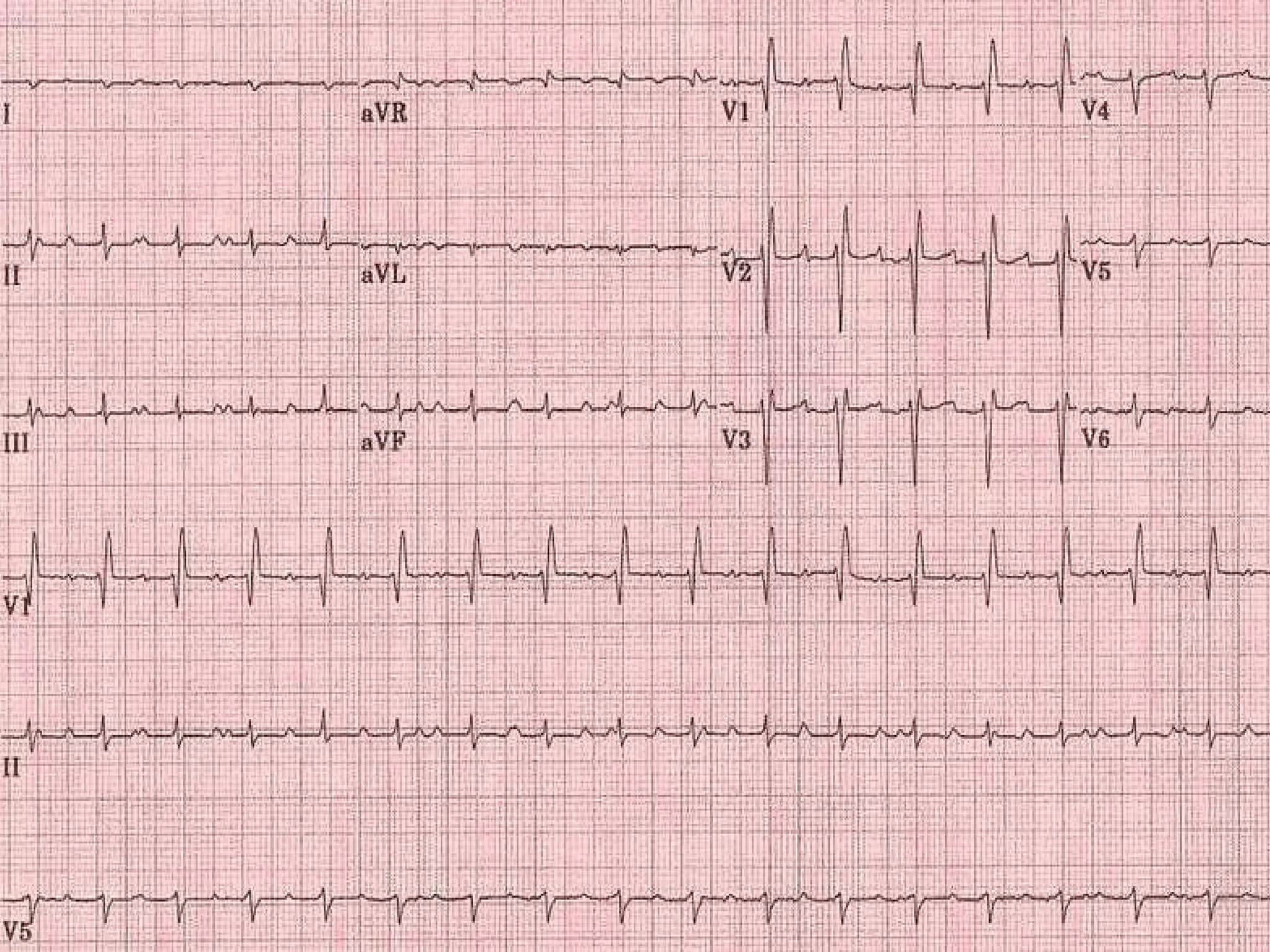
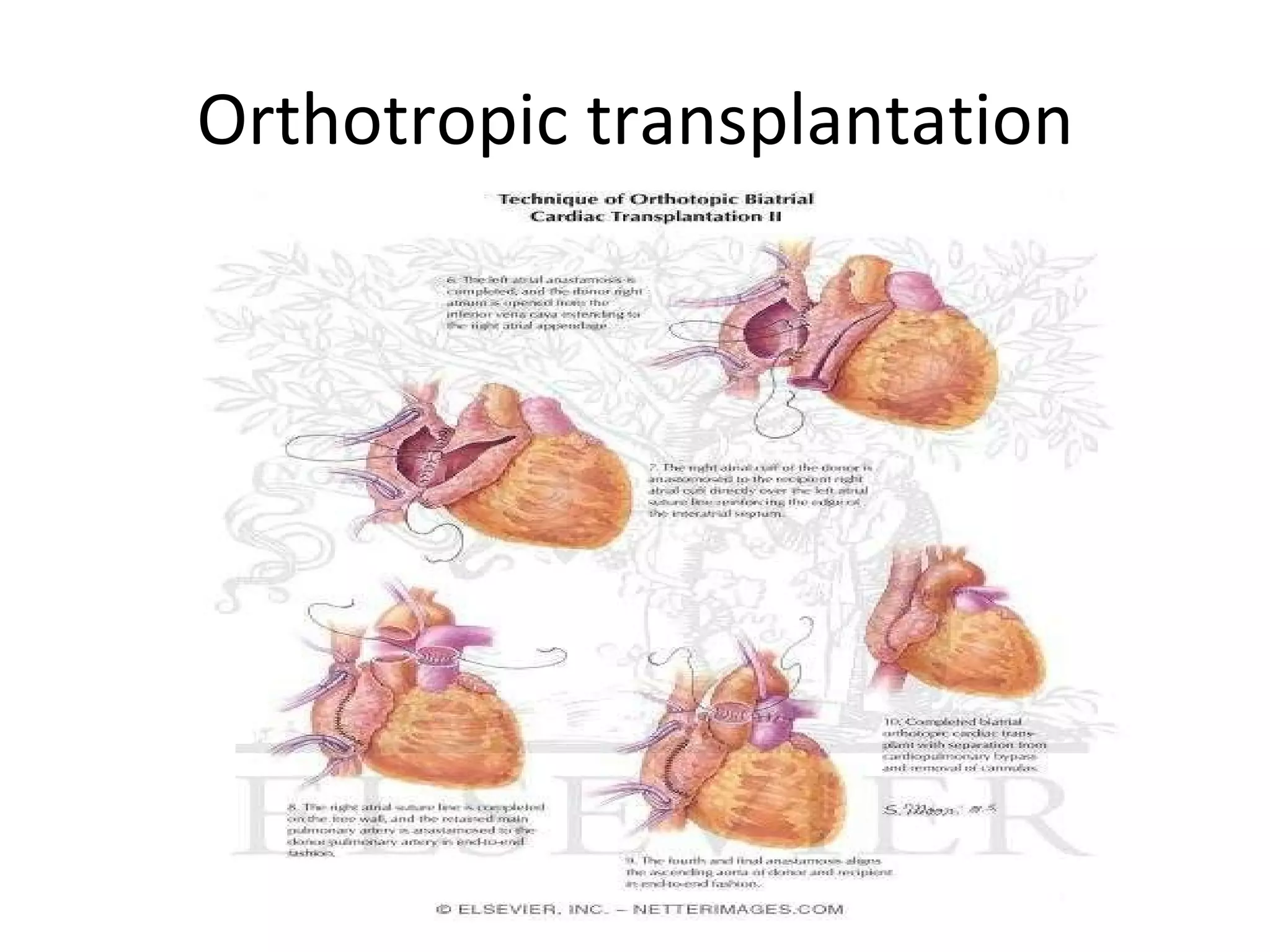
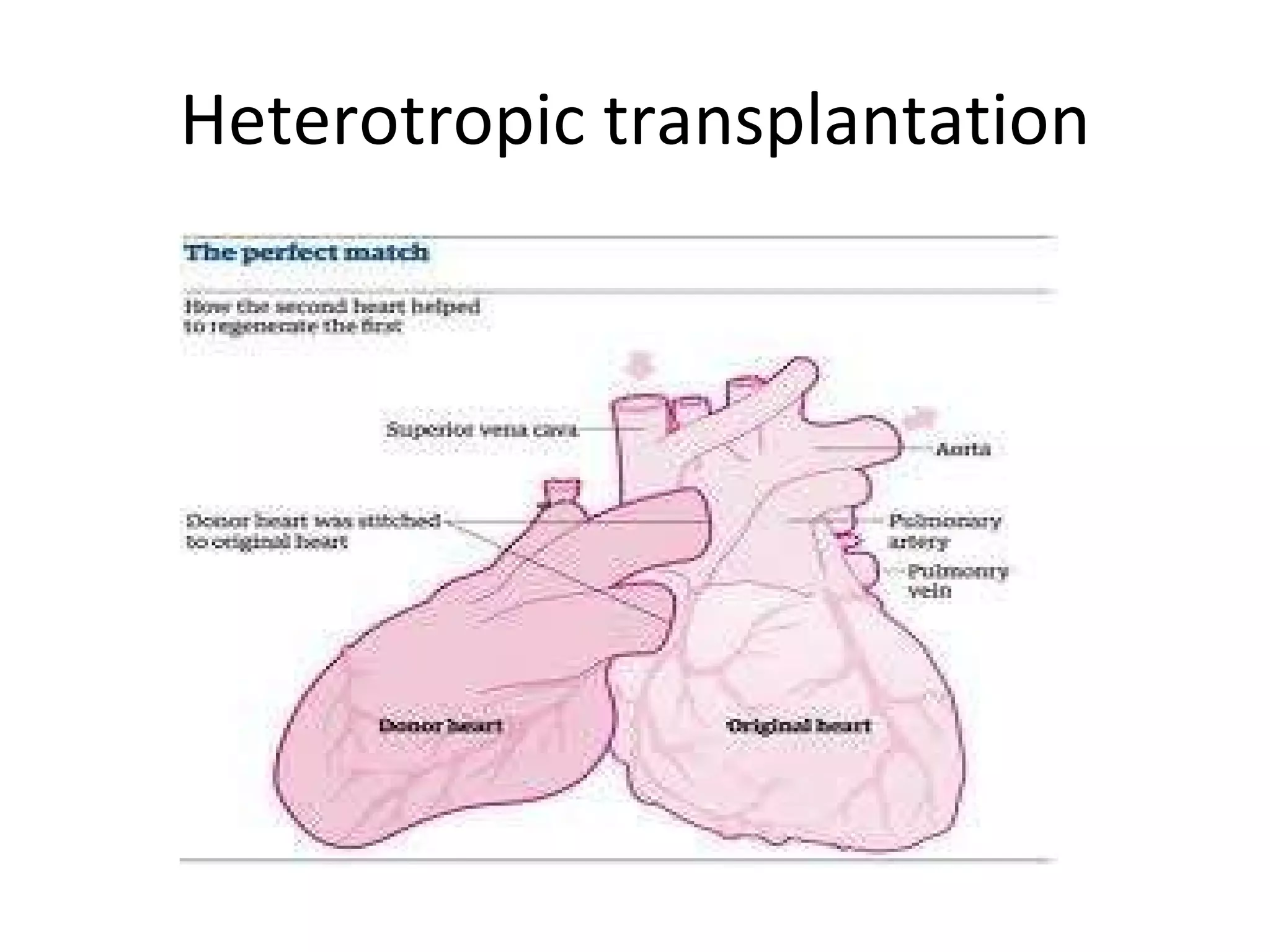
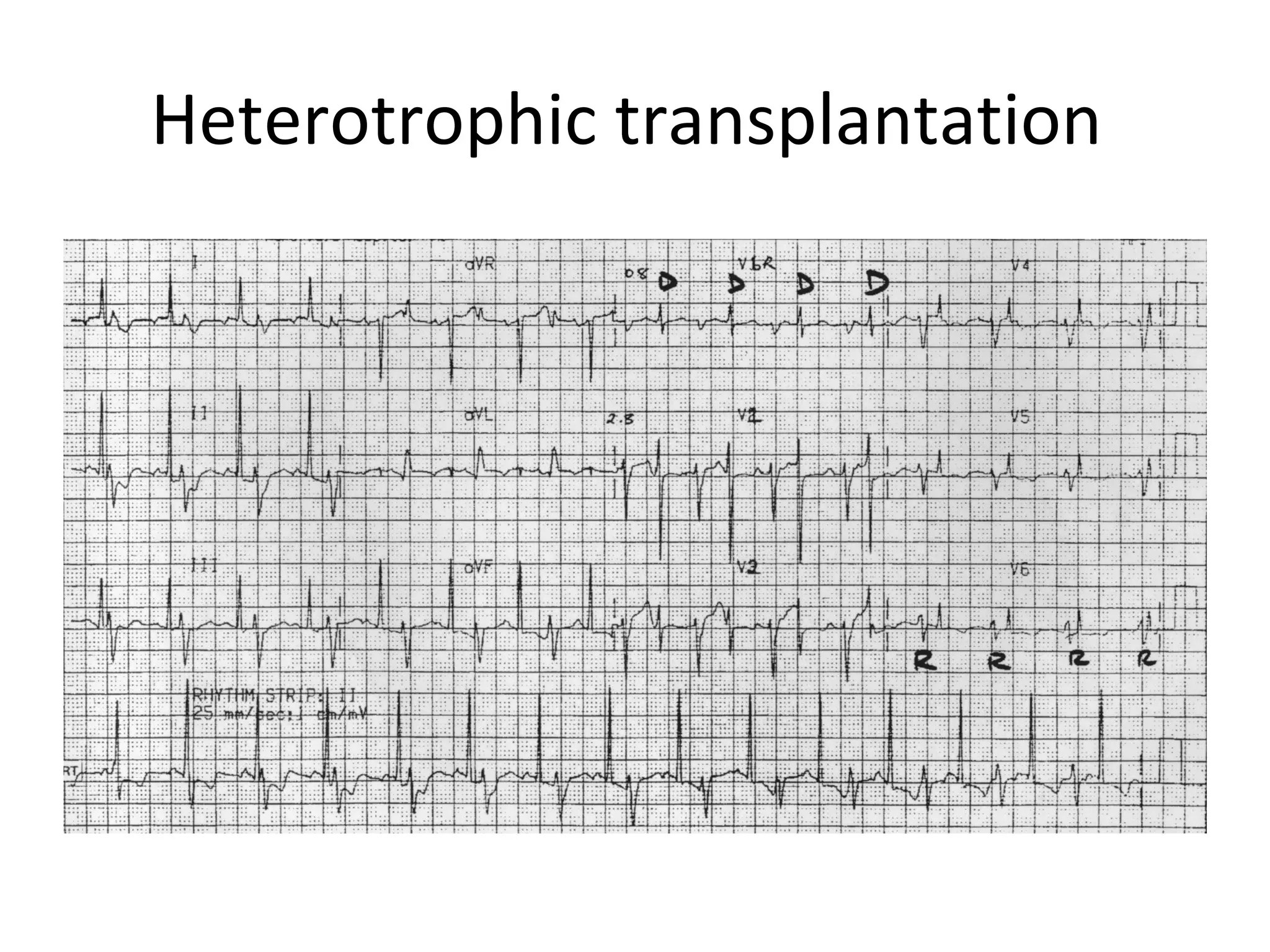
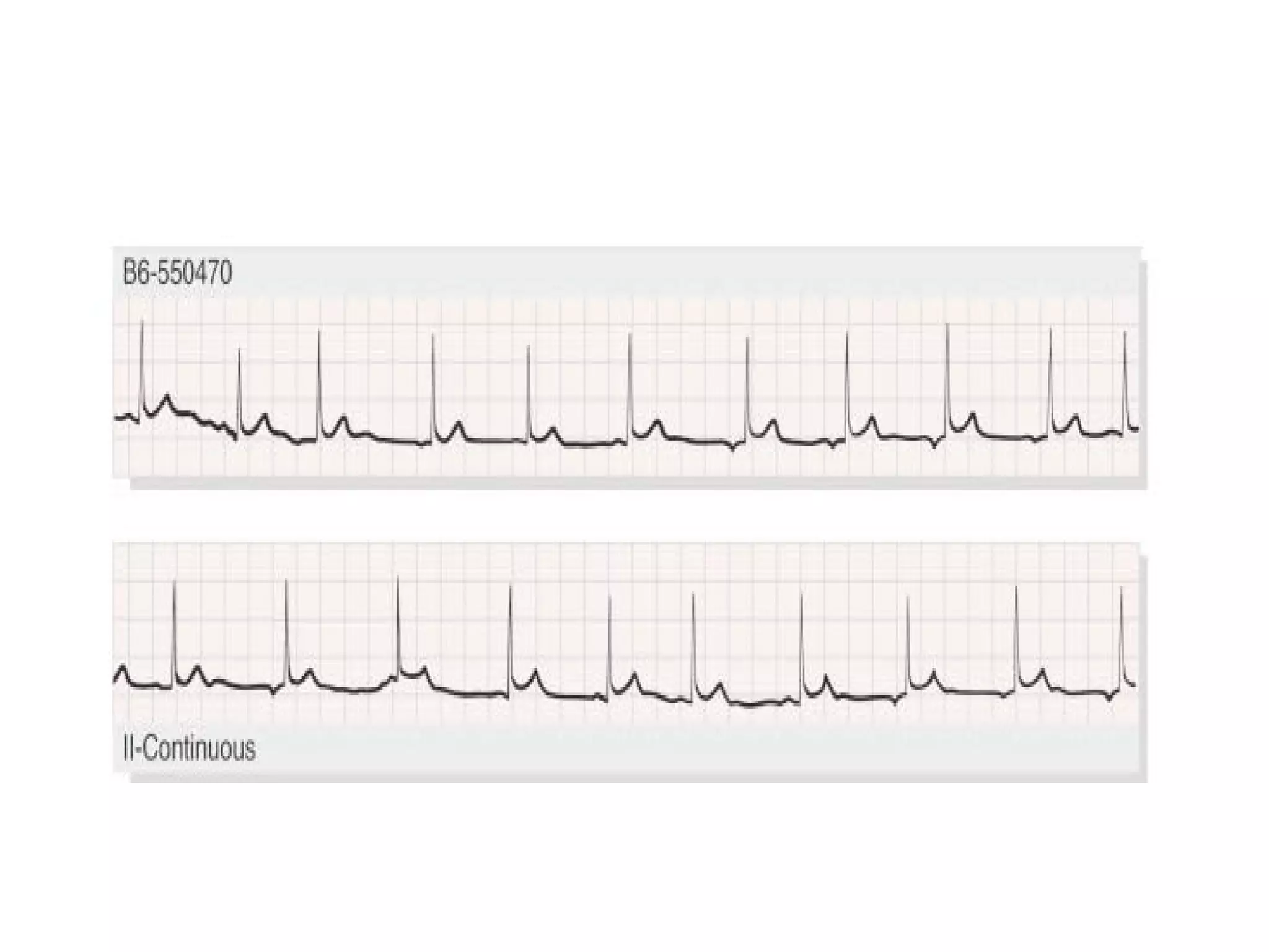
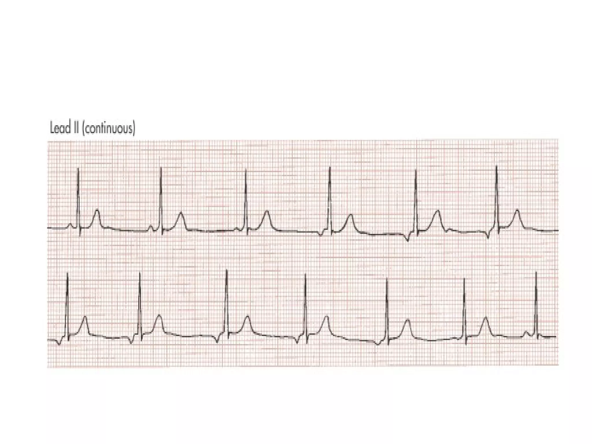
ECG OF THE WEEK DR.Prof.K.H.Noorul Ameen’s unit(M4) Dr.S.Dhileepan
 
History 70 yr old male, k/c HT,CAD for 20 years, Had cardiac transplantation 6 months back.
ECG shows HR-140/min, Rhythm –regular, PR interval -0.28 sec, Axis  +60-90 degree, Incomplete RBBB Two sets of P waves, independent to each other and one being always conducted.
DIAGNOSIS ATRIAL DISSOCIATION
Atrial dissociation First reported by Wenkeback in 1906. It has been suggested that in atrial dissociation each atrium beats separately and independently as a result of blockage or interruption of an interatrial pathway (Bachmann's bundle). The sinus pacemaker controls one atrium and the ectopic pacemaker controls the other atrium. The two pacemakers do not interfere with each other as a result of interatrial block or presence of preferential exit block
Differential diagnosis 1)Atrial parasystole-conducted, - normal P wave. 2)Artefact –rhythmic muscular twitchings, electrical disturbance from adjacent person.
causes RHD SHT CCF Sudden neurogenic stimuli Uremia Diphtheria
Atrial dissociation in transplanted heart Heart transplantation- orthotropic, - heterotropic .  In orthotropic transplantation posterior wall of recipient heart is retained but denervated. It produces impulse but not conducted as it is blocked by suture line. In heterotrophic transplantation 2 sets of complexes are produced.
Orthotropic transplantation
Heterotropic transplantation
Heterotrophic transplantation
 
 
ECG shows Varying PP,PR,RR interval, P wave inverted and gradually become normal when heart rate speeds up.
Wandering pacemaker
causes Phyisiologic-deep sleep Young adults with high vagal tone Digitalis  Sick sinus syndrome
WANDERING PACEMAKER.  This variant of sinus arrhythmia involves passive transfer of the dominant pacemaker focus from the sinus node to latent pacemakers that have the next highest degree of automaticity located in other atrial sites (usually lower in the crista terminalis) or in AV junctional tissue. The change occurs in a gradual fashion over the duration of several beats; thus, only one pacemaker at a time controls the rhythm, in sharp contrast to AV dissociation. Precursor of MAT
 
THANK YOU

ECG: Atrial Dissociation

  • 1.
    ECG OF THEWEEK DR.Prof.K.H.Noorul Ameen’s unit(M4) Dr.S.Dhileepan
  • 2.
  • 3.
    History 70 yrold male, k/c HT,CAD for 20 years, Had cardiac transplantation 6 months back.
  • 4.
    ECG shows HR-140/min,Rhythm –regular, PR interval -0.28 sec, Axis +60-90 degree, Incomplete RBBB Two sets of P waves, independent to each other and one being always conducted.
  • 5.
  • 6.
    Atrial dissociation Firstreported by Wenkeback in 1906. It has been suggested that in atrial dissociation each atrium beats separately and independently as a result of blockage or interruption of an interatrial pathway (Bachmann's bundle). The sinus pacemaker controls one atrium and the ectopic pacemaker controls the other atrium. The two pacemakers do not interfere with each other as a result of interatrial block or presence of preferential exit block
  • 7.
    Differential diagnosis 1)Atrialparasystole-conducted, - normal P wave. 2)Artefact –rhythmic muscular twitchings, electrical disturbance from adjacent person.
  • 8.
    causes RHD SHTCCF Sudden neurogenic stimuli Uremia Diphtheria
  • 9.
    Atrial dissociation intransplanted heart Heart transplantation- orthotropic, - heterotropic . In orthotropic transplantation posterior wall of recipient heart is retained but denervated. It produces impulse but not conducted as it is blocked by suture line. In heterotrophic transplantation 2 sets of complexes are produced.
  • 10.
  • 11.
  • 12.
  • 13.
  • 14.
  • 15.
    ECG shows VaryingPP,PR,RR interval, P wave inverted and gradually become normal when heart rate speeds up.
  • 16.
  • 17.
    causes Phyisiologic-deep sleepYoung adults with high vagal tone Digitalis Sick sinus syndrome
  • 18.
    WANDERING PACEMAKER. This variant of sinus arrhythmia involves passive transfer of the dominant pacemaker focus from the sinus node to latent pacemakers that have the next highest degree of automaticity located in other atrial sites (usually lower in the crista terminalis) or in AV junctional tissue. The change occurs in a gradual fashion over the duration of several beats; thus, only one pacemaker at a time controls the rhythm, in sharp contrast to AV dissociation. Precursor of MAT
  • 19.
  • 20.