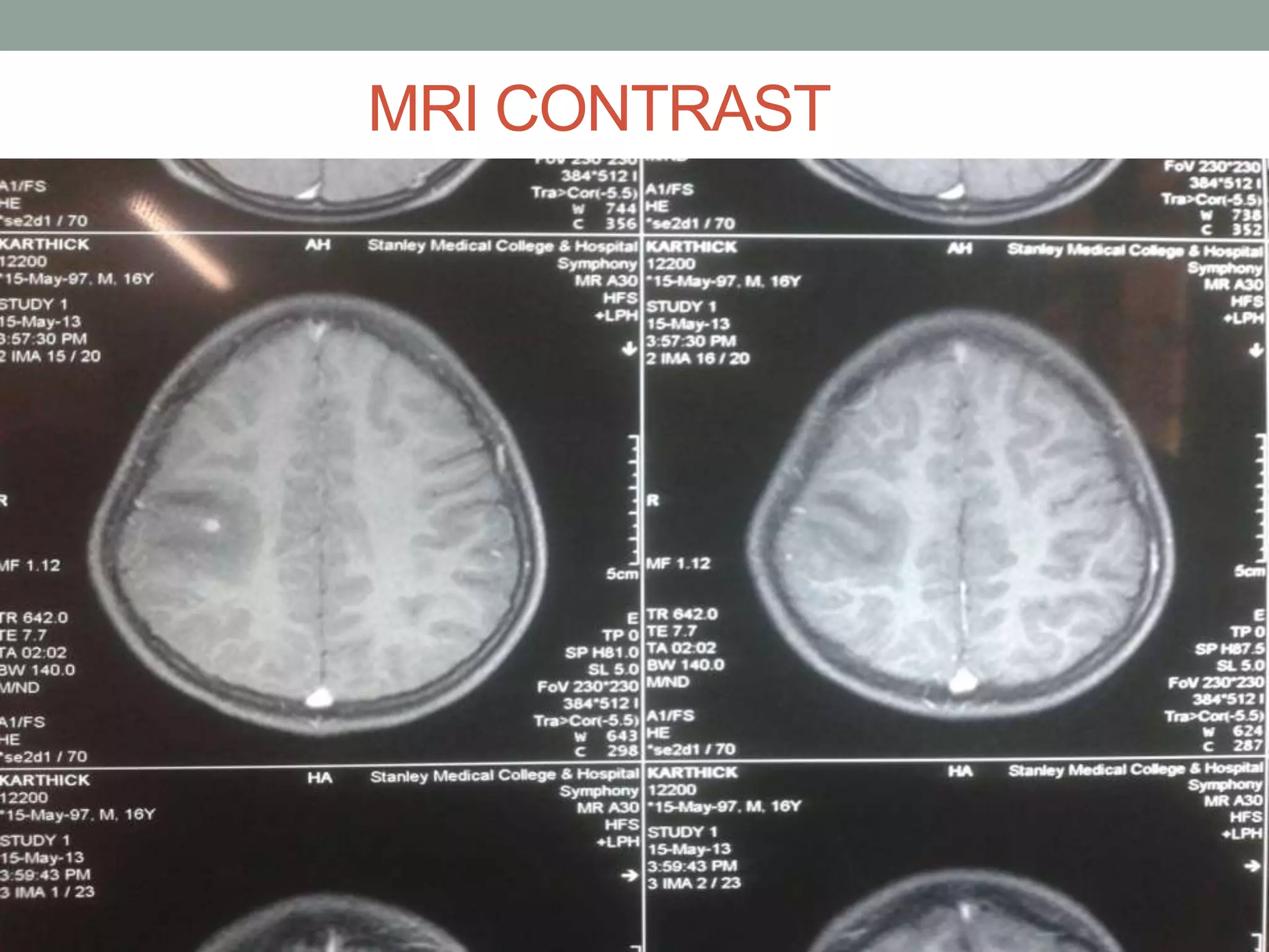
IMAGE OF THE WEEK
Prof.Dr.P.Vijayaraghavan’s Unit
Presented by
Dr.T. Jaya Packiam
CASE SUMMARY
A 16 years old boy admitted with complaints of Abnormal
movement left thumb and left eyelid more than 1 hour.
Patient takes mixed diet
General examination and systemic examination were
unremarkable.
Routine investigations CBC LFT RFT ECG CXR were also
normal.
T1 WEIGHTED MRI
T2 WEIGHTED MRI
MRI FLAIR
MRI CONTRAST
DIFFERENTIAL DIAGNOSIS
Infections- neurocysticercosis, tuberculomas ,
toxoplasmosis, cryptococcosis
histoplasmosis,candida albicans
Metastases
Abscesses
Granulomas
 Resolving hematomas
Primary cns lymphomas
Multiple sclerosis
Vascular malformations
Thrombosed aneurysms
Tuberculomas NEUROCYSTICERCOSIS
1.Usually larger>2CM and usually multiple Smaller<2CM,may be single or multiple
2.Associated with basal meningitis or
hydrocephalus.
NOT ASSOCIATED.
3.They are more common in posterior
fossa .
Most commonly occur at the gray-white
junction.
4.Look for CF of tb elsewhere lungs,lymph
nodes,intestine.
Look for occular involvement,muscle
involvement or subcutaneous nodules
5.T2 weighted MRI shows
Hypointense.
scolex will be absent.
Midline shift will be present.
Conglomerate enhancement
T2 weighted MRI shows
Hyperintense.
scolex will be present.
No Midline shift.
Isolated ring enhancement.
6.MR spectroscopy may show lipid peaks
with tuberculoma.
MR spectroscope may show multiple
amino acid peaks.
MRI FINDINGS IN
NEUROCYSTICERCOSIS
T1W1 T2W2 MRI FLAIR
VESICULAR-ISOINTENSE TO
CSF,DISCRETE ECCENTRIC
SCOLEX(HYPERINTENSE)
ISOINTENSE TO
CSF.DISCRETE
ECCENTRIC SCOLEX.
NO EDEMA.
ISO INTENSE TO CSF.
DISCRETE
ECCENTRIC SCOLEX
NO EDEMA
COLLOIDAL VESICULAR-CYST
MILDLY HYPERINTENSE TO
CSF
HYPERINTENSE TO
CSF,MILD TO MARKED
EDEMA
HYPERINTENSE TO
CSF,MILD TO MARKED
EDEMA
GRANULAR NODULAR-
THICKENED RETRACTED CYST
WALL.EDEMA DECREASES.
NODULAR CALCIFIED-
DIFFICULT TO DETECT
THICKENED
RETRACTED CYST
WALL .EDEMA
REDUCED.
SHRUNKEN
CALCIFIED LESION
DISCUSSION
• Neurocysticercosis is the most common parasitic disease
of CNS.
• Intraparenchymal parasitic infection caused by pork
Tapeworm Tinea solium.
• 4 pathogenic stages
• -Vesicular(larva alive)
• -colloidal vesicular(degenerating larva)
• -granular nodular(healing)
• -nodular calcified.(healed)
Cut section of brain affected by NCC
GENERAL FEATURES
Location: cysterns>parenchymal>ventricles
-parenchymal cyst often hemispheric at grey white
junction.
-Intraventricles often isolated
-Basal cisterns cysts may be racemose(Multiple Grapelike)
-Size of the cyst 1-2cm and scolex will be 1-4mm.
-Lesions may be different stages in same patient.
CINICAL FINDINGS
New onset seizures with or without generalisation
May involve brain parenchyma – seizure/FND
Subarachnoid space / Ventricular Space – May mimic
Intraspinal tumors.
Cysticercosis can be diagnosed several
ways.
• Travels
• Eating
• MRI or CT brain scans
• Blood tests-ELISA
TREATMENT
• ANTI CONVULSANT THERAPY:
CAN BE STOPPED ONCE CT SHOWS RESOLUTION
OF LESION
• ANTIHELMINTHIC THERAPY:
• ALBENDAZOLE 15MG/KG/DAY IN 2DOSES FOR 8DAYS
• PRAZIQUANTEL 50MG/KG/DAY TDS FOR 15 DAYS
TO REDUCE THE INFLAMMATORY RESPONSE OF
DEGENERATING PARASITES.
• PREDNISOLONE/DEXAMETHASONE.
THANK YOU
Thank you..........

IMAGING: NEUROCYSTICERCOSIS

  • 1.
    IMAGE OF THEWEEK Prof.Dr.P.Vijayaraghavan’s Unit Presented by Dr.T. Jaya Packiam
  • 2.
    CASE SUMMARY A 16years old boy admitted with complaints of Abnormal movement left thumb and left eyelid more than 1 hour. Patient takes mixed diet General examination and systemic examination were unremarkable. Routine investigations CBC LFT RFT ECG CXR were also normal.
  • 3.
  • 4.
  • 5.
  • 6.
  • 7.
    DIFFERENTIAL DIAGNOSIS Infections- neurocysticercosis,tuberculomas , toxoplasmosis, cryptococcosis histoplasmosis,candida albicans Metastases Abscesses Granulomas  Resolving hematomas Primary cns lymphomas Multiple sclerosis Vascular malformations Thrombosed aneurysms
  • 8.
    Tuberculomas NEUROCYSTICERCOSIS 1.Usually larger>2CMand usually multiple Smaller<2CM,may be single or multiple 2.Associated with basal meningitis or hydrocephalus. NOT ASSOCIATED. 3.They are more common in posterior fossa . Most commonly occur at the gray-white junction. 4.Look for CF of tb elsewhere lungs,lymph nodes,intestine. Look for occular involvement,muscle involvement or subcutaneous nodules 5.T2 weighted MRI shows Hypointense. scolex will be absent. Midline shift will be present. Conglomerate enhancement T2 weighted MRI shows Hyperintense. scolex will be present. No Midline shift. Isolated ring enhancement. 6.MR spectroscopy may show lipid peaks with tuberculoma. MR spectroscope may show multiple amino acid peaks.
  • 9.
    MRI FINDINGS IN NEUROCYSTICERCOSIS T1W1T2W2 MRI FLAIR VESICULAR-ISOINTENSE TO CSF,DISCRETE ECCENTRIC SCOLEX(HYPERINTENSE) ISOINTENSE TO CSF.DISCRETE ECCENTRIC SCOLEX. NO EDEMA. ISO INTENSE TO CSF. DISCRETE ECCENTRIC SCOLEX NO EDEMA COLLOIDAL VESICULAR-CYST MILDLY HYPERINTENSE TO CSF HYPERINTENSE TO CSF,MILD TO MARKED EDEMA HYPERINTENSE TO CSF,MILD TO MARKED EDEMA GRANULAR NODULAR- THICKENED RETRACTED CYST WALL.EDEMA DECREASES. NODULAR CALCIFIED- DIFFICULT TO DETECT THICKENED RETRACTED CYST WALL .EDEMA REDUCED. SHRUNKEN CALCIFIED LESION
  • 10.
    DISCUSSION • Neurocysticercosis isthe most common parasitic disease of CNS. • Intraparenchymal parasitic infection caused by pork Tapeworm Tinea solium. • 4 pathogenic stages • -Vesicular(larva alive) • -colloidal vesicular(degenerating larva) • -granular nodular(healing) • -nodular calcified.(healed)
  • 12.
    Cut section ofbrain affected by NCC
  • 13.
    GENERAL FEATURES Location: cysterns>parenchymal>ventricles -parenchymalcyst often hemispheric at grey white junction. -Intraventricles often isolated -Basal cisterns cysts may be racemose(Multiple Grapelike) -Size of the cyst 1-2cm and scolex will be 1-4mm. -Lesions may be different stages in same patient.
  • 14.
    CINICAL FINDINGS New onsetseizures with or without generalisation May involve brain parenchyma – seizure/FND Subarachnoid space / Ventricular Space – May mimic Intraspinal tumors.
  • 15.
    Cysticercosis can bediagnosed several ways. • Travels • Eating • MRI or CT brain scans • Blood tests-ELISA
  • 16.
    TREATMENT • ANTI CONVULSANTTHERAPY: CAN BE STOPPED ONCE CT SHOWS RESOLUTION OF LESION • ANTIHELMINTHIC THERAPY: • ALBENDAZOLE 15MG/KG/DAY IN 2DOSES FOR 8DAYS • PRAZIQUANTEL 50MG/KG/DAY TDS FOR 15 DAYS TO REDUCE THE INFLAMMATORY RESPONSE OF DEGENERATING PARASITES. • PREDNISOLONE/DEXAMETHASONE.
  • 17.

Editor's Notes

  • #16 Neurocysticercosis diagnosed by MRI or CT brain scans. Blood tests are not always accurate – could develop antibodies for parasite, but not have the infection. Surgery can confirm diagnosis. Research article: greater that 50% of individuals diagnosed with neurocysticercosis by CT scans test negative for the disease after enzyme-linked immunoelectrotransfer blot assay (EITB) is done. This is because it is assumed that NC is main caused of symptomatic epilepsy in developing countries.