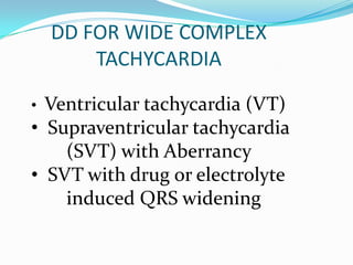
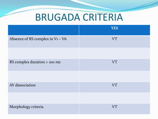
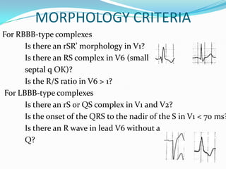
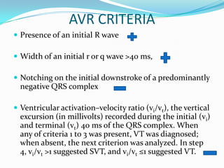
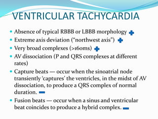
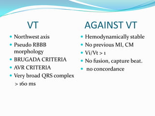
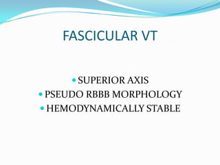
K.M.JEYABALAJI
Dr.P. VIJAYARAGAVAN’S UNIT
HISTORY
 A 22 yr male patient came with complaints
 of
   Acute onset breathlessness
   Palpitation
   Profuse sweating
   Vague chest discomfort
                  For past 1 hour
EXAMINATION
 Dyspnoeic, tachypnoeic,
 Pulse- 180/ min REGULAR
 BP- 90/ 60 mmhg
 JVP- ---
 CVS- s1,s2 heard
 RS – NVBS
 P/A- soft
 CNS- NFND
ADMISSION ECG
CHEST LEADS
FINDINGS
 TACHYCARDIA
 REGULAR RHYTM
 RATE- 200/min
 AXIS – EXTREME NORTH ( northwest)
 WIDE QRS COMPLEX
 RBBB PATTERN IN V1
DD FOR WIDE COMPLEX
        TACHYCARDIA

• Ventricular tachycardia (VT)
• Supraventricular tachycardia
    (SVT) with Aberrancy
• SVT with drug or electrolyte
    induced QRS widening
APPROACH
     WIDE COMPLEX TACHYCARDIA

        REGULAR/IRREGULAR

          AV DISSOCIATION

CLASSICAL BUNDLE BRANCH MORPHOLOGY

         BRUGADA CRITERIA

           AVR CRITERIA
BRUGADA CRITERIA
                                   YES

Absence of RS complex in V1 – V6   VT


RS complex duration > 100 ms       VT


AV dissociation                    VT


Morphology criteria                VT
BRUGADA CRITERIA
MORPHOLOGY CRITERIA
For RBBB-type complexes
      Is there an rSR’ morphology in V1?
      Is there an RS complex in V6 (small
      septal q OK)?
      Is the R/S ratio in V6 > 1?
For LBBB-type complexes
      Is there an rS or QS complex in V1 and V2?
      Is the onset of the QRS to the nadir of the S in V1 < 70 ms?
      Is there an R wave in lead V6 without a
      Q?
AVR CRITERIA
 Presence of an initial R wave

 Width of an initial r or q wave >40 ms,

 Notching on the initial downstroke of a predominantly
  negative QRS complex

 Ventricular activation–velocity ratio (vi/vt), the vertical
  excursion (in millivolts) recorded during the initial (vi)
  and terminal (vt) 40 ms of the QRS complex. When
  any of criteria 1 to 3 was present, VT was diagnosed;
  when absent, the next criterion was analyzed. In step
  4, vi/vt >1 suggested SVT, and vi/vt ≤1 suggested VT.
VENTRICULAR TACHYCARDIA
 Absence of typical RBBB or LBBB morphology
 Extreme axis deviation (“northwest axis”)
 Very broad complexes (>160ms)
 AV dissociation (P and QRS complexes at different
  rates)
 Capture beats — occur when the sinoatrial node
  transiently ‘captures’ the ventricles, in the midst of AV
  dissociation, to produce a QRS complex of normal
  duration.
 Fusion beats — occur when a sinus and ventricular
  beat coincides to produce a hybrid complex.
 Positive or negative concordance throughout the chest
  leads, i.e. leads V1-6 show entirely positive (R) or
  entirely negative (QS) complexes, with no RS
  complexes seen.
 Brugada’s sign– The distance from the onset of the
  QRS complex to the nadir of the S-wave is > 100ms
 Josephson’s sign – Notching near the nadir of the S-
  wave
VT
CAPTURE BEAT                         FUSION BEAT


               BRUGADA SIGN , JOSEPHSON SIGN
NEGATIVE CONCORDANCE POSITIVE CONCORDANCE
SVT WITH ABBERANCY
 • Any SVT can be conducted with aberrancy:
 – Sinus Tachycardia
 – Atrial tachycardia
 – Atrial flutter
 – Atrioventricular nodal reentrant tachycardia
      (AVNRT)
 – Junctional Tachycardia
 – Orthodromic Atrioventricular Reentrant Tachycardia
      (AVRT)
VT                   AGAINST VT
 Northwest axis            Hemodynamically stable
 Pseudo RBBB               No previous MI, CM
  morphology                Vi/Vt > 1
 BRUGADA CRITERIA          No fusion, capture beat.
 AVR CRITERIA              no concordance
 Very broad QRS complex
  > 160 ms
FASCICULAR VT

      SUPERIOR AXIS
 PSEUDO RBBB MORPHOLOGY
 HEMODYNAMICALLY STABLE
TAKE HOME MESSAGE
 No criteria is 100% sensitive nor specific
 Never go blindly by ECG
 Give equal imortance to history, clinical
  presentation,
 Vitals
 If you are 100% sure that it is SVT, then
  proceed.
 Having even 1% doubt, then treat it as VT
THANK YOU

Broad complex tachycardia

  • 1.
  • 2.
    HISTORY  A 22yr male patient came with complaints of  Acute onset breathlessness  Palpitation  Profuse sweating  Vague chest discomfort For past 1 hour
  • 3.
    EXAMINATION  Dyspnoeic, tachypnoeic, Pulse- 180/ min REGULAR  BP- 90/ 60 mmhg  JVP- ---  CVS- s1,s2 heard  RS – NVBS  P/A- soft  CNS- NFND
  • 4.
  • 5.
  • 6.
    FINDINGS  TACHYCARDIA  REGULARRHYTM  RATE- 200/min  AXIS – EXTREME NORTH ( northwest)  WIDE QRS COMPLEX  RBBB PATTERN IN V1
  • 7.
    DD FOR WIDECOMPLEX TACHYCARDIA • Ventricular tachycardia (VT) • Supraventricular tachycardia (SVT) with Aberrancy • SVT with drug or electrolyte induced QRS widening
  • 8.
    APPROACH WIDE COMPLEX TACHYCARDIA REGULAR/IRREGULAR AV DISSOCIATION CLASSICAL BUNDLE BRANCH MORPHOLOGY BRUGADA CRITERIA AVR CRITERIA
  • 9.
    BRUGADA CRITERIA YES Absence of RS complex in V1 – V6 VT RS complex duration > 100 ms VT AV dissociation VT Morphology criteria VT
  • 10.
  • 11.
    MORPHOLOGY CRITERIA For RBBB-typecomplexes Is there an rSR’ morphology in V1? Is there an RS complex in V6 (small septal q OK)? Is the R/S ratio in V6 > 1? For LBBB-type complexes Is there an rS or QS complex in V1 and V2? Is the onset of the QRS to the nadir of the S in V1 < 70 ms? Is there an R wave in lead V6 without a Q?
  • 12.
    AVR CRITERIA  Presenceof an initial R wave  Width of an initial r or q wave >40 ms,  Notching on the initial downstroke of a predominantly negative QRS complex  Ventricular activation–velocity ratio (vi/vt), the vertical excursion (in millivolts) recorded during the initial (vi) and terminal (vt) 40 ms of the QRS complex. When any of criteria 1 to 3 was present, VT was diagnosed; when absent, the next criterion was analyzed. In step 4, vi/vt >1 suggested SVT, and vi/vt ≤1 suggested VT.
  • 14.
    VENTRICULAR TACHYCARDIA  Absenceof typical RBBB or LBBB morphology  Extreme axis deviation (“northwest axis”)  Very broad complexes (>160ms)  AV dissociation (P and QRS complexes at different rates)  Capture beats — occur when the sinoatrial node transiently ‘captures’ the ventricles, in the midst of AV dissociation, to produce a QRS complex of normal duration.  Fusion beats — occur when a sinus and ventricular beat coincides to produce a hybrid complex.
  • 15.
     Positive ornegative concordance throughout the chest leads, i.e. leads V1-6 show entirely positive (R) or entirely negative (QS) complexes, with no RS complexes seen.  Brugada’s sign– The distance from the onset of the QRS complex to the nadir of the S-wave is > 100ms  Josephson’s sign – Notching near the nadir of the S- wave
  • 16.
    VT CAPTURE BEAT FUSION BEAT BRUGADA SIGN , JOSEPHSON SIGN
  • 17.
  • 18.
    SVT WITH ABBERANCY • Any SVT can be conducted with aberrancy: – Sinus Tachycardia – Atrial tachycardia – Atrial flutter – Atrioventricular nodal reentrant tachycardia (AVNRT) – Junctional Tachycardia – Orthodromic Atrioventricular Reentrant Tachycardia (AVRT)
  • 19.
    VT AGAINST VT  Northwest axis  Hemodynamically stable  Pseudo RBBB  No previous MI, CM morphology  Vi/Vt > 1  BRUGADA CRITERIA  No fusion, capture beat.  AVR CRITERIA  no concordance  Very broad QRS complex > 160 ms
  • 20.
    FASCICULAR VT  SUPERIOR AXIS  PSEUDO RBBB MORPHOLOGY  HEMODYNAMICALLY STABLE
  • 21.
    TAKE HOME MESSAGE No criteria is 100% sensitive nor specific  Never go blindly by ECG  Give equal imortance to history, clinical presentation,  Vitals  If you are 100% sure that it is SVT, then proceed.  Having even 1% doubt, then treat it as VT
  • 22.