Downloaded 1,767 times
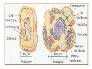
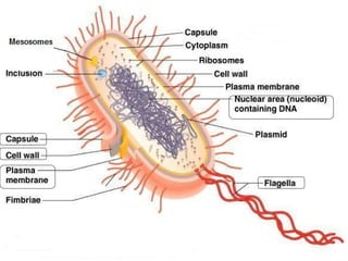
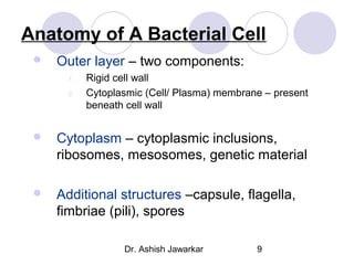
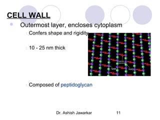
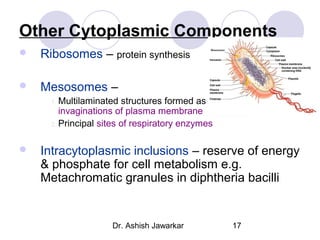
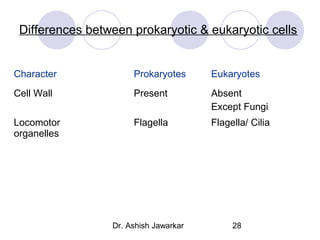
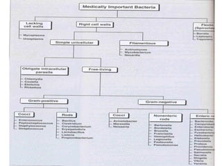
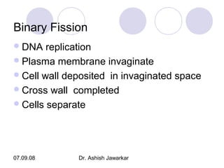
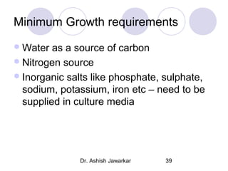

This document outlines the morphology and classification of bacteria, distinguishing between prokaryotic and eukaryotic cells. It details the structure of bacterial cells, including cell walls, membranes, and various organelles, and emphasizes the importance of cell wall characteristics in bacterial classification. Additionally, it touches on bacterial growth, reproduction through binary fission, and the minimum growth requirements for different bacterial types.


![CASE_PRESENTATION_ON_subdural_hematoma(SDH)[1 FINAL PPT]-1.pptx](https://cdn.slidesharecdn.com/ss_thumbnails/casepresentationonsubduralhematomasdh1finalppt-1-260129172522-d405d375-thumbnail.jpg?width=640&height=640&fit=bounds)


