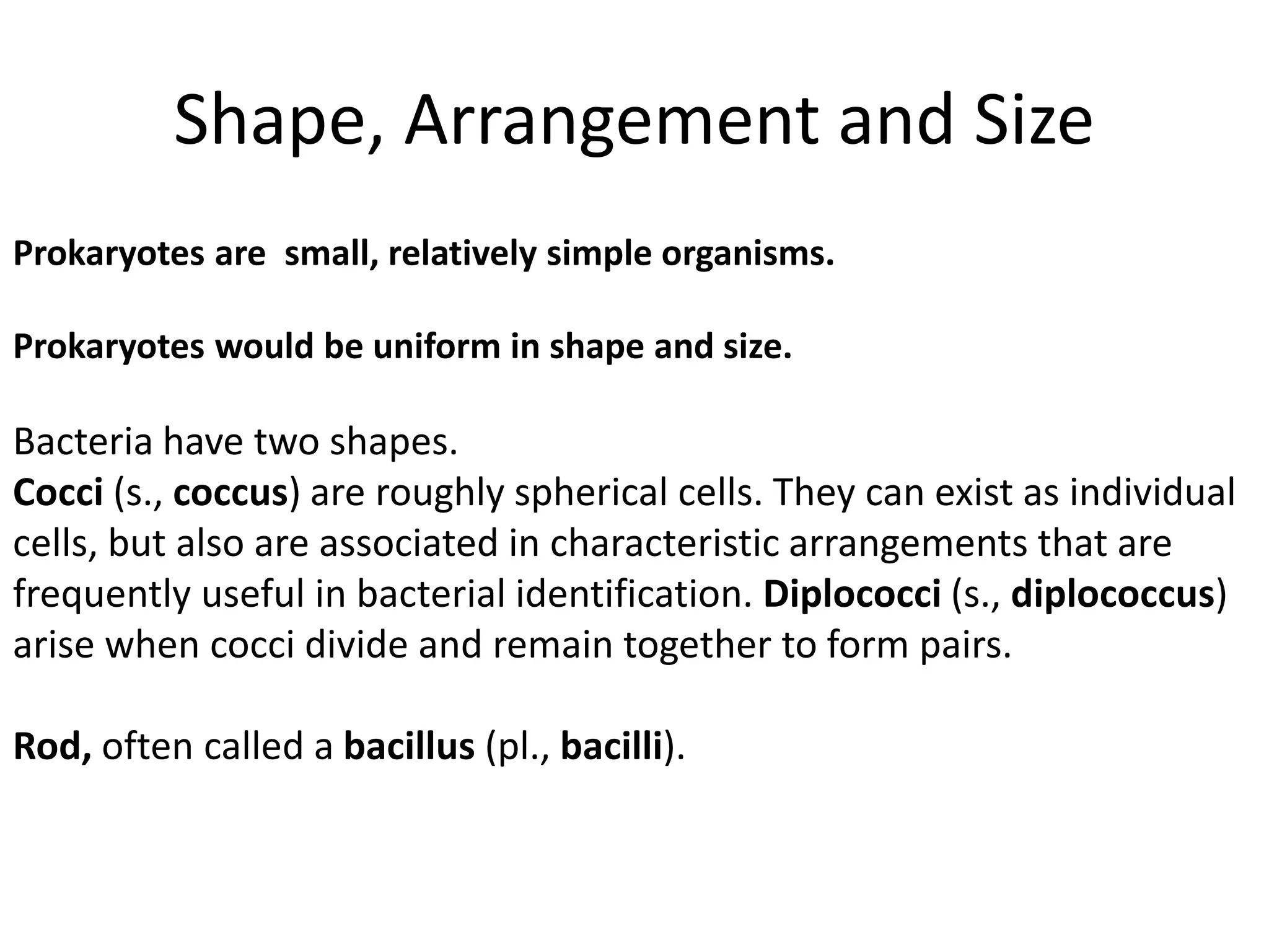
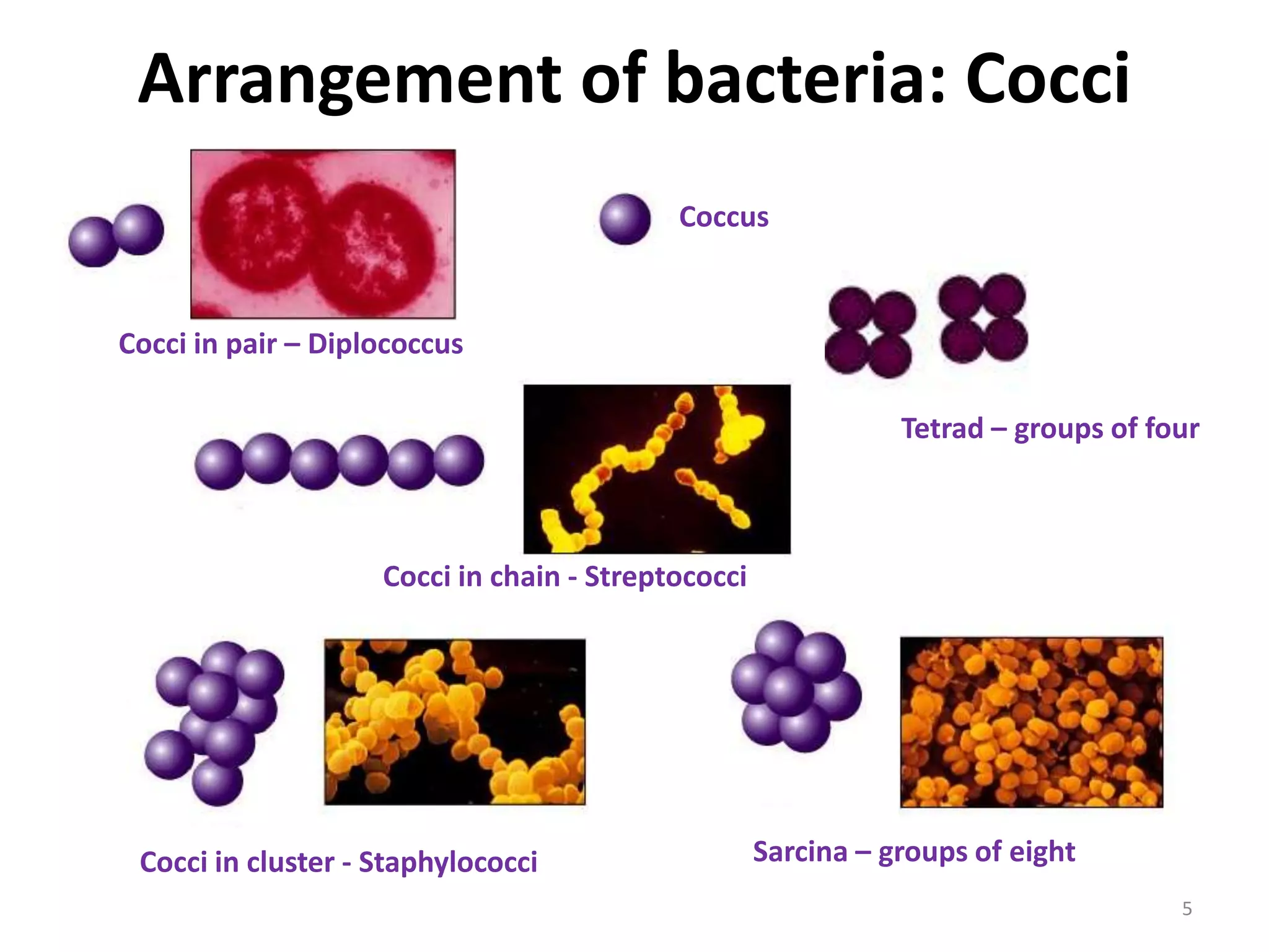
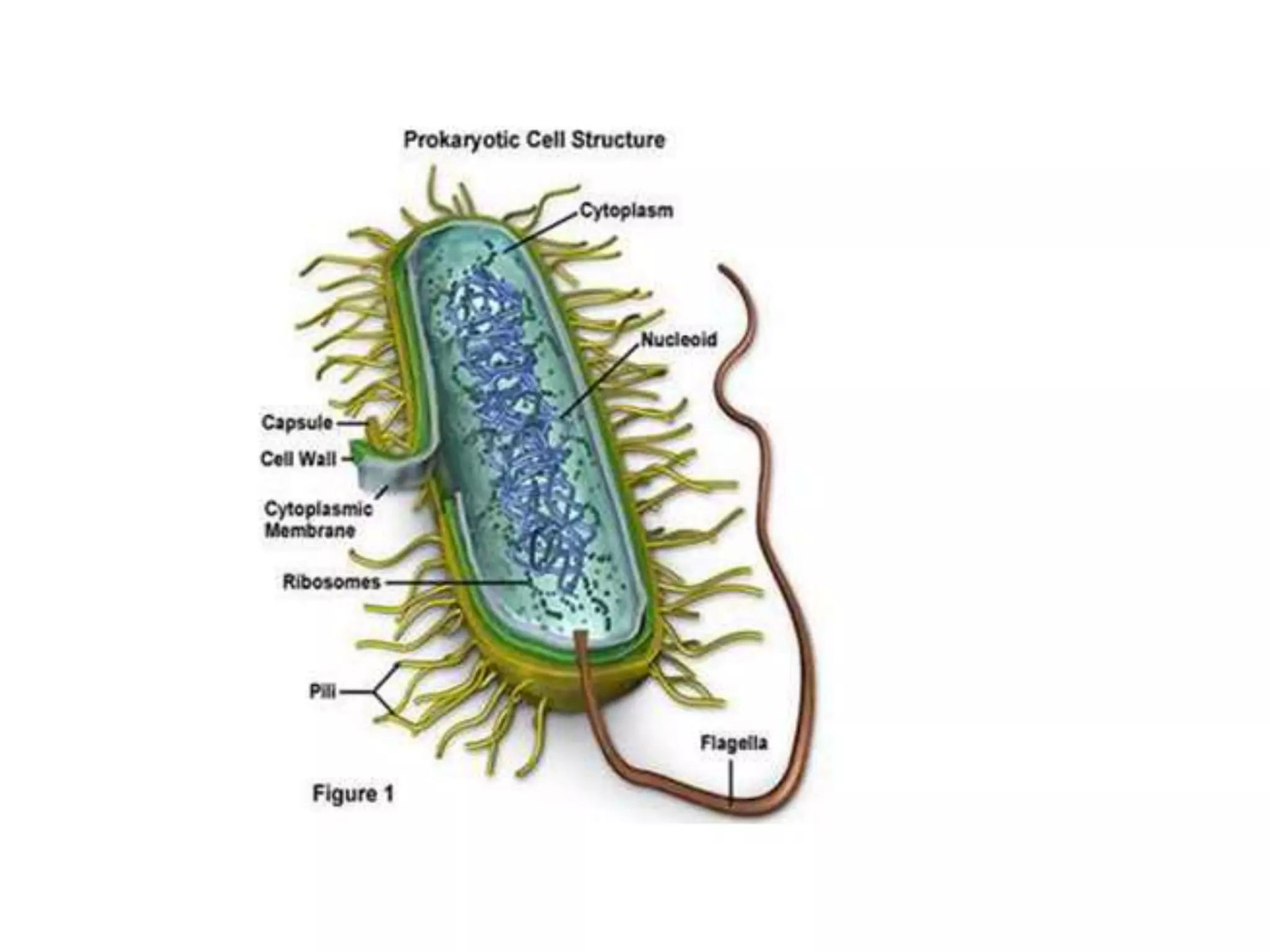
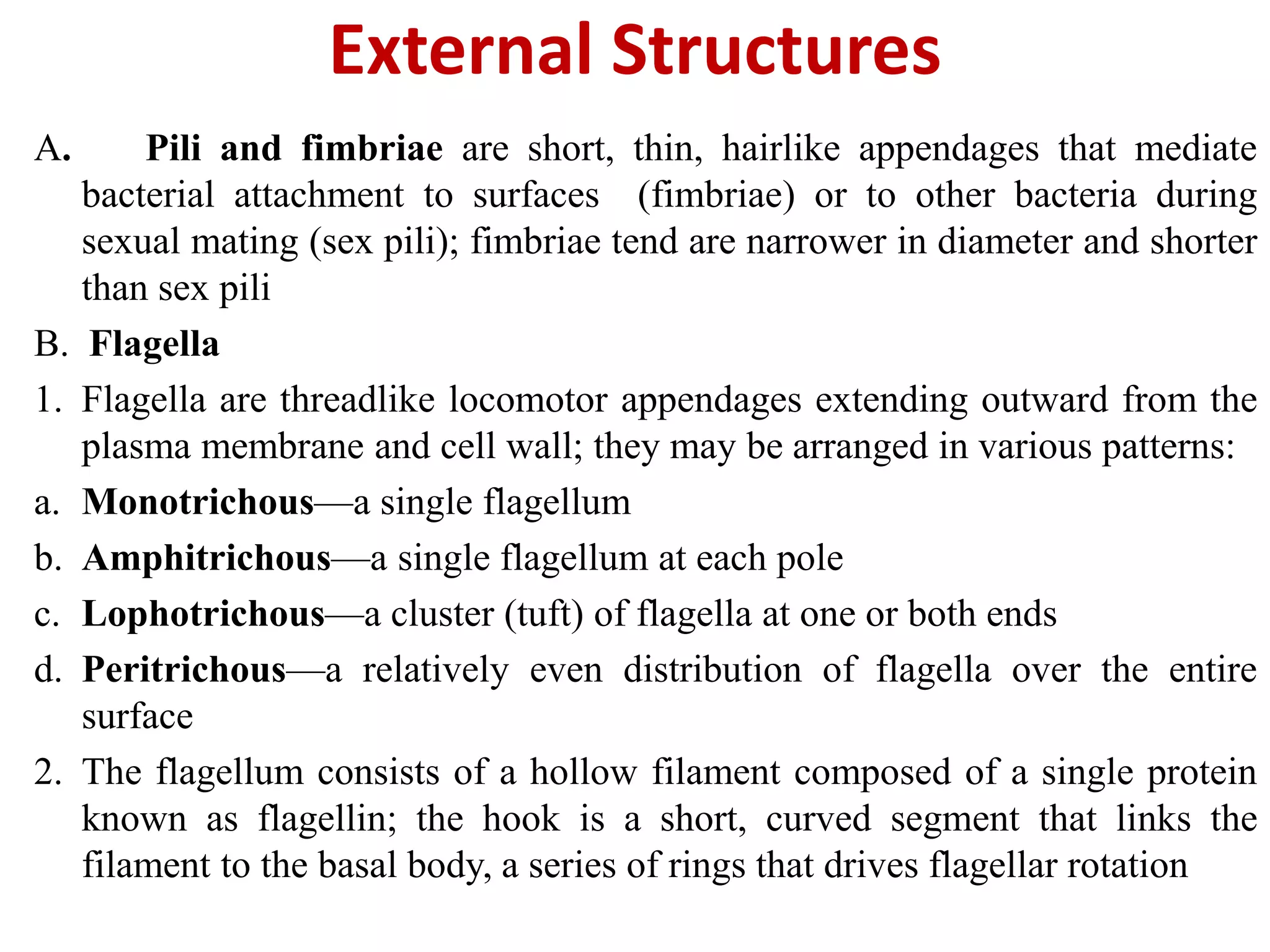
2. Shape, Arrangement and Size
Prokaryotes are small, relatively simple organisms.
Prokaryotes would be uniform in shape and size.
Bacteria have two shapes.
Cocci (s., coccus) are roughly spherical cells. They can exist as individual
cells, but also are associated in characteristic arrangements that are
frequently useful in bacterial identification. Diplococci (s., diplococcus)
arise when cocci divide and remain together to form pairs.
Rod, often called a bacillus (pl., bacilli).
4. Other shapes of bacteria
Comma shaped
Spirochetes
Spirilla
Most bacteria retain a particular shape; a few
are pleiomorphic
5. Arrangement of bacteria: Cocci
Cocci in pair – Diplococcus
Sarcina – groups of eight
Tetrad – groups of four
Cocci in chain - Streptococci
Cocci in cluster - Staphylococci
Coccus
5
6. Size
S. No. Bacteria Size
1 Escherichia coli 1.1 to 1.5 wide by
2.0to 6.0 µm
2 Mycoplasma 0.3µm in diameter
3 spirochaetes 500µm in length
4 Oscillatoria 7 µm in diameter
Streptococcus 0.8 to 1.0µm
5 Acanthurus
nigrofuscus (intestinal
600 by 80 µm
6 Epulopiscium
fishelsoni
600 by 80 µm
7 Thiomargrita
namibiensis
Nanobacteria range from around 0.2 µm to less than 0.05 µm
7. Prokaryotic Cell Organization
Plasma
Membrane Selectively permeable barrier, mechanical, boundary of cell, nutrient and waste, transport,
location of many metabolic, processes (respiration, photosynthesis), detection of
environmental cues for chemotaxis,
Gas Vacuole Buoyancy for floating in Aquatic environments.
Ribosomes Protein synthesis
Inclusion
Bodies Storage of carbon, phosphate, and other substances
Nucleiod Localization of genetic material (DNA)
Cell wall Gives bacteria shape and protection from lysis in dilute solutions
Periplasmic
Space Contains hydrolytic enzymes and binding. Proteins for nutrient processing and uptake
Capsule and
slime layer Resistance to phagocytosis, adherence to surfaces
Fimbriae and
pili Attachment to surfaces, bacterial mating
Flagella Movement
Endospore Survival under harsh environmental conditions
Bacterial and archaeal cell share common cell organization . Bacterial and archaeal
Cells are surrounded by a cell envelope with complex cell walls; they lack many
internal features common in eukaryotic cells
8.
9. Procaryotic Cell Membranes
• Membranes are an absolute requirement for all living
organisms
• Cell able to acquire nutrients and eliminate wastes but
they also have to maintain their interior in a constant,
highly organized state in the face of external changes.
• The cell envelope includes the plasma membrane, cell
wall, and other external layers of the cell .
10. •Bacterial Plasma Membranes
The plasma membrane serves several functions:
•It retains the cytoplasm and separates the cell from its environment.
• It serves as a selectively permeable barrier.
•It contains transport systems used for nutrient uptake, waste excretion, and pro
secretion .
•It is the location of a variety of crucial metabolic processes including respiratio
photosynthesis, lipid synthesis, and cell wall synthesis.
•It contains special receptor molecules that enable detection of and response to
chemicals in the surroundings
11. The fluid mosaic model of membrane structure
This model, proposed by Singer and Nicholson, states
that membranes are lipid bilayers with floating proteins.
Cell membranes are very thin (5–10 nm thick); the lipids
are amphipathic, having hydrophilic (interact with water)
head groups and long hydrophobic (insoluble in water) tails;
the head groups face out of the membrane while the tails are
buried in the membrane to form bilayers.
Two types of proteins are associated with the lipid bilayer of
the membrane: peripheral (loosely associated and easily
removed) and integral (embedded within the membrane
and not easily removed)
13. Bacterial lipids
• The plasma membrane of bacteria consists of a
phospholipid bilayer with hydrophilic surfaces
and a hydrophobic interior; bacterial
membranes lack sterols, but many contain
sterol-like molecules called hopanoids that
help stabilize the membrane
• Bacteria do not have membranous organelles,
but can have internal membrane systems with
specialized functions such as photosynthesis or
respiration
14. Bacterial Cell Walls
• The cell wall is a rigid structure that lies outside the
plasma membrane; it creates characteristic shapes for
the bacteria and protects from osmotic lysis and
toxins, often increasing pathogenicity.
• Overview of bacterial cell wall structure
– The cell walls of most bacteria contain
peptidoglycan (murein).
– The cell walls of gram-positive bacteria and gram-
negative bacteria differ greatly, but both have the
periplasmic space between the cell wall material
and the plasma membrane
16. Peptidoglycan structure
– Peptidoglycan is a polysaccharide polymer composed of two
sugar derivatives with peptide linkers; the polysaccharide
polymer is a linear chain of alternating N-acetylglucosamine
(NAG) and N-acetylmuramic acid(NAM) moieties
– Polysaccharide chains of peptidoglycan are cross-linked via a
peptide interbridge attached to the sugar backbone via a short
peptide chain; these peptides contain some amino acids not
found in proteins; cross-linking adds strength to the
peptidoglycan mesh
– Variations in peptidoglycan structure are seen in certain bacterial
groups and can be diagnostic
– Archaea do not have peptidoglycan, they have pseudomurein
17. They consist of a thick wall composed of many layers of
peptidoglycan and large amounts of teichoic acids .
Techoic acids are polymers with a glycerol and phosphate
backbone that span the cell wall and likely enhance its
structural stability .
The periplasmic space of gram-positive cells is usually thin
and contains only a few secreted proteins (exoenzymes).
Many gram-positive bacteria also have a layer of proteins
(S-layer proteins) on the outer.
Surface of the peptidoglycan that have a role in wall
synthesis and virulence.
Acid-fast bacteria include mycolic acids in their cell walls.
Gram-positive cell walls
19. Gram-negative cell walls
The gram-negative cell wall is more complex than the gram-
positive cell wall; and has a thin layer of peptidoglycan
surrounded by an outer membrane .
The periplasmic space is often wide and contains many different
proteins; some are involved with energy conservation or nutrient
acquisition .
The outer membrane is composed of lipids, lipoproteins, and
lipopolysaccharides (LPS); lipoprotein attaches the outer
membrane to the peptidoglycan .
LPS are large complex molecules composed of lipid A, core
polysaccharides, and O antigen carbohydrate side.
The thinner, less cross-linked peptidoglycan layer of gram-
negative bacteria does not retain the stain, and thus more readily
decolorized when treated with alcohol
21. Cell walls and osmotic protection
• The cell wall prevents swelling and lysis of bacteria in
hypotonic solutions; in hypertonic habitats, the plasma
membrane shrinks away from the cell wall in a process
known as plasmolysis.
• Bacteria without cell walls (by removal with lysozyme
or through peptidoglycan synthesis inhibition by
penicillin) called spheroplasts are osmotically sensitive.
• Mycoplasmas lack a cell wall and tend to be
pleomorphic.
23. Cell Envelope Layers Outside the Cell Wall
• Capsules and slime layers
• Capsules and slime layers (also known as glycocalyx) are
layers of polysaccharides lying outside the cell wall; they
protect the bacteria from phagocytosis, desiccation, viral
infection, and hydrophobic toxic materials such as
detergents; they also aid bacterial attachment to
surfaces and gliding motility
• Capsules are well organized, whereas slime layers are
diffuse and unorganized
24. S-layers
• S-layers are regularly structured layers of
protein or glycoprotein outside of the cell wall
• S-layers protect against ion and pH fluctuations,
osmotic stress, hydrolytic enzymes; can help
maintain cell shape and envelope rigidity,
promote cell adhesion, protect against host
defenses
• S-layers may be the only structural component
to many of the archaea
25. Bacterial Cytoplasm
A. The cytoplasmic matrix is the substance bounded by the plasma
membrane; it is often packed with ribosomes and inclusion
bodies
B. The prokaryotic cytoskeleton has homologs of the elements seen
in eukaryotes, filaments of actin, tubulin, and intermediate
filament proteins
C. Intracytoplasmic membranes are often involved in energy
metabolism in nitrifying and photosynthetic bacteria
26. Inclusions
• Many inclusions are granules of organic or inorganic material that
are stockpiled by the cell for future use; some are not bounded by a
membrane, but others are enclosed by a single-layered membrane .
• Storage inclusions include glycogen (carbon storage), poly--
hydroxybutyrate (carbon storage), polyphosphate granules (energy
and phosphorus storage), sulfur globules (wastes), and cyanophycin
granules (nitrogen storage in cyanobacteria)
• Carboxysomes are microcompartments that accumulate carbon
dioxide and the enzyme ribulose-1,5-bisphosophate carboxylase
• Gas vacuoles are composed of hollow protein sacs that are filled with
gases and used for buoyancy control in aquatic environments;
magnetosomes are magnetite granules that provide orientation in
the Earth's magnetic field
27. Microcompartments
• These are not used just for storing substances
for later use by the cell.
• Relatively large polyhedrom formed by one or
more different proteins.
• Best studied are the carboxysomes, found in
many of the cyanobacteria and other CO2
fixing bacteria.
28. Bacterial Ribosomes
• Ribosomes are the site of protein synthesis
(translation)
• They are complex structures consisting of
protein and rRNA (ribosomal RNA)
• Bacterial and archaeal ribosomes are 70S with 50S
and 30S subunits; although differences are apparent
in ribosomal protein and rRNA components,
ribosomes are similar across cells from all three
Domains (eukaryotic cells have 80S ribosomes)
29. • The procaryotic chromosome is located in an
irregularly shaped region called the nucleoid.
• Procaryotes contain a single circle of double-
stranded deoxyribonucleic acid (DNA).
• The nucleoid is visible in the light microscope
and electron microscope after staining with
the Feulgen stain, which specifically reacts
with DNA.
The Nucleoid
30. Plasmids
• Many bacteria possess plasmids in addition to
their chromo-some.
• These are double-stranded DNA molecules,
usually circular.
• Plasmid can exist and replicate independently of
the chromosome or may be integrated with it.
• Plasmids are not re-quired for host growth and
reproduction, although they may carry genes that
give their bacterial host a selective advantage.
31. External Structures
A. Pili and fimbriae are short, thin, hairlike appendages that mediate
bacterial attachment to surfaces (fimbriae) or to other bacteria during
sexual mating (sex pili); fimbriae tend are narrower in diameter and shorter
than sex pili
B. Flagella
1. Flagella are threadlike locomotor appendages extending outward from the
plasma membrane and cell wall; they may be arranged in various patterns:
a. Monotrichous—a single flagellum
b. Amphitrichous—a single flagellum at each pole
c. Lophotrichous—a cluster (tuft) of flagella at one or both ends
d. Peritrichous—a relatively even distribution of flagella over the entire
surface
2. The flagellum consists of a hollow filament composed of a single protein
known as flagellin; the hook is a short, curved segment that links the
filament to the basal body, a series of rings that drives flagellar rotation
32. Bacterial Motility and Chemotaxis
• A. Motility
1. Motility in prokaryotes is not aimless; responses are made to
temperature, light, oxygen, Osmotic pressure, and gravity;
chemotaxis is directed movement of bacteria either toward a
chemical attractant or away from a chemical repellent
2. Prokaryotic flagella rotate to create motion (like a propeller); the
direction of flagellar rotation determines the nature of bacterial
movement: counterclockwise rotation causes forward motion (called
a run) and clockwise rotation disrupts forward motion (resulting in a
tumble)
3. The basal body is the motor that drives the flagellum; it is powered
by a proton motive force
4. Prokaryotes can move by other mechanisms: in spirochetes, axial
fibrils cause movement by flexing and spinning; other
prokaryotes exhibit twitching or gliding motility, a mechanism
involving pili by which they move along solid surfaces with the help
of the slime layer
33. Chemotaxis
1. The concentrations of attractants and repellents are detected by
chemoreceptors in the periplasmic space or the plasma membrane
2. Directional travel toward a chemoattractant is caused by lowering the
frequency of tumbles (twiddles), thereby lengthening the runs when
traveling up the gradient, but allowing tumbling to occur at normal
frequency when traveling down the gradient
3.Directional travel away from a chemorepellent involves similar but
opposite responses
34. Bacterial Endospores
A. The bacterial endospore is a special, resistant, dormant structure
formed by some bacteria; it enables them to resist harsh
environmental conditions
B. Endospore structure is complex, consisting of an outer covering
called the exosporium, a spore coat beneath the exosporium, the
cortex beneath the spore coat, and the spore cell wall, which is
inside the cortex and surrounds the core
C. Endospore formation (sporulation) normally commences when
growth ceases because of lack of nutrients; it is a complex,
multistage process
D. Transformation of dormant endospores into active vegetative cells is
also a complex, multistage process that includes activation
(preparation) of the endospore, germination (breaking of the
endospore’s dormant state), and outgrowth (emergence of the new
vegetative cell)