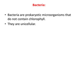
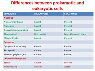
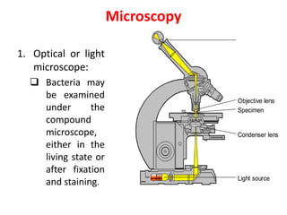
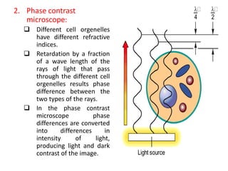
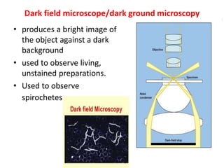
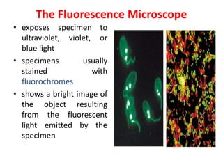
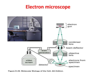
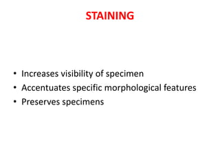
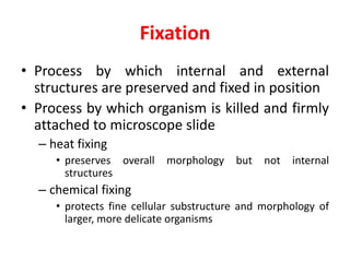
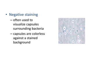
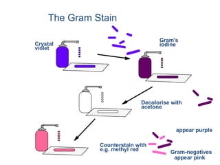
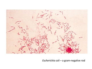
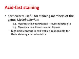
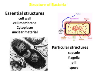
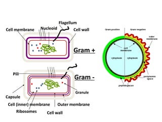
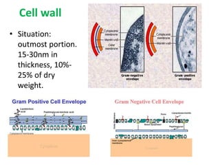
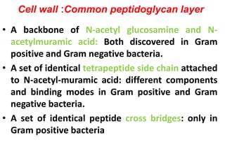
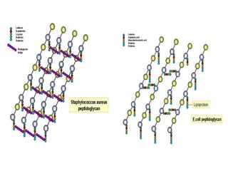
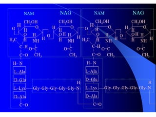
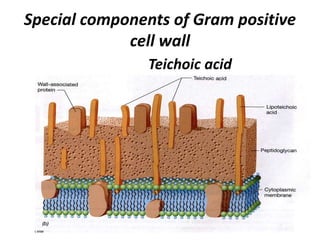
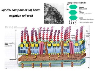
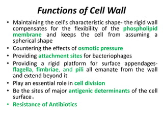
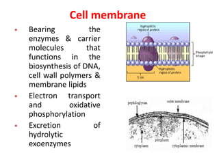
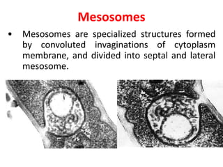
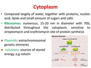
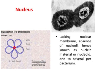
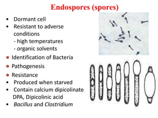
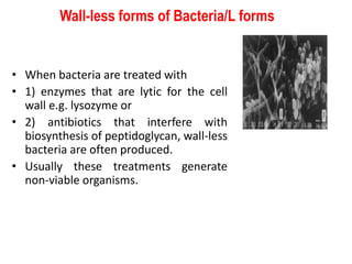
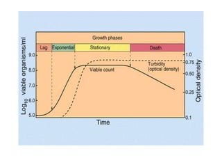
Introduction to bacteriology, morphology & staining
DR ADILRAZA
DEPARTMENTOF MICROBIOLOGY,
JNMC.AMU
Medical Microbiology:
• It is the study of microbes that infect humans,
the disease they cause, their diagnosis,
prevention and treatment. It also deals with
the response of the human host to microbial
and their antigens.
• Branches of Microbiology:
– Bacteriology
– Virology
– Mycology
– Immunology
– Parasitology
– Mycobacteriology
Bacteria:
• Bacteria are prokaryotic microorganisms that
do not contain chlorophyll.
• They are unicellular.
Differences between prokaryotic and
eukaryotic cells
CHARACTER PROKARYOTES EUKARYOTES
NUCLEUS
Nuclear membrane Absent Present
Nucleolus Absent Present
Deoxyribonucleoprotein Absent Present
Chromosome One circular More than one ( linear)
Mitotic division Absent Present
Cytoplasm
Cytoplasmic streaming Absent Present
Pinocytosis Absent Present
Mitocho, golgi App, ER Absent Present
Chemical composition
Sterols Absent Present
Muramic acid Present Absent
Size of Bacteria
• The unit of measurement used in bacteriology is the
MICRON (micrometre, µM).
• 1 micron (µ), or micrometre (µM)= 10-6 M or one
thousands of a millimetre.
• 1 millimicron (mµ ) or nanometre (nm)= 10-9M= one
thousands of a micron or one millionth of a
millimetre.
• 1 Angstrom unit (Å)= one tenth of a nanometre.
Cocci: sphere, 1μm
Bacilli: rods , 0.5-1 μm in width -3 μm in length
Spiral bacteria: 1~3 μm in length and 0.3-0.6 μm in width
Microscopy
1. Optical or light
microscope:
 Bacteria may
be examined
under the
compound
microscope,
either in the
living state or
after fixation
and staining.
Ocular lens
Specimen
Objective lens
Light source
Condenser lens
2. Phase contrast
microscope:
 Different cell orgenelles
have different refractive
indices.
 Retardation by a fraction
of a wave length of the
rays of light that pass
through the different cell
orgenelles results phase
difference between the
two types of the rays.
 In the phase contrast
microscope phase
differences are converted
into differences in
intensity of light,
producing light and dark
contrast of the image.


2
Light source


4
• produces a bright image of
the object against a dark
background
• used to observe living,
unstained preparations.
• Used to observe
spirochetes.
Dark field microscope/dark ground microscopy
The Fluorescence Microscope
• exposes specimen to
ultraviolet, violet, or
blue light
• specimens usually
stained with
fluorochromes
• shows a bright image of
the object resulting
from the fluorescent
light emitted by the
specimen
Electron microscope
STAINING
• Increases visibility of specimen
• Accentuates specific morphological features
• Preserves specimens
Fixation
• Process by which internal and external
structures are preserved and fixed in position
• Process by which organism is killed and firmly
attached to microscope slide
– heat fixing
• preserves overall morphology but not internal
structures
– chemical fixing
• protects fine cellular substructure and morphology of
larger, more delicate organisms
Simple Staining
• Simple staining
– a single staining agent is used
– basic dyes are frequently used
• dyes with positive charges
• e.g., crystal violet
• Negative staining
– often used to
visualize capsules
surrounding bacteria
– capsules are colorless
against a stained
background
Differential Staining
• divides microorganisms into groups based on
their staining properties
– e.g., Gram stain
– e.g., acid-fast stain
18
Gram staining
• most widely used differential staining
procedure
• divides Bacteria into two groups based on
differences in cell wall structure
Crystal
violet
Gram's
iodine
Decolorise with
acetone
Counterstain with
e.g. methyl red
appear purple
Gram-negatives
appear pink
The Gram Stain
Escherichia coli – a gram-negative rod
Acid-fast staining
• particularly useful for staining members of the
genus Mycobacterium
e.g., Mycobacterium tuberculosis – causes tuberculosis
e.g., Mycobacterium leprae – causes leprosy
– high lipid content in cell walls is responsible for
their staining characteristics
Structure of Bacteria
Particular structures
capsule
flagella
pili
spore
Essential structures
cell wall
cell membrane
Cytoplasm
nuclear material
Gram +
Gram -
Cell wall
Cell (inner) membrane Outer membrane
Ribosomes
Granule
Cell wall
NucleoidCell membrane
Capsule
Flagellum
Pili
Cell wall
• Situation:
outmost portion.
15-30nm in
thickness, 10%-
25% of dry
weight.
Cell wall :Common peptidoglycan layer
• A backbone of N-acetyl glucosamine and N-
acetylmuramic acid: Both discovered in Gram
positive and Gram negative bacteria.
• A set of identical tetrapeptide side chain attached
to N-acetyl-muramic acid: different components
and binding modes in Gram positive and Gram
negative bacteria.
• A set of identical peptide cross bridges: only in
Gram positive bacteria
Special components of Gram positive
cell wall
Teichoic acid
Special components of Gram
negative cell wall
Functions of Cell Wall
• Maintaining the cell's characteristic shape- the rigid wall
compensates for the flexibility of the phospholipid
membrane and keeps the cell from assuming a
spherical shape
• Countering the effects of osmotic pressure
• Providing attachment sites for bacteriophages
• Providing a rigid platform for surface appendages-
flagella, fimbriae, and pili all emanate from the wall
and extend beyond it
• Play an essential role in cell division
• Be the sites of major antigenic determinants of the cell
surface。
• Resistance of Antibiotics
Cell membrane
• Bearing the
enzymes & carrier
molecules that
functions in the
biosynthesis of DNA,
cell wall polymers &
membrane lipids
• Electron transport
and oxidative
phosphorylation
• Excretion of
hydrolytic
exoenzymes
Mesosomes
• Mesosomes are specialized structures formed
by convoluted invaginations of cytoplasm
membrane, and divided into septal and lateral
mesosome.
Cytoplasm
• Composed largely of water, together with proteins, nucleic
acid, lipids and small amount of sugars and salts
• Ribosomes: numerous, 15-20 nm in diameter with 70S;
distributed throughout the cytoplasm; sensitive to
streptomycin and erythromycin site of protein synthesis
 Plasmids: extrachromosomal
genetic elements
 Inclusions: sources of stored
energy, e,g volutin
Nucleus
• Lacking nuclear
membrane, absence
of nucleoli, hence
known as nucleic
material or nucleoid,
one to several per
bacterium.
Capsules and slime layers
• Many bacteria secrete a viscid material
around the cell surface, when this is organized
into a shapely defined structure, as in
pneumococcus, (Capsule).
• When is loosely attached (slime layer).
• Capsule is antigenic in nature.
Flagella
Monotrichate/Amphitrichate/Lophotrichate/Peritrichate
 Identification of Bacteria
 Pathogenesis
 Motility of bacteria
 Made up of flagellin
Pili
• Pili are hair-like projections of the
cell , They are known to be
receptors for certain bacterial
viruses. Chemical nature is pilin
• Classification and Function
a. Common pili or fimbriae: fine ,
rigid numerous, related to
bacterial adhesion
b. Sex pili: longer and coarser,
only 1-4, related to bacterial
conjugation
Endospores (spores)
• Dormant cell
• Resistant to adverse
conditions
- high temperatures
- organic solvents
 Identification of Bacteria
 Pathogenesis
 Resistance
• Produced when starved
• Contain calcium dipicolinate
DPA, Dipicolinic acid
• Bacillus and Clostridium
Wall-less forms of Bacteria/L forms
• When bacteria are treated with
• 1) enzymes that are lytic for the cell
wall e.g. lysozyme or
• 2) antibiotics that interfere with
biosynthesis of peptidoglycan, wall-less
bacteria are often produced.
• Usually these treatments generate
non-viable organisms.
Bacterial growth curve
• Lag phase
• Log (logarithmic) or exponential phase
• Stationary phase
• Phase of decline
Phases of Growth
• Lag
– Adapt to nutrients
• Log
– Active growth
• Stationary
– Death = Growth rate
• Death
– Nutrients consumed
– pH too low (why?)
• Optimize curves in production
Points to remember:
• Staining
• Fixation
• CW of bacteria
– Techoic acid
– LPS
• Classification of flagella
• Spores
• Pili
• Bacterial growth curve
Introduction to Bacteriology, Morphology & staining

Introduction to Bacteriology, Morphology & staining

  • 1.
    Introduction to bacteriology,morphology & staining DR ADILRAZA DEPARTMENTOF MICROBIOLOGY, JNMC.AMU
  • 2.
    Medical Microbiology: • Itis the study of microbes that infect humans, the disease they cause, their diagnosis, prevention and treatment. It also deals with the response of the human host to microbial and their antigens.
  • 3.
    • Branches ofMicrobiology: – Bacteriology – Virology – Mycology – Immunology – Parasitology – Mycobacteriology
  • 4.
    Bacteria: • Bacteria areprokaryotic microorganisms that do not contain chlorophyll. • They are unicellular.
  • 5.
    Differences between prokaryoticand eukaryotic cells CHARACTER PROKARYOTES EUKARYOTES NUCLEUS Nuclear membrane Absent Present Nucleolus Absent Present Deoxyribonucleoprotein Absent Present Chromosome One circular More than one ( linear) Mitotic division Absent Present Cytoplasm Cytoplasmic streaming Absent Present Pinocytosis Absent Present Mitocho, golgi App, ER Absent Present Chemical composition Sterols Absent Present Muramic acid Present Absent
  • 6.
    Size of Bacteria •The unit of measurement used in bacteriology is the MICRON (micrometre, µM). • 1 micron (µ), or micrometre (µM)= 10-6 M or one thousands of a millimetre. • 1 millimicron (mµ ) or nanometre (nm)= 10-9M= one thousands of a micron or one millionth of a millimetre. • 1 Angstrom unit (Å)= one tenth of a nanometre.
  • 7.
    Cocci: sphere, 1μm Bacilli:rods , 0.5-1 μm in width -3 μm in length Spiral bacteria: 1~3 μm in length and 0.3-0.6 μm in width
  • 8.
    Microscopy 1. Optical orlight microscope:  Bacteria may be examined under the compound microscope, either in the living state or after fixation and staining. Ocular lens Specimen Objective lens Light source Condenser lens
  • 9.
    2. Phase contrast microscope: Different cell orgenelles have different refractive indices.  Retardation by a fraction of a wave length of the rays of light that pass through the different cell orgenelles results phase difference between the two types of the rays.  In the phase contrast microscope phase differences are converted into differences in intensity of light, producing light and dark contrast of the image.  2 Light source  4
  • 10.
    • produces abright image of the object against a dark background • used to observe living, unstained preparations. • Used to observe spirochetes. Dark field microscope/dark ground microscopy
  • 11.
    The Fluorescence Microscope •exposes specimen to ultraviolet, violet, or blue light • specimens usually stained with fluorochromes • shows a bright image of the object resulting from the fluorescent light emitted by the specimen
  • 12.
  • 13.
    STAINING • Increases visibilityof specimen • Accentuates specific morphological features • Preserves specimens
  • 14.
    Fixation • Process bywhich internal and external structures are preserved and fixed in position • Process by which organism is killed and firmly attached to microscope slide – heat fixing • preserves overall morphology but not internal structures – chemical fixing • protects fine cellular substructure and morphology of larger, more delicate organisms
  • 15.
    Simple Staining • Simplestaining – a single staining agent is used – basic dyes are frequently used • dyes with positive charges • e.g., crystal violet
  • 16.
    • Negative staining –often used to visualize capsules surrounding bacteria – capsules are colorless against a stained background
  • 17.
    Differential Staining • dividesmicroorganisms into groups based on their staining properties – e.g., Gram stain – e.g., acid-fast stain
  • 18.
    18 Gram staining • mostwidely used differential staining procedure • divides Bacteria into two groups based on differences in cell wall structure
  • 19.
    Crystal violet Gram's iodine Decolorise with acetone Counterstain with e.g.methyl red appear purple Gram-negatives appear pink The Gram Stain
  • 20.
    Escherichia coli –a gram-negative rod
  • 21.
    Acid-fast staining • particularlyuseful for staining members of the genus Mycobacterium e.g., Mycobacterium tuberculosis – causes tuberculosis e.g., Mycobacterium leprae – causes leprosy – high lipid content in cell walls is responsible for their staining characteristics
  • 22.
    Structure of Bacteria Particularstructures capsule flagella pili spore Essential structures cell wall cell membrane Cytoplasm nuclear material
  • 23.
    Gram + Gram - Cellwall Cell (inner) membrane Outer membrane Ribosomes Granule Cell wall NucleoidCell membrane Capsule Flagellum Pili
  • 24.
    Cell wall • Situation: outmostportion. 15-30nm in thickness, 10%- 25% of dry weight.
  • 25.
    Cell wall :Commonpeptidoglycan layer • A backbone of N-acetyl glucosamine and N- acetylmuramic acid: Both discovered in Gram positive and Gram negative bacteria. • A set of identical tetrapeptide side chain attached to N-acetyl-muramic acid: different components and binding modes in Gram positive and Gram negative bacteria. • A set of identical peptide cross bridges: only in Gram positive bacteria
  • 28.
    Special components ofGram positive cell wall Teichoic acid
  • 29.
    Special components ofGram negative cell wall
  • 30.
    Functions of CellWall • Maintaining the cell's characteristic shape- the rigid wall compensates for the flexibility of the phospholipid membrane and keeps the cell from assuming a spherical shape • Countering the effects of osmotic pressure • Providing attachment sites for bacteriophages • Providing a rigid platform for surface appendages- flagella, fimbriae, and pili all emanate from the wall and extend beyond it • Play an essential role in cell division • Be the sites of major antigenic determinants of the cell surface。 • Resistance of Antibiotics
  • 31.
    Cell membrane • Bearingthe enzymes & carrier molecules that functions in the biosynthesis of DNA, cell wall polymers & membrane lipids • Electron transport and oxidative phosphorylation • Excretion of hydrolytic exoenzymes
  • 32.
    Mesosomes • Mesosomes arespecialized structures formed by convoluted invaginations of cytoplasm membrane, and divided into septal and lateral mesosome.
  • 33.
    Cytoplasm • Composed largelyof water, together with proteins, nucleic acid, lipids and small amount of sugars and salts • Ribosomes: numerous, 15-20 nm in diameter with 70S; distributed throughout the cytoplasm; sensitive to streptomycin and erythromycin site of protein synthesis  Plasmids: extrachromosomal genetic elements  Inclusions: sources of stored energy, e,g volutin
  • 34.
    Nucleus • Lacking nuclear membrane,absence of nucleoli, hence known as nucleic material or nucleoid, one to several per bacterium.
  • 35.
    Capsules and slimelayers • Many bacteria secrete a viscid material around the cell surface, when this is organized into a shapely defined structure, as in pneumococcus, (Capsule). • When is loosely attached (slime layer). • Capsule is antigenic in nature.
  • 36.
    Flagella Monotrichate/Amphitrichate/Lophotrichate/Peritrichate  Identification ofBacteria  Pathogenesis  Motility of bacteria  Made up of flagellin
  • 38.
    Pili • Pili arehair-like projections of the cell , They are known to be receptors for certain bacterial viruses. Chemical nature is pilin • Classification and Function a. Common pili or fimbriae: fine , rigid numerous, related to bacterial adhesion b. Sex pili: longer and coarser, only 1-4, related to bacterial conjugation
  • 39.
    Endospores (spores) • Dormantcell • Resistant to adverse conditions - high temperatures - organic solvents  Identification of Bacteria  Pathogenesis  Resistance • Produced when starved • Contain calcium dipicolinate DPA, Dipicolinic acid • Bacillus and Clostridium
  • 40.
    Wall-less forms ofBacteria/L forms • When bacteria are treated with • 1) enzymes that are lytic for the cell wall e.g. lysozyme or • 2) antibiotics that interfere with biosynthesis of peptidoglycan, wall-less bacteria are often produced. • Usually these treatments generate non-viable organisms.
  • 41.
    Bacterial growth curve •Lag phase • Log (logarithmic) or exponential phase • Stationary phase • Phase of decline
  • 43.
    Phases of Growth •Lag – Adapt to nutrients • Log – Active growth • Stationary – Death = Growth rate • Death – Nutrients consumed – pH too low (why?) • Optimize curves in production
  • 44.
    Points to remember: •Staining • Fixation • CW of bacteria – Techoic acid – LPS • Classification of flagella • Spores • Pili • Bacterial growth curve