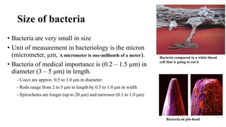
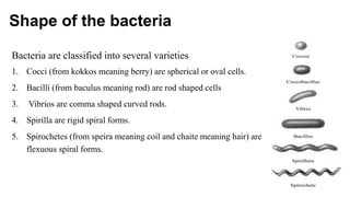
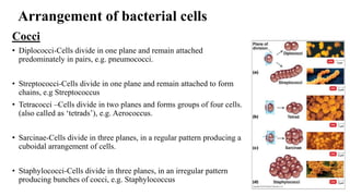
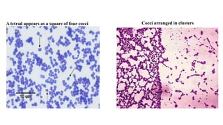
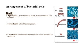
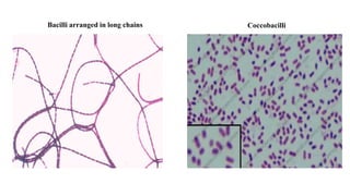
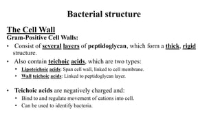
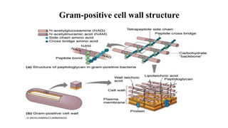
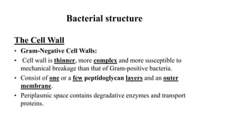
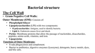
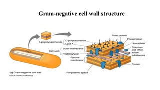
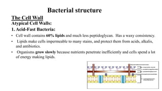
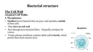
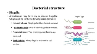
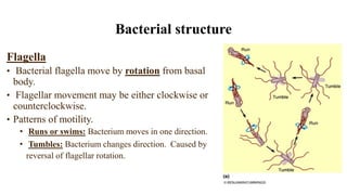
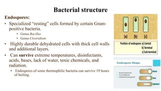
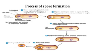
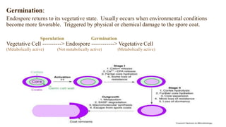
BACTERIA
(morphology and classification)
By
SAMIRA FATTAH HAMID
Ph.D. Medical Bacteriology
College of Health Sciences
Hawler Medical University
Bacteria
• Widely distributed. It can be found in soil, air, water, and living
bodies.
• Some bacteria can cause diseases for human, animals and plants.
• Some bacteria are harmless (i.e. live in human bodies as normal flora)
• The major characteristics of bacteria are based on their size, shape and
arrangements
Size of bacteria
• Bacteria are very small in size
• Unit of measurement in bacteriology is the micron
(micrometer, µm, A micrometer is one-millionth of a meter).
• Bacteria of medical importance is (0.2 – 1.5 µm) in
diameter (3 – 5 µm) in length.
- Cocci are approx. 0.5 to 1.0 μm in diameter.
- Rods range from 2 to 5 μm in length by 0.5 to 1.0 μm in width
- Spirochetes are longer (up to 20 μm) and narrower (0.1 to 1.0 μm)
Bacteria compared to a white blood
cell that is going to eat it
Shape of the bacteria
Bacteria are classified into several varieties
1. Cocci (from kokkos meaning berry) are spherical or oval cells.
2. Bacilli (from baculus meaning rod) are rod shaped cells
3. Vibrios are comma shaped curved rods.
4. Spirilla are rigid spiral forms.
5. Spirochetes (from speira meaning coil and chaite meaning hair) are
flexuous spiral forms.
Arrangement of bacterial cells
Cocci
• Diplococci-Cells divide in one plane and remain attached
predominately in pairs, e.g. pneumococci.
• Streptococci-Cells divide in one plane and remain attached to form
chains, e.g Streptococcus
• Tetracocci –Cells divide in two planes and forms groups of four cells.
(also called as ‘tetrads’), e.g. Aerococcus.
• Sarcinae-Cells divide in three planes, in a regular pattern producing a
cuboidal arrangement of cells.
• Staphylococci-Cells divide in three planes, in an irregular pattern
producing bunches of cocci, e.g. Staphylococcus
A tetrad appears as a square of four cocci Cocci arranged in clusters
Arrangement of bacterial cells
Bacilli
• Diplobacilli: A pair of attached bacilli. Remain attached after
dividing.
• Streptobacilli: Chainlike arrangement.
• Coccobacilli: Intermediate shape between coccus and bacillus.
Oval rods.
Bacilli arranged in long chains Coccobacilli
Bacterial structure
The Cell Wall
Gram-Positive Cell Walls:
• Consist of several layers of peptidoglycan, which form a thick, rigid
structure.
• Also contain teichoic acids, which are two types:
• Lipoteichoic acids: Span cell wall, linked to cell membrane.
• Wall teichoic acids: Linked to peptidoglycan layer.
• Teichoic acids are negatively charged and:
• Bind to and regulate movement of cations into cell.
• Can be used to identify bacteria.
Gram-positive cell wall structure
The Cell Wall
• Gram-Negative Cell Walls:
• Cell wall is thinner, more complex and more susceptible to
mechanical breakage than that of Gram-positive bacteria.
• Consist of one or a few peptidoglycan layers and an outer
membrane.
• Periplasmic space contains degradative enzymes and transport
proteins.
Bacterial structure
The Cell Wall
• Gram-Negative Cell Walls:
Outer Membrane (OM): Consists of:
• Phospholipid bilayer
• Lipopolysaccharides (LPS) with two components:
• O polysaccharides: Antigens, used to identify bacteria.
• Lipid A: Endotoxin causes fever and shock.
• Porins: Membrane proteins that allow the passage of nucleotides, disaccharides,
peptides, amino acids, vitamins, and iron.
• Lipoproteins
Functions of Outer Membrane:
• Evade phagocytosis and complement.
• Barrier to antibiotics, digestive enzymes (lysozyme), detergents, heavy metals, dyes,
and bile salts.
Bacterial structure
Gram-negative cell wall structure
The Cell Wall
Atypical Cell Walls:
1. Acid-Fast Bacteria:
• Cell wall contains 60% lipids and much less peptidoglycan. Has a waxy consistency.
• Lipids make cells impermeable to many stains, and protect them from acids, alkalis,
and antibiotics.
• Organisms grow slowly because nutrients penetrate inefficiently and cells spend a lot
of energy making lipids.
Bacterial structure
The Cell Wall
Atypical Cell Walls:
2. Mycoplasmas:
• Smallest known bacteria that can grow and reproduce outside
of host cells.
• They have no cell wall.
• Pass through most bacterial filters. Originally mistaken for
viruses.
• Unique plasma membrane contains lipids called sterols, which
protect them from osmotic lysis.
Bacterial structure
• Flagella
• A bacterium may have one or several flagella,
which can be in the following arrangements:
• Monotrichous: Single polar flagellum at one end.
• Lophotrichous: Two or more flagella at one end.
• Amphitrichous: Two or more polar flagella, on
each end.
• Peritrichous: Many flagella over entire cell
surface.
Bacterial structure
Flagella
• Bacterial flagella move by rotation from basal
body.
• Flagellar movement may be either clockwise or
counterclockwise.
• Patterns of motility.
• Runs or swims: Bacterium moves in one direction.
• Tumbles: Bacterium changes direction. Caused by
reversal of flagellar rotation.
Bacterial structure
• Axial Filaments (Endoflagella):
• Bundles of fibers that are anchored at ends of the cell beneath the
outer sheath.
• Spiral around the cells.
• Have similar structure to flagella.
• Rotation of endoflagella produces a corkscrew motion.
• May enable bacteria to penetrate body tissues.
• Found in spirochetes.
Bacterial structure
Endospores:
• Specialized “resting” cells formed by certain Gram-
positive bacteria.
• Genus Bacillus
• Genus Clostridium
• Highly durable dehydrated cells with thick cell walls
and additional layers.
• Can survive extreme temperatures, disinfectants,
acids, bases, lack of water, toxic chemicals, and
radiation.
• Endospores of some thermophilic bacteria can survive 19 hours
of boiling.
Bacterial structure
Process of spore formation
Sporulation
• May be part of normal life cycle or triggered by adverse environmental
conditions.
• Endospores do not carry out metabolic reactions, unlike normal
vegetative cells.
• Endospores can remain dormant for thousands of years.
Germination:
Endospore returns to its vegetative state. Usually occurs when environmental conditions
become more favorable. Triggered by physical or chemical damage to the spore coat.
Sporulation Germination
Vegetative Cell ----------> Endospore ------------> Vegetative Cell
(Metabolically active) (Not metabolically active) (Metabolically active)
Next lecture
BACTERIA
growth and metabolism

Bacteria morphology and Classification

  • 1.
    BACTERIA (morphology and classification) By SAMIRAFATTAH HAMID Ph.D. Medical Bacteriology College of Health Sciences Hawler Medical University
  • 2.
    Bacteria • Widely distributed.It can be found in soil, air, water, and living bodies. • Some bacteria can cause diseases for human, animals and plants. • Some bacteria are harmless (i.e. live in human bodies as normal flora) • The major characteristics of bacteria are based on their size, shape and arrangements
  • 3.
    Size of bacteria •Bacteria are very small in size • Unit of measurement in bacteriology is the micron (micrometer, µm, A micrometer is one-millionth of a meter). • Bacteria of medical importance is (0.2 – 1.5 µm) in diameter (3 – 5 µm) in length. - Cocci are approx. 0.5 to 1.0 μm in diameter. - Rods range from 2 to 5 μm in length by 0.5 to 1.0 μm in width - Spirochetes are longer (up to 20 μm) and narrower (0.1 to 1.0 μm) Bacteria compared to a white blood cell that is going to eat it
  • 4.
    Shape of thebacteria Bacteria are classified into several varieties 1. Cocci (from kokkos meaning berry) are spherical or oval cells. 2. Bacilli (from baculus meaning rod) are rod shaped cells 3. Vibrios are comma shaped curved rods. 4. Spirilla are rigid spiral forms. 5. Spirochetes (from speira meaning coil and chaite meaning hair) are flexuous spiral forms.
  • 5.
    Arrangement of bacterialcells Cocci • Diplococci-Cells divide in one plane and remain attached predominately in pairs, e.g. pneumococci. • Streptococci-Cells divide in one plane and remain attached to form chains, e.g Streptococcus • Tetracocci –Cells divide in two planes and forms groups of four cells. (also called as ‘tetrads’), e.g. Aerococcus. • Sarcinae-Cells divide in three planes, in a regular pattern producing a cuboidal arrangement of cells. • Staphylococci-Cells divide in three planes, in an irregular pattern producing bunches of cocci, e.g. Staphylococcus
  • 6.
    A tetrad appearsas a square of four cocci Cocci arranged in clusters
  • 7.
    Arrangement of bacterialcells Bacilli • Diplobacilli: A pair of attached bacilli. Remain attached after dividing. • Streptobacilli: Chainlike arrangement. • Coccobacilli: Intermediate shape between coccus and bacillus. Oval rods.
  • 8.
    Bacilli arranged inlong chains Coccobacilli
  • 9.
    Bacterial structure The CellWall Gram-Positive Cell Walls: • Consist of several layers of peptidoglycan, which form a thick, rigid structure. • Also contain teichoic acids, which are two types: • Lipoteichoic acids: Span cell wall, linked to cell membrane. • Wall teichoic acids: Linked to peptidoglycan layer. • Teichoic acids are negatively charged and: • Bind to and regulate movement of cations into cell. • Can be used to identify bacteria.
  • 10.
  • 11.
    The Cell Wall •Gram-Negative Cell Walls: • Cell wall is thinner, more complex and more susceptible to mechanical breakage than that of Gram-positive bacteria. • Consist of one or a few peptidoglycan layers and an outer membrane. • Periplasmic space contains degradative enzymes and transport proteins. Bacterial structure
  • 12.
    The Cell Wall •Gram-Negative Cell Walls: Outer Membrane (OM): Consists of: • Phospholipid bilayer • Lipopolysaccharides (LPS) with two components: • O polysaccharides: Antigens, used to identify bacteria. • Lipid A: Endotoxin causes fever and shock. • Porins: Membrane proteins that allow the passage of nucleotides, disaccharides, peptides, amino acids, vitamins, and iron. • Lipoproteins Functions of Outer Membrane: • Evade phagocytosis and complement. • Barrier to antibiotics, digestive enzymes (lysozyme), detergents, heavy metals, dyes, and bile salts. Bacterial structure
  • 13.
  • 14.
    The Cell Wall AtypicalCell Walls: 1. Acid-Fast Bacteria: • Cell wall contains 60% lipids and much less peptidoglycan. Has a waxy consistency. • Lipids make cells impermeable to many stains, and protect them from acids, alkalis, and antibiotics. • Organisms grow slowly because nutrients penetrate inefficiently and cells spend a lot of energy making lipids. Bacterial structure
  • 15.
    The Cell Wall AtypicalCell Walls: 2. Mycoplasmas: • Smallest known bacteria that can grow and reproduce outside of host cells. • They have no cell wall. • Pass through most bacterial filters. Originally mistaken for viruses. • Unique plasma membrane contains lipids called sterols, which protect them from osmotic lysis. Bacterial structure
  • 16.
    • Flagella • Abacterium may have one or several flagella, which can be in the following arrangements: • Monotrichous: Single polar flagellum at one end. • Lophotrichous: Two or more flagella at one end. • Amphitrichous: Two or more polar flagella, on each end. • Peritrichous: Many flagella over entire cell surface. Bacterial structure
  • 17.
    Flagella • Bacterial flagellamove by rotation from basal body. • Flagellar movement may be either clockwise or counterclockwise. • Patterns of motility. • Runs or swims: Bacterium moves in one direction. • Tumbles: Bacterium changes direction. Caused by reversal of flagellar rotation. Bacterial structure
  • 18.
    • Axial Filaments(Endoflagella): • Bundles of fibers that are anchored at ends of the cell beneath the outer sheath. • Spiral around the cells. • Have similar structure to flagella. • Rotation of endoflagella produces a corkscrew motion. • May enable bacteria to penetrate body tissues. • Found in spirochetes. Bacterial structure
  • 19.
    Endospores: • Specialized “resting”cells formed by certain Gram- positive bacteria. • Genus Bacillus • Genus Clostridium • Highly durable dehydrated cells with thick cell walls and additional layers. • Can survive extreme temperatures, disinfectants, acids, bases, lack of water, toxic chemicals, and radiation. • Endospores of some thermophilic bacteria can survive 19 hours of boiling. Bacterial structure
  • 20.
  • 21.
    Sporulation • May bepart of normal life cycle or triggered by adverse environmental conditions. • Endospores do not carry out metabolic reactions, unlike normal vegetative cells. • Endospores can remain dormant for thousands of years.
  • 22.
    Germination: Endospore returns toits vegetative state. Usually occurs when environmental conditions become more favorable. Triggered by physical or chemical damage to the spore coat. Sporulation Germination Vegetative Cell ----------> Endospore ------------> Vegetative Cell (Metabolically active) (Not metabolically active) (Metabolically active)
  • 23.