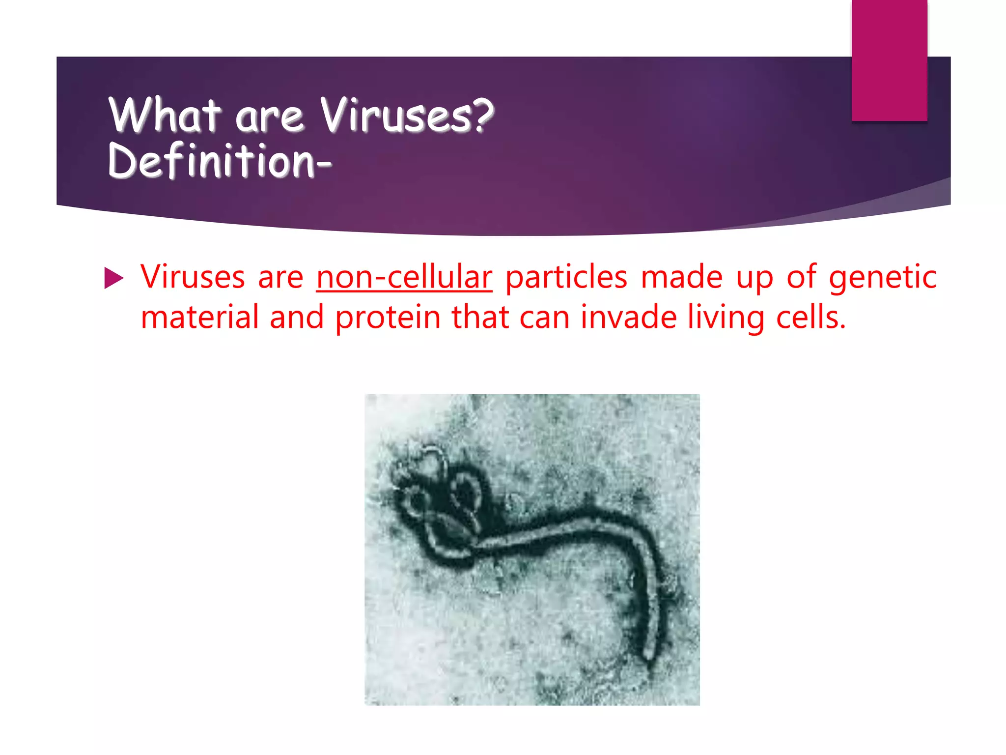
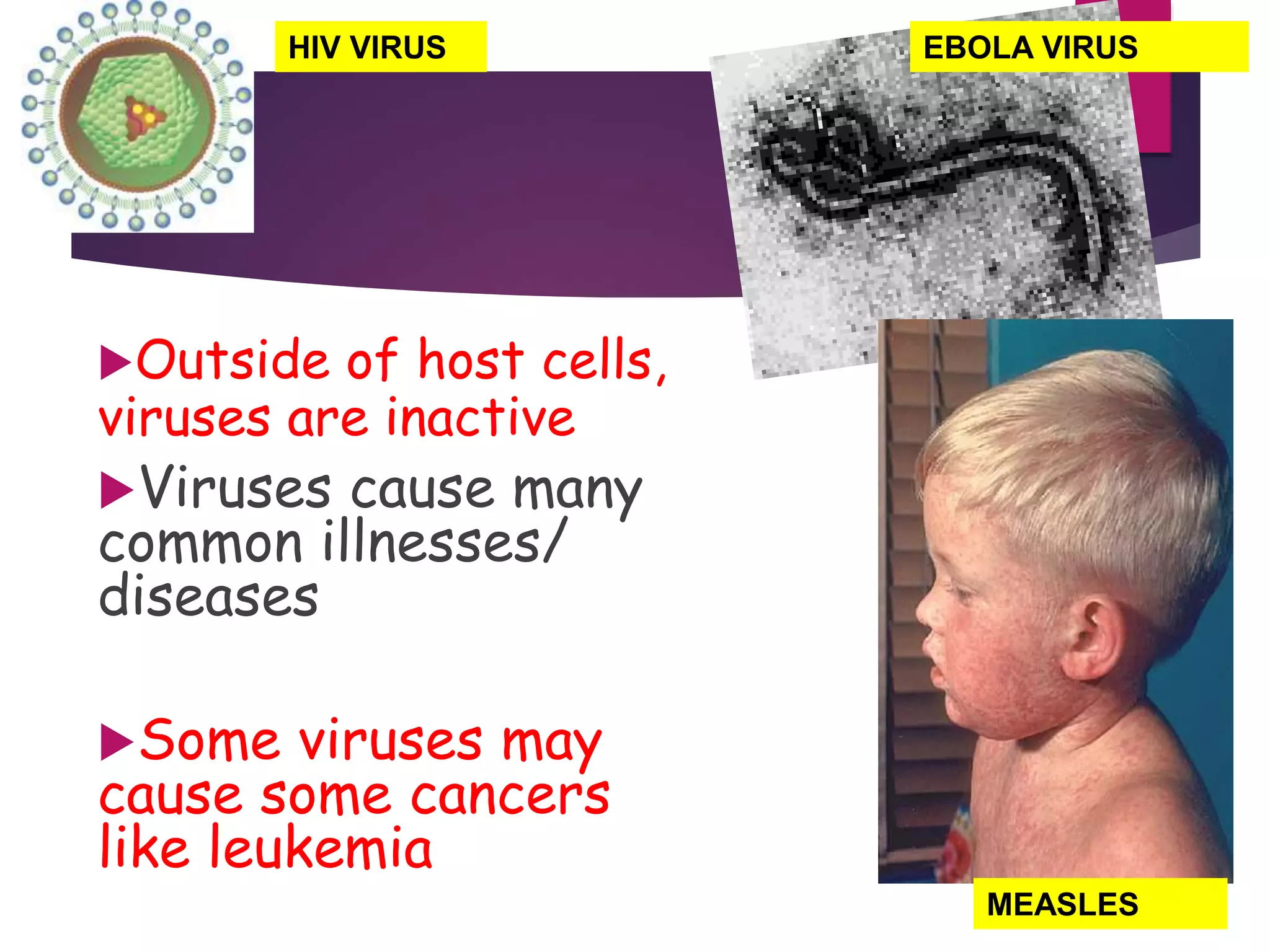
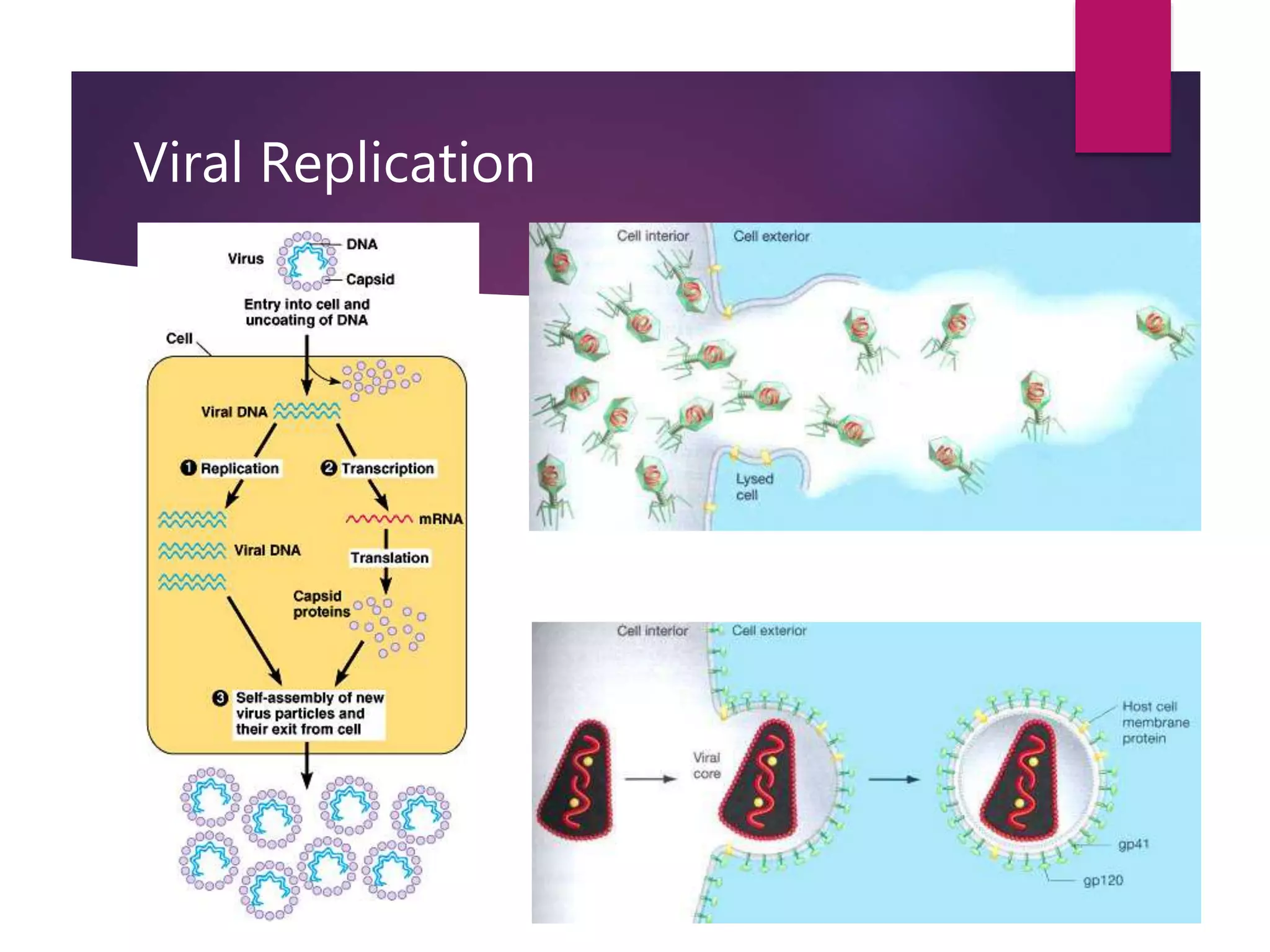
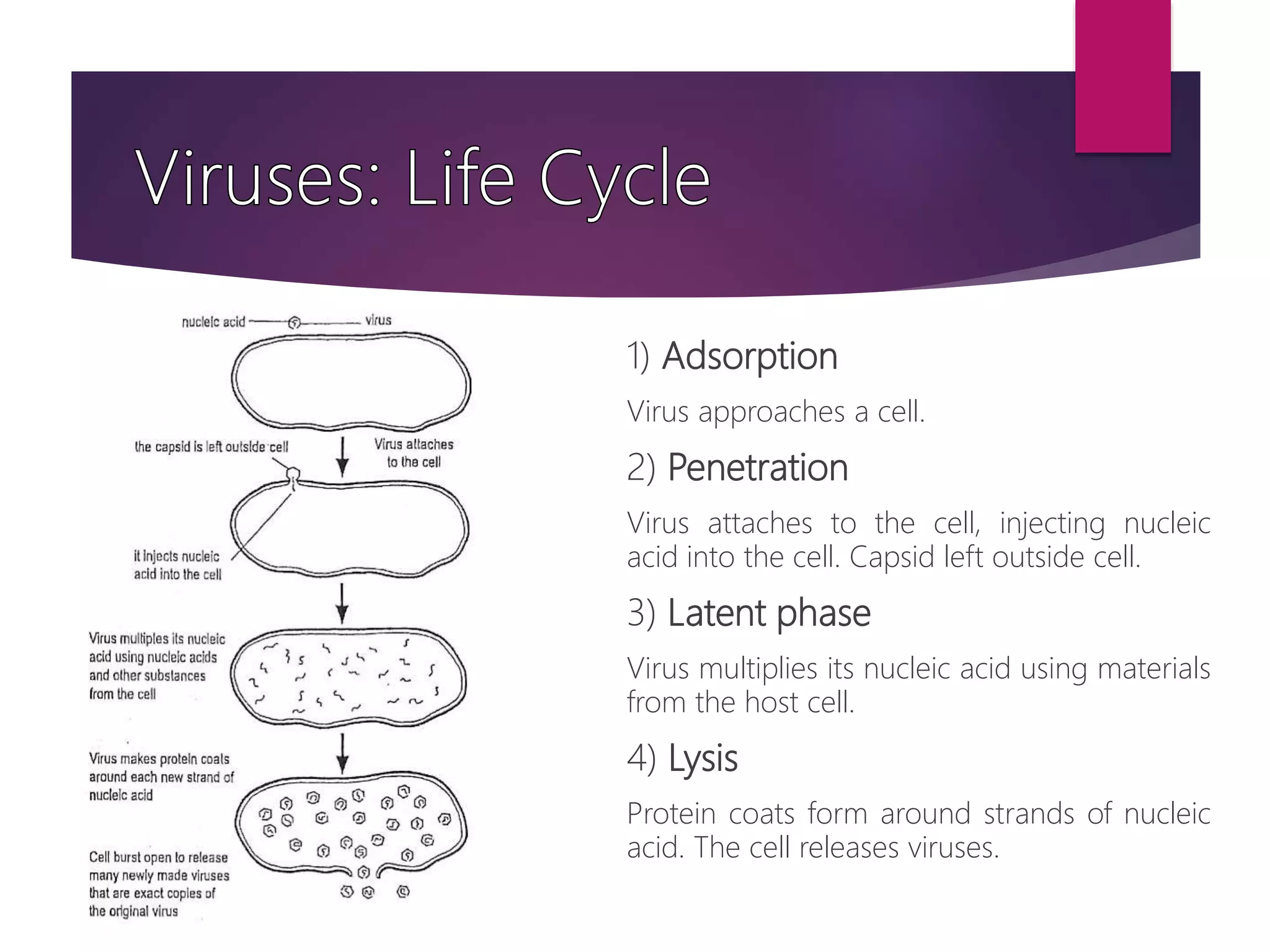
1) Viruses are non-living infectious particles that contain genetic material and a protein coat called a capsid. 2) Viruses can only replicate inside a host cell by injecting their genetic material and using the host cell's machinery. 3) Viruses exist in two states - as active viruses when infecting a host cell, or dormant virions when not in contact with a host.
Infectious diseases are mainly caused by
microbes.
These are small microorganisms which are
invisible with the naked eye.
They mainly include bacteria, virus, fungi
and parasites.
The symptoms caused by infection depends
on
the location.
Nature of the infection
Type of the microbe
Infectious diseases are mainly caused by
microbes.
These are small microorganisms which are
invisible with the naked eye.
They mainly include bacteria, virus, fungi
and parasites.
The symptoms caused by infection depends
on
the location.
Nature of the infection
Type of the microbe
Introduction
Classification of virus
Size of virus
Structure of Virus
Morphology of Virus
Effect of Physical and chemical agents on virus.
Life cycle of bacteriophages
Cultivation of viruses
Introduction to virology for Medical studentsNCRIMS, Meerut
Introduction to virology for MBBS students
A virus is an obligate intracellular parasite containing genetic material surrounded by protein
Virus particles can only be observed by an electron microscope
Most viruses range in sizes from 20 – 250 nanometers
Viruses are obligate intracellular parasites
Viruses are non-living entities
Viruses cannot make energy or proteins independent of a host cell (Depends on host cell for replication)
Viral genome are either RNA or DNA but not both.
Viruses have a naked capsid or envelope with attached proteins
Do not possess cellular organization
Viruses do not have the genetic capability to multiply by division.
They are NOT cultiviable on ordinary media.
Much smaller than bacteria
“Filterable agents” – can pass through filters that can hold back bacteria
Vary widely in size:
Largest – poxvirus (300nm)
Smallest – parvovirus (20nm)
General Characters and Classification of Viruses. Includes ICTV classification and Baltimore classification of viruses. A brief explanation of the Viral structure and Lifecycle.
This presentation gives a detail overview on Viruses - Morphology and Classification. The presentation is helpful for students of B. Pharm Second Year and those who wants to gain basic knowledge about Viruses.
Subject - Microbiology
Education Material about Virus Presentation Template
If you want to buy this presentation template, please visit http://madlis.com
Good design gets out of the way of the content you are sharing. It helps your audience focus on the content itself instead of the design.
But, it's no secret that most people dislike giving presentations. The dread of public speaking consistently ranks among the greatest fears in public surveys.
This presentation slides can help you reduce the anxiety involved with giving a presentation. Well-designed slides not only build your own confidence, they make your key points clearer to the audience.
Salas, V. (2024) "John of St. Thomas (Poinsot) on the Science of Sacred Theol...Studia Poinsotiana
I Introduction
II Subalternation and Theology
III Theology and Dogmatic Declarations
IV The Mixed Principles of Theology
V Virtual Revelation: The Unity of Theology
VI Theology as a Natural Science
VII Theology’s Certitude
VIII Conclusion
Notes
Bibliography
All the contents are fully attributable to the author, Doctor Victor Salas. Should you wish to get this text republished, get in touch with the author or the editorial committee of the Studia Poinsotiana. Insofar as possible, we will be happy to broker your contact.
DERIVATION OF MODIFIED BERNOULLI EQUATION WITH VISCOUS EFFECTS AND TERMINAL V...Wasswaderrick3
In this book, we use conservation of energy techniques on a fluid element to derive the Modified Bernoulli equation of flow with viscous or friction effects. We derive the general equation of flow/ velocity and then from this we derive the Pouiselle flow equation, the transition flow equation and the turbulent flow equation. In the situations where there are no viscous effects , the equation reduces to the Bernoulli equation. From experimental results, we are able to include other terms in the Bernoulli equation. We also look at cases where pressure gradients exist. We use the Modified Bernoulli equation to derive equations of flow rate for pipes of different cross sectional areas connected together. We also extend our techniques of energy conservation to a sphere falling in a viscous medium under the effect of gravity. We demonstrate Stokes equation of terminal velocity and turbulent flow equation. We look at a way of calculating the time taken for a body to fall in a viscous medium. We also look at the general equation of terminal velocity.
What is greenhouse gasses and how many gasses are there to affect the Earth.moosaasad1975
What are greenhouse gasses how they affect the earth and its environment what is the future of the environment and earth how the weather and the climate effects.
This presentation explores a brief idea about the structural and functional attributes of nucleotides, the structure and function of genetic materials along with the impact of UV rays and pH upon them.
Slide 1: Title Slide
Extrachromosomal Inheritance
Slide 2: Introduction to Extrachromosomal Inheritance
Definition: Extrachromosomal inheritance refers to the transmission of genetic material that is not found within the nucleus.
Key Components: Involves genes located in mitochondria, chloroplasts, and plasmids.
Slide 3: Mitochondrial Inheritance
Mitochondria: Organelles responsible for energy production.
Mitochondrial DNA (mtDNA): Circular DNA molecule found in mitochondria.
Inheritance Pattern: Maternally inherited, meaning it is passed from mothers to all their offspring.
Diseases: Examples include Leber’s hereditary optic neuropathy (LHON) and mitochondrial myopathy.
Slide 4: Chloroplast Inheritance
Chloroplasts: Organelles responsible for photosynthesis in plants.
Chloroplast DNA (cpDNA): Circular DNA molecule found in chloroplasts.
Inheritance Pattern: Often maternally inherited in most plants, but can vary in some species.
Examples: Variegation in plants, where leaf color patterns are determined by chloroplast DNA.
Slide 5: Plasmid Inheritance
Plasmids: Small, circular DNA molecules found in bacteria and some eukaryotes.
Features: Can carry antibiotic resistance genes and can be transferred between cells through processes like conjugation.
Significance: Important in biotechnology for gene cloning and genetic engineering.
Slide 6: Mechanisms of Extrachromosomal Inheritance
Non-Mendelian Patterns: Do not follow Mendel’s laws of inheritance.
Cytoplasmic Segregation: During cell division, organelles like mitochondria and chloroplasts are randomly distributed to daughter cells.
Heteroplasmy: Presence of more than one type of organellar genome within a cell, leading to variation in expression.
Slide 7: Examples of Extrachromosomal Inheritance
Four O’clock Plant (Mirabilis jalapa): Shows variegated leaves due to different cpDNA in leaf cells.
Petite Mutants in Yeast: Result from mutations in mitochondrial DNA affecting respiration.
Slide 8: Importance of Extrachromosomal Inheritance
Evolution: Provides insight into the evolution of eukaryotic cells.
Medicine: Understanding mitochondrial inheritance helps in diagnosing and treating mitochondrial diseases.
Agriculture: Chloroplast inheritance can be used in plant breeding and genetic modification.
Slide 9: Recent Research and Advances
Gene Editing: Techniques like CRISPR-Cas9 are being used to edit mitochondrial and chloroplast DNA.
Therapies: Development of mitochondrial replacement therapy (MRT) for preventing mitochondrial diseases.
Slide 10: Conclusion
Summary: Extrachromosomal inheritance involves the transmission of genetic material outside the nucleus and plays a crucial role in genetics, medicine, and biotechnology.
Future Directions: Continued research and technological advancements hold promise for new treatments and applications.
Slide 11: Questions and Discussion
Invite Audience: Open the floor for any questions or further discussion on the topic.
The ability to recreate computational results with minimal effort and actionable metrics provides a solid foundation for scientific research and software development. When people can replicate an analysis at the touch of a button using open-source software, open data, and methods to assess and compare proposals, it significantly eases verification of results, engagement with a diverse range of contributors, and progress. However, we have yet to fully achieve this; there are still many sociotechnical frictions.
Inspired by David Donoho's vision, this talk aims to revisit the three crucial pillars of frictionless reproducibility (data sharing, code sharing, and competitive challenges) with the perspective of deep software variability.
Our observation is that multiple layers — hardware, operating systems, third-party libraries, software versions, input data, compile-time options, and parameters — are subject to variability that exacerbates frictions but is also essential for achieving robust, generalizable results and fostering innovation. I will first review the literature, providing evidence of how the complex variability interactions across these layers affect qualitative and quantitative software properties, thereby complicating the reproduction and replication of scientific studies in various fields.
I will then present some software engineering and AI techniques that can support the strategic exploration of variability spaces. These include the use of abstractions and models (e.g., feature models), sampling strategies (e.g., uniform, random), cost-effective measurements (e.g., incremental build of software configurations), and dimensionality reduction methods (e.g., transfer learning, feature selection, software debloating).
I will finally argue that deep variability is both the problem and solution of frictionless reproducibility, calling the software science community to develop new methods and tools to manage variability and foster reproducibility in software systems.
Exposé invité Journées Nationales du GDR GPL 2024
Earliest Galaxies in the JADES Origins Field: Luminosity Function and Cosmic ...Sérgio Sacani
We characterize the earliest galaxy population in the JADES Origins Field (JOF), the deepest
imaging field observed with JWST. We make use of the ancillary Hubble optical images (5 filters
spanning 0.4−0.9µm) and novel JWST images with 14 filters spanning 0.8−5µm, including 7 mediumband filters, and reaching total exposure times of up to 46 hours per filter. We combine all our data
at > 2.3µm to construct an ultradeep image, reaching as deep as ≈ 31.4 AB mag in the stack and
30.3-31.0 AB mag (5σ, r = 0.1” circular aperture) in individual filters. We measure photometric
redshifts and use robust selection criteria to identify a sample of eight galaxy candidates at redshifts
z = 11.5 − 15. These objects show compact half-light radii of R1/2 ∼ 50 − 200pc, stellar masses of
M⋆ ∼ 107−108M⊙, and star-formation rates of SFR ∼ 0.1−1 M⊙ yr−1
. Our search finds no candidates
at 15 < z < 20, placing upper limits at these redshifts. We develop a forward modeling approach to
infer the properties of the evolving luminosity function without binning in redshift or luminosity that
marginalizes over the photometric redshift uncertainty of our candidate galaxies and incorporates the
impact of non-detections. We find a z = 12 luminosity function in good agreement with prior results,
and that the luminosity function normalization and UV luminosity density decline by a factor of ∼ 2.5
from z = 12 to z = 14. We discuss the possible implications of our results in the context of theoretical
models for evolution of the dark matter halo mass function.
(May 29th, 2024) Advancements in Intravital Microscopy- Insights for Preclini...Scintica Instrumentation
Intravital microscopy (IVM) is a powerful tool utilized to study cellular behavior over time and space in vivo. Much of our understanding of cell biology has been accomplished using various in vitro and ex vivo methods; however, these studies do not necessarily reflect the natural dynamics of biological processes. Unlike traditional cell culture or fixed tissue imaging, IVM allows for the ultra-fast high-resolution imaging of cellular processes over time and space and were studied in its natural environment. Real-time visualization of biological processes in the context of an intact organism helps maintain physiological relevance and provide insights into the progression of disease, response to treatments or developmental processes.
In this webinar we give an overview of advanced applications of the IVM system in preclinical research. IVIM technology is a provider of all-in-one intravital microscopy systems and solutions optimized for in vivo imaging of live animal models at sub-micron resolution. The system’s unique features and user-friendly software enables researchers to probe fast dynamic biological processes such as immune cell tracking, cell-cell interaction as well as vascularization and tumor metastasis with exceptional detail. This webinar will also give an overview of IVM being utilized in drug development, offering a view into the intricate interaction between drugs/nanoparticles and tissues in vivo and allows for the evaluation of therapeutic intervention in a variety of tissues and organs. This interdisciplinary collaboration continues to drive the advancements of novel therapeutic strategies.
Richard's aventures in two entangled wonderlandsRichard Gill
Since the loophole-free Bell experiments of 2020 and the Nobel prizes in physics of 2022, critics of Bell's work have retreated to the fortress of super-determinism. Now, super-determinism is a derogatory word - it just means "determinism". Palmer, Hance and Hossenfelder argue that quantum mechanics and determinism are not incompatible, using a sophisticated mathematical construction based on a subtle thinning of allowed states and measurements in quantum mechanics, such that what is left appears to make Bell's argument fail, without altering the empirical predictions of quantum mechanics. I think however that it is a smoke screen, and the slogan "lost in math" comes to my mind. I will discuss some other recent disproofs of Bell's theorem using the language of causality based on causal graphs. Causal thinking is also central to law and justice. I will mention surprising connections to my work on serial killer nurse cases, in particular the Dutch case of Lucia de Berk and the current UK case of Lucy Letby.
2. A virus is an infectious agent made up of
nucleic acid (DNA or RNA) wrapped in a
protein coat called a capsid.
Viruses have no nucleus, no organelles, no cytoplasm or
cell membrane—Non-cellular
vs
This is why it does NOT belong to any kingdom.
3. Are Viruses Living or Non-
living?
Biologists consider viruses to be non-living because:
Are not cells
Do not grow or respond to their surroundings
Cannot make food, take in food, or produce wastes
Viruses do not respond to stimuli.
They can only multiply if in another living cell
4. Viruses have either DNA or RNA but NOT both.
Viruses with RNA that transcribe
into DNA are called retroviruses.
HIV Infected Cell
A flea is a parasite to a dog
and is harmful to the dog.
Viruses are parasites—an
organism that depends entirely
upon another living organism (a
host) for its existence in such a
way that it harms that organism.
5. What are Viruses?
Definition-
Viruses are non-cellular particles made up of genetic
material and protein that can invade living cells.
9. How Big is a Virus?
Viruses are very small – smaller than the smallest cell.
Porcine circovirus type 1 has a capsid diameter of only 17nm
i.e. 0.017µm
Mimivirus: Protein filaments measuring 100 nm project from the
surface of the capsid, bringing the total length of the virus up to
600nm (0.6µm) and a capsid diameter of 400nm (0.4µm).
It is the third-largest virus, preceded by the recently discovered
Megavirus chilensis and Pandoravirus.
10.
11. Characteristics
Non living structures
Non-cellular
Contain a protein coat called the capsid
Have a nucleic acid core containing DNA or RNA (one or the
other - not both)
Capable of replication only when inside a HOST cell
Exist in two distinct states: a virus (when active) and a virion
(when dormant and not in contact with a host cell).
Can also remain dormant within an organism (latency).
A viriod (NOT VIRION) is an infectious RNA particle that
resembles a virus – but is smaller.
12. Prions has protein only, no DNA or RNA (cause of
mad cow disease and Creutfeldt-Jacob disease in
humans)—affects the brain and is always fatal
No DNA or RNA!
Prions are made up of harmless proteins that are found in
mammals and birds. But these proteins are in abnormal
form and once they enter human brain, they are capable of
severe brain infections. Normally these prions are ingested
but they also get formed through mutation of a gene that
contains this protein.
13. Characteristics
Some viruses are
enclosed in an protective
envelope
Some viruses may have
spikes to help attach to
the host cell
Most viruses infect only
SPECIFIC host cells
CAPSID
ENVELOPE
DNA
SPIKES
14. Certain viruses can only attack certain
cell types. They are said to be specific.
It’s like the pieces of a puzzle. The
ends have to match up so only
certain pieces fit.
Surface Markers
Receptor Sites
Example: The rabies virus only attacks brain or nervous cells.
Virus
Cell
16. Outside of host cells,
viruses are inactive
Viruses cause many
common illnesses/
diseases
Some viruses may
cause some cancers
like leukemia
EBOLA VIRUS
HIV VIRUS
MEASLES
17. What do Viruses look like?
Viruses are unusual and different from other things in
nature.
Viruses come in a variety of shapes
Some may be helical shape like the Ebola virus
Some may be polyhedral shapes like the influenza virus
Others have more complex shapes like bacteriophages
18. Viruses can have different shapes:
Polyhedral
cubical
e.g. adenovirus
Helical
spiral cylinder
e.g. tobacco virus
Complex
tadpole-like
e.g. bacteriophage
27. Viral Taxonomy
Order names end in -virales
Family names end in –viridae
Subfamily -virinae
Genus names end in -virus
Viral Species: A group of viruses sharing the same
genetic information and ecological niche (host).
Common names are used for species
Subspecies are designated by a number
32. HOST SPECIFICITY
All kingdoms can be infected by viruses
Viruses are kingdom specific but they may or may
not be species specific
Spread is specific to the type of virus
33. PARASITISM
Viruses are parasites.
i.e. it depends upon another living
organism for its existence in such a
way that it harms that organism.
37. Capsid (protein coat)
– inside contains either
RNA or DNA
Bacteriophage—viruses that infect bacteria
38.
39. 1) Adsorption
Virus approaches a cell.
2) Penetration
Virus attaches to the cell, injecting nucleic
acid into the cell. Capsid left outside cell.
3) Latent phase
Virus multiplies its nucleic acid using materials
from the host cell.
4) Lysis
Protein coats form around strands of nucleic
acid. The cell releases viruses.
42. Lysogenic Cycle
All phage species can undergo a lytic
cycle
Phages capable of only the lytic cycle
are called virulent
The alternative to the lytic cycle is
called the lysogenic cycle: no progeny
particles are produced, the infected
bacterium survives, and a phage DNA
is transmitted to each bacterial
progeny cell when the cell divides
Those phages that are also capable of
the lysogenic cycle are called
temperate
44. Cylces
Lysogenic Cycle
Viral DNA
May stay inactive in host for long periods of time
Long lasting
Example Mono or chickenpox
Lytic Cylce
Short and can be over come
Example flu virus
45. Host cells are affected in three ways:
The host cells may be destroyed. They may swell and burst, e.g. as with
nerve cells infected with the rabies virus.
The host cells may not be able to function correctly, e.g. ciliated
epithelial cells infected with the influenza virus.
The virus can interact with the host cell’s chromosomes causing a
mutation, e.g. warts and cancer cells.
The immune system becomes activated in order to fight the
infection. This may lead to fever, tiredness or an opportunistic
disease.
46. Vaccine
Is a weaken form of the virus
To expose your immune system to the virus which will allow
your body to better fight off the virus when exposed to the full
blow virus.
47.
48. RNA or DNA core (center),
protein coat (capsid)
Copies itself only inside
host cell--REPLICATION
DNA or RNA
NO
NO
NO
NO
Cell membrane, cytoplasm,
genetic material, organelles
Asexual or Sexual
DNA and RNA
YES—Multicellular Organisms
YES
YES
YES
Structure
Reproduction
Genetic Material
Growth and
Development
Response to
Environment
Change over time
Obtain and
Use Energy