Downloaded 429 times
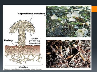
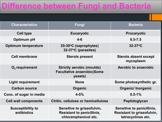
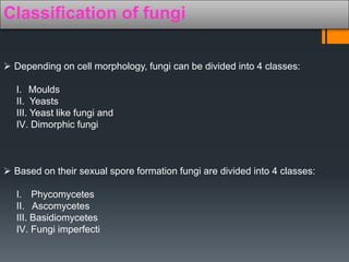
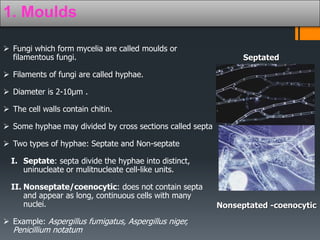


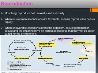


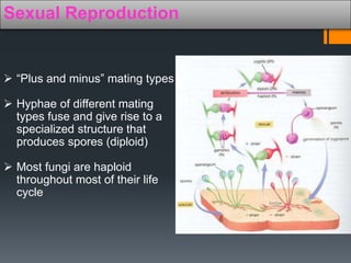


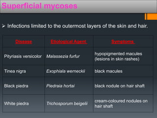
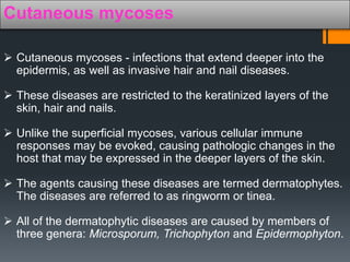
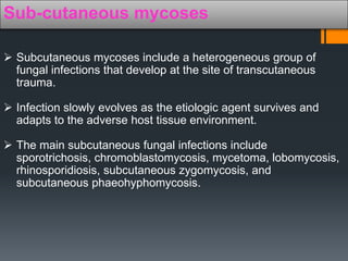


This document provides an overview of fungi. It discusses that fungi are eukaryotic organisms classified in the kingdom Fungi. They can exist as molds, yeasts, or dimorphic fungi. Fungi have cell walls containing chitin and reproduce asexually through spores or budding or sexually through the fusion of hyphae. The document outlines the morphological characteristics, differences from bacteria, classification systems based on cell structure and reproduction, and types of fungal infections like superficial, cutaneous, subcutaneous, and systemic mycoses.


