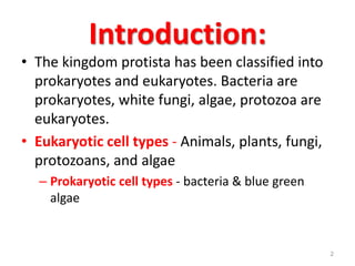
SlideShare a Scribd company logo
1 of 106
Bacteriology
Basics
Morphology, Classification, Staining
Methods
IMRAN TARIQ
(ASSISTANT PROFESSOR)
Introduction:
ā€¢ The kingdom protista has been classified into
prokaryotes and eukaryotes. Bacteria are
prokaryotes, white fungi, algae, protozoa are
eukaryotes.
ā€¢ Eukaryotic cell types - Animals, plants, fungi,
protozoans, and algae
ā€“ Prokaryotic cell types - bacteria & blue green
algae
2
Antioni van Leeuwenhoek
ā€¢ Leeuwenhoek is
called "the inventor
of the microscope"
ā€¢ Created a ā€œsimpleā€
microscope that could
magnify to about 275x,
and
published drawings of
microorganisms in 1683
3
Eukaryotes have organelles
ā€¢ Much larger; more complex than prokaryotes
ā€¢ Processes compartmentalized into organelles
ā€“ Nucleus
ā€“ Protein synthesis (ribosomes, RER, Golgi)
ā€“ Mitochondria; chloroplasts
ā€“ Lysosomes
ā€“ Plasma membranes have different modifications
ā€“ Cytoskeleton
4
Schematic of typical animal (eukaryotic) cell, showing subcellular
components.
Organelles: (1) nucleolus (2) nucleus (3) ribosome (4) vesicle
(5) rough endoplasmic reticulum (ER) (6) Golgi apparatus (7) Cytoskeleton
(8) smooth ER (9) mitochondria (10) vacuole
(11) cytoplasm (12) lysosome (13) centrioles
5
Differences between prokaryotic & eukaryotic cells
Character Prokaryotes Eukaryotes
Nucleus Nuclear
membrane
Absent Present
Nucleolus Absent Present
Chromosome One circular One or more paired
and linear
Cell division Binary fission Mitosis
Cytoplasmic
membrane
Structure and
Composition
fluid phospholipid
bilayer, lacks sterols
fluid phospholipid
bilayer containing
sterols
Function Incapable of
endocytosis
(phagocytosis and
pinocytosis) and
exocytosis
Capable of
endocytosis and
exocytosis
6
Differences between prokaryotic & eukaryotic
cells
Character Prokaryotes Eukaryotes
Cytoplasm Mitochondria Absent Present
Lysosomes Absent Present
Golgi apparatus Absent Present
Endoplasmic
reticulum
Absent Present
Vacuoles Absent Present
Ribosomes 70 S 80 S
7
Differences between prokaryotic &
eukaryotic cells
Character Prokaryotes Eukaryotes
Cell Wall Present Animals & Protozoans ā€“
Absent
Plants, Fungi & Algae -
Present
Composition Peptidoglycan ā€“
complex
carbohydrate
Cellulose or chitin
Locomotor
organelles
Flagella Flagella/ Cilia
8
Prokaryotic Cells
ā€¢ Much smaller (microns) and more simple
than eukaryotes
ā€¢ Prokaryotes are molecules surrounded by
a membrane and cell wall.
ā€¢ They lack a true nucleus and donā€™t have
membrane bound organelles like
mitochondria, etc.
ā€¢ Large surface-to-volume ratio : nutrients
can easily and rapidly reach any part of
the cells interior 9
Size of Bacteria
ā€¢ Unit of measurement in
bacteriology is the micron
(micrometre, Āµm)
ā€¢ Bacteria of medical importance
ā€“0.2 ā€“ 1.5 Āµm in diameter
ā€“3 ā€“ 5 Āµm in length
10
11
Eukaryotic cell Prokaryotic cell
Gram +
Gram -
Cell wall
Rough endoplasmic
reticulum
Mitochondria
(e.g. animal)
Nucleoid
Nucleus
Cell membrane
Cytoplasm
Flagellum
Cell (inner) membrane Outer membrane
Ribosomes
Granule
Cell wall
Capsule
Pili
Shapes of Bacteria
ā€¢ When viewed under the microscope, most
clinically significant bacteria appear in three
basic shapes, they are ; Cocci (from Kokkes
meaning berry) are spherical or oral. Bacilli
(from bacillus meaning rod) are rod shaped.
Spiral Other then three basic bacterial shapes
some bacterial are square, filaments and large
number produces buds, lancet (spneumoid
bean ā€“ shaped (Neisseria gonnorhea).
12
Shapes of Bacteria
ā€¢ Cocci ā€“ spherical/ oval shaped major groups
ā€¢ Bacilli ā€“ rod shaped
ā€¢ Vibrios ā€“ comma shaped
ā€¢ Spirilla ā€“ rigid spiral forms
ā€¢ Spirochetes ā€“ flexible spiral forms
ā€¢ Actinomycetes ā€“ branching filamentous
bacteria
ā€¢ Mycoplasmas ā€“ lack cell wall
13
Bacteria Have One of Three Cellular
Shapes
ā€¢ Rods (bacilli)
ā€¢ Coccoid-Shaped
ā€¢ Spirilla
14
ā€¢Cocci These spherical bacteria vary in
diameter between 0.5 ļ­ m - 2 ļ­ m. They occur
as single cells (micro cocci)
ā€¢ Pairs of cocci (Diplococci)
ā€¢ Chains (Streptococci)
ā€¢ Packets (Staphylococci)
ā€¢ Cube ā€“ shaped cellular packets (Sarcinae )
15
Arrangement of bacteria: Cocci
Cocci in pair ā€“ Diplococcus
Sarcina ā€“ groups of eight
Tetrad ā€“ groups of four
Cocci in chain - Streptococci
Cocci in cluster - Staphylococci
Coccus
16
Spherical Bacteria
Bacilli
ā€¢ This morphotype consists of cylindrical
cells, which are either large or small rods.
The bacilli are usually 0.2 ā€“ 1 ļ­ m in
diameter, with lengths of approximately
10 ļ­ m being common. Most rods occur
single, but some form long chains called
as stretptobaccilli.
18
Arrangement of bacteria: Bacilli
19
Rod-Shaped Bacteria
Other shapes of bacteria
ā€¢ Spiral: In reality, the spirilla are bacilli twisted
into helix. the rigid spirilla are surrounded by a
cell wall and are represented by genera
spirillum and vibrio. Flexible spirilla with more
than one way are typical of the spirochaetes,
represented by the genera treponema,
spirocheta, leptespira, borrelia and cristispira.
Actinomycetes are branching filamentous
bacteria, so called because of fancied
resembled to the radiating rays of sun when
seen in tissue lesion (from actis meaning ray
and mykes meaning fungus).
21
Other shapes of bacteria
Comma shaped
Spirochetes
Spirilla
22
Spiral-Shaped Bacteria
Spirochete:
Borrelia
burgdorferi
Bacterial Morphology
24
Anatomy of a Bacterial Cell
25
Anatomy of A Bacterial Cell
ā€¢ Outer layer ā€“ two components:
1. Rigid cell wall
2. Cytoplasmic (Cell/ Plasma) membrane ā€“
present beneath cell wall
ā€¢ Cytoplasm ā€“ cytoplasmic inclusions,
ribosomes, mesosomes and nucleus
ā€¢ Additional structures ā€“ plasmid, slime
layer, capsule, flagella, fimbriae (pili),
spores
26
Typical shapes of bacteria
Most bacteria retain a particular shape; a few
are pleiomorphic
27
Typical prokaryotic structures
Working from the outside inā€¦
28
Cell Envelope
ā€¢ The cell envelope is all the
layers from the cell
membrane outward,
including the cell wall, the
periplasmic space, the outer
membrane, and the capsule.
ā€“ All free-living bacteria
have a cell wall
ā€“ periplasmic space and
outer membrane are
found in Gram-negatives
ā€“ the capsule is only found
in some strains
29
Structure & Function
of Cell Components
CELL WALL
ā€¢ Outermost layer, encloses cytoplasm
1. Confers shape and rigidity
2. 10 - 25 nm thick
3. Composed of complex polysaccharides
(peptidoglycan/ mucopeptide) - formed by N
acetyl glucosamine (NAG) & N acetyl muramic
acid (NAM) alternating in chains,
held by peptide chains.
31
Cell Wall
4. Carries bacterial antigens ā€“ important in virulence &
immunity
5. Chemical nature of the cell wall helps to divide bacteria
into two broad groups ā€“ Gram positive & Gram negative
6. Gram +ve bacteria have simpler chemical nature than
Gram ā€“ve bacteria.
7. Cell wall also serves as primary sieve in the transport
molecules into the cell. It also functions as an anchor for
the capsule, as well as pilli, fimbriae, flagella. It is primary
receptor for the absorption of specific viruses and
bacteriophages
8. Several antibiotics may interfere with cell wall synthesis
e.g. Penicillin, Cephalosporins
32
Peptidoglycan structure
ā€¢ The polymer contains two sugar derivatives ,
N ā€“ acetyl glucosamine N ā€“ acetyl muramic
acid alternating in chains which are
crosslinked by peptide chains. Gram positive
cell wall Normally thick, homogeneous cell
wall of gram positive bacteria composed
primarily of peptidoglycan. However gram
positive cell walls, usually contain large
amounts of teichoic acids, polymers of
glycerol joined by phosphate group. Lipids and
proteins are usually absent. 33
Gram positive cell wall
The Gram-positive cell wall is composed of a thick, multilayered
peptidoglycan sheath outside of the cytoplasmic membrane. Teichoic
acids are linked to and embedded in the peptidoglycan, and
lipoteichoic acids extend into the cytoplasmic membrane
34
Gram negative cell wall
The Gram-negative cell wall is composed of an outer membrane
linked to thin, mainly single-layered peptidoglycan by lipoproteins.
The peptidoglycan is located within the periplasmic space that is
created between the outer and inner membranes. The outer
membrane includes porins, which allow the passage of small
hydrophilic molecules across the membrane, and
lipopolysaccharide molecules that extend into extracellular space.
35
Functions of teichoic acids
ā€¢ They may contribute to the overall negative change
density of cell surface. This negative surface charge
and lipoteichoic acids are strong adhesions,
especially in the attachment to mammalian tissues,
and adherence to red blood cells, tooth surfaces and
other positively charged surfaces.
36
Bacterial
Cell
Walls
37
Summary of the differences between
Gram positive & Gram negative bacteria
Property of bacteria Gram Positive Gram Negative
Thickness of wall 20-80 nm 10 nm
Number of layers in wall 1 2
Peptidoglycan content >50% 10-20%
Teichoic acid in wall + -
Lipid & lipoprotein content 0-3% 58%
Protein content 0% 9%
Lipopolysaccharide 0 13%
Sensitive to penicillin Yes Less sensitive
Digested by lysozyme Yes Weakly
38
39
Outer Membrane
Gram negative bacteria
ā€¢ major permeability barrier
ā€¢ space between inner and outer
membrane
ā€“Periplasmic space
ļ¶store degradative enzymes
ā€¢ Gram positive bacteria
ā€¢ no Periplasmic space
Cytoplasmic (Plasma) membrane
ā€¢ The cytoplasmic membrane is a layer (5-10 nm) lining the
inner surface of the cell wall separating it from the
cytoplasm. It consists of three layers ā€“ outer, middle and
inner layer, chemically, both inner and outer layers are
composed of 70%, protein, the middle (hydrophobic)
layer contains 30% lipid in addition to fatty acids,
phospholipids, transmembrane proteins (permeases)
with small amounts of carbohydrates
ā€¢ In summary, functionally, the cytoplasmic membrane is
one of most important components of the bacterial cell,
which is site of anabolic and catabolic metabolism and
regulates the transport of molecules into without of the
cell. 40
Transport Across the Cell Membrane
ā€¢ Basic rule: things spontaneously
move from high concentration
to low concentration (downhill).
This process is called diffusion.
ā€“ Getting many molecules into the
cell is simply a matter of opening
up a protein channel of the proper
size and shape. The molecules
then move into the cell by
diffusing down the concentration
gradient. Passive transport, or
facilitated diffusion.
ā€¢ To get things to move from low
to high (uphill), you need to add
energy: the molecules must be
pumped into the cell. Pumps
are driven by ATP energy.
Active transport. 41
cytoplasmic membrane
42
Other Cytoplasmic Components
ā€¢ The bacterial cytoplasm consists of central
nuclear region surrounded by ribosomeā€™s
mixed in a sea of enzymes, cofactors, amino
acids, and vitamin, as well as multitude of
other bone and molecules. It differs from
eukaryotic cytoplasm in not exhibiting
internal mobility and the absence of
endoplasmic reticulum and mitochondria.
These serve as building blocks for cellular
biosynthesis and energy production. The
cytoplasm contains ribosomeā€™s, mesosomes,
inclusions and vacuoles. 43
Ribosomeā€™s
ā€¢ The ribosomeā€™s are the most abundant
inclusion in bacterial cytoplasm. They are
called 70S ribosomes. It consists of 40%
protein and 60% RNA. Eukaryotic ribosomeā€™s
are larger than bacterial ribosomeā€™s. Three
types of RNA exist ribosomal RNA (rRNA),
Transfer RNA (tRNA), messenger RNA
(mRNA)).
44
Mesosomes (Chondroids):
ā€¢ These are vesicular, convoluted or
multilaminated structures formed as
invaginations of the plasma membrane into
cytoplasmā€™s. They are more prominent in gram
positive bacteria. They are principal sites of
respiratory enzymes in bacteria and are
analogous to the mitochondria of eukaryotes.
Mesosomes are often seen in relation to the
nuclear body and site of synthesis of cross wall
septa, suggesting that they coordinate nuclear
and cytoplasmic division during binary fission.
45
Nucleus
ā€¢ Bacterial nuclei can be demonstrated by acid or ribo
nuclease hydrolysis and subsequent staining for
nuclear material. Bacterial nuclei have no nuclear
membrane or nucleolus. The nuclear DNA is not
associated with basic protein. The genome consists
of single molecule of DNA arranged in the form of
circle. The bacterial chromosome is haploid and
replicates by simple fission instead of by mitosis as in
higher cells. Bacteria may posses extranuclear
genetic elements consisting of DNA. These
cytoplasmic carries of genetic information are
termed as plasmid or episomes. These may account
for drug resistance which may constitute a survival
advantage . 46
Plasmid ā€“
ā€“ Extra nuclear genetic elements consisting
of DNA
ā€“ Transmitted to daughter cells during
binary fission
ā€“ May be transferred from one bacterium
to another
ā€“ Not essential for life of the cell
ā€“ Confer certain properties e.g. drug
resistance, toxicity 47
Bacterial storage granules and
inclusion :
ā€¢ These are aggregates of various compounds
that are normally involved in storing energy
reserves or building blocks for the cell. During
their growth and metabolism, bacteria
accumulate variety of cytoplasmic storage
products, as a result of the environment and
metabolism by the cell of large number of
carbohydrates, lipids, proteins. Two major
types of inorganic inclusion bodies are seen.
Polyphosphate granules or volutin granules
Magnetosome. 48
Inclusions of
Bacteria
granulose
Additional Organelles
Capsule & Slime layer ā€“
ā€“ Capsule ā€“ usually of uniform consistency
and integrity
ā€“ Slime layer ā€“ are of ill defined and
loosely formed
ā€“ Viscous layer secreted around the cell
wall.
ā€“ Polysaccharide / polypeptide in nature
Capsule ā€“ sharply defined structure,
antigenic in nature
ā€¢ Protects bacteria from lytic
enzymes
ā€¢ Inhibits phagocytosis
ā€¢ Stained by negative staining
using India Ink
50
Flagella ā€“
ā€¢ Flagella and Motility Flagella are organelles of motility / organs
of locomotion. Each flagellum consists of three distinct parts,
ā€¢ The filament ā€“ external to the cell and connected to the hook
at the cell surface.
ā€¢ Hook ā€“ connects the basal body to the distal portion of
filament.
ā€¢ The basal body ā€“ which is embedded in the cytoplasmic
membrane and portions of cell wall. Gram positive - 1 rings.
Gram negative ā€“ 2 rings.
ā€¢ Chemically, flagella are made up of protein flagellin similar to
keratin or myosin. Flagellin structure of each bacterial species is
sufficiently unique to confer immunologic specificity and useful
in microbiological identification of specific microorganisms.
51
In Gram-negative Bacteria
5/03/2012 52
Masdiana Padaga
In Gram-positive Bacteria
5/03/2012 53
Masdiana Padaga
A TYPICAL FLAGELLA
54
Types of flagellar arrangement
ā€¢ 1. Flagella arranged all round the cell
(peritrichous) eg. Typhoid
ā€¢ 2. Monotrichous ā€“ single flagella situated at
one end eg. Vibrio cholera
ā€¢ 3. Lopotrichous ā€“ Tufts of flagella at one end
eg. Spirilla
ā€¢ 4. Amphitrichous ā€“ Flagella at the both poles
55
Types of flagellar arrangement
Polar/ Monotrichous ā€“ single
flagellum at one pole
Lophotrichous ā€“ tuft of flagella at one
pole
Peritrichous ā€“ flagella all over
Amphitrichous ā€“ flagella at both
poles
Amphilophotrichous ā€“ tuft of flagella
at both ends
56
Peritrichous monotrichous
(or amphi, or lophotrichous
Cocci do not have flagella
57
Fimbriae/ Pili
ā€“ Both Pilli and Fimbriae are attached to the outer most
surface of cell. These hair like appendages, 10-20Āµ
long. They are confined to gram negative bacteria and
to selected gram positive bacteria (i.e, streptococcus,
actinomyces). The pilli are distinguished from flagella
in being approximately one half their diameter as well
as being distinctively straight or stiff in appearance.
ā€“ Made up of proteins called pilins.
ā€“ Pili can be of two types ā€“
ļƒ˜ Common pili ā€“ short & abundant
ļƒ˜ Sex pili - small number (one to six), very long pili,
helps in conjugation (process of transfer of DNA)
58
Functions of Pilli or Fimbriae
ā€¢ These structures function primarily in the
transfer of genetic material between the
bacteria, as well as adherence. Pilli that help
in transfer of genetic material are referred as F
or sex Pilli. F Pilli form physical bridges
between the donor and recipient cells known
as conjugation bridges, which permit the
transfer of DNA between them. The Pilli are
also bacterophage receptors, specific
bacteriophage will absorb to the type of the
Pilli, which are then retracted into the cell. 59
60
Spore
ā€¢ Some bacteria have the ability to form highly resistant resting
stages called spores. Each bacterium forms one spore, which
on germination forms a single vegetative cell. An bacterial
spores are formed inside the parent cell, they are called
endospores. Sporulation occurs after a period of vegetative
growth and is presumed to be related to the depletion of
exogenous nutrient. Sporulation is initiated by the appearance
of clear area, usually near one end of the cell, which gradually
becomes more opaque to form forespore. The fully developed
spore has at its core the nuclear body, surrounded by the
spore wall, a delicate membrane from which the cell wall of
the future vegetative bacterium will develop.
61
Spores
ā€“
ā€“ Highly resistant resting
stages formed during
adverse environment
(depletion of nutrients)
ā€“ Formed inside the parent
cell, hence called
Endospores
ā€“ Very resistant to heat,
radiation and drying and
can remain dormant for
hundreds of years.
ā€¢ They can be destroyed by
autoclaving at 120Ā°C for 15
minutes
62
Shape & position of bacterial spore
Oval central
Spherical central
Oval sub terminal
Oval sub terminal
Oval terminal
Spherical terminal
Free spore
Non bulging
Bulging
63
Spores
ā€¢ Some bacteria can form very tough
spores, which are metabolically
inactive and can survive a long time
under very harsh conditions.
ā€“Allegedly, some bacterial
spores that were embedded
in amber or salt deposits for
25 million years have been
revived. These experiments
are viewed skeptically by
many scientists.
64
Spores
ā€¢ Spores can also survive very high or low temperatures and
high UV radiation for extended periods. This makes them
difficult to kill during sterilization.
ā€¢ Anthrax
ā€¢ Bacillus species including anthracis (anthrax) and cereus
(endotoxin causes ~5% of food poisoning)
ā€¢ Clostridium species including tetani (tetanus), perfringens
(gangrene), and botulinum (botulism: food poisoning from
improperly canned food)
65
Bacterial Growth and
Multiplication
ā€¢ Transverse or Binary cell division
Growth of bacteria can be separated into two
distinct stages 1. An increase in cell mass such as
elongation of a bacillus, or an increase in size or
volume such as in occurs
2. The division of these cells into two daughter cells
The most common mechanism of bacterial cell
division which can be described as Binary or
transverse division.
66
Reproduction
ā€¢ Prokaryotic cell division is
binary fission.
ā€“ Single DNA molecule that
first replicates.
ā€“ Attaches each copy to a
different part of the cell
membrane.
ā€“ Cell begins to pull apart.
ā€“ Following cytokinesis, there
are then two cells of
identical genetic
composition.
67
ā€¢ Other mechanism of bacteria cell division
Bacteria such as hyphomicrobium,
corynebacterium and mycobacteria divide by
budding. The budding cell originates as an
outgrowth of the original, or mother cell, which
eventually reach a size equal to that of the
mother cell. the bud then separates to undertake
an independent existence several of the
actinomycetes form filamentous cells that
reproduce by the formation of spores from a
chain of cells or by a simple fragmentation of the
original filament into new cells.
68
The bacterial growth curve
ā€¢ When bacterium is seeded into a suitable liquid medium
and incubated its growth shows a definite course. If
bacterial consists are made at intervals after inoculation
and plated in relation to time, a growth curve is obtained.
The curve shows the following phases.
ā€¢ Lag phase
ā€¢ This phase of growth occurs when cells are transferred
from one medium to another or one environment to
another It is a phase of adjustment and represents the
period required for the adaptation of the cells to new
environment. The cells in this phase often increase in total
volume almost two or threefold, but they do not divide.
These cells are rapidly synthesizing DNA, new protein and
new enzymes as a prerequisite to division
69
Exponential phase
ā€¢ ā€“ or log phase In this phase, cells are dividing
at both a constant geometric rate as well as
maximum rate. Cellular component such as
RNA, protein, dry weight, and cell wall
polymers are also increasing at a constant
rate, they are much smaller in diameter than
cells in the lag phase. The exponential growth
phase usually comes to an end because of
depletion of essential nutrients, depletion of
oxygen in an acrostic culture or the
accumulation of toxic products. 70
Stationary phase
ā€¢ During this phase, there is a rapid decrease in the rate
of cell division. Eventually, the total number of dividing
cells will equal the number of dying cells, a true
stationery cell population occurs. The cells then direct
their resources towards survival energy generation is
harnessed to maintain osmotic barriers, mobility, and
the repair and synthesis of essential macromolecules.
The energy required to maintain cells in stationery
phase is called Maintenance energy and is obtained
from the degradation of cellular storage products.
After all of the storage products are exhausted, the
cells then degrade their cellular components, which
ultimately leads to death phase.
71
Death Phase
ā€¢ In this stage, the cells reproduce more slowly,
and death overtakes them in increasing
numbers. The cells eventually enter the
logarithmic death phase, during which the
decrease in the number of viatie cells occurs
at a regular, unchanging rate, a rate that
approximates that of the exponential growth
phase but is of negative slope.
72
73
Bacterial Taxonomy: Classification
ā€¢ Orderly arrangement : Kingdom ā€“
Division ā€“ Class ā€“ Order ā€“ Family ā€“ Tribe ā€“ Genus ā€“
Species
ļ± Phylogenetic classification ā€“ represents a branching
tree like arrangement. One characteristic being used
for division at each branch or level
ļ± Molecular or Genetic classification ā€“ based on the
degree of genetic relatedness of different organisms
ļ± Intraspecies classification ā€“ based on biochemical
properties (biotypes), antigenic features (serotypes),
bacteriophage susceptibility (phage types)
74
Bacterial Taxonomy: Nomenclature
ā€¢ Two kinds of name are given to
bacteria
ā€“Casual / common name ā€“ for local use,
varies from country to country
e.g. ā€œtyphoid bacillusā€
ā€“Scientific / International Name ā€“ same
all over world, consists of two words (in
Italics)
e.g. Salmonella typhi, Staphylococcus 75
Microscopy helps to Measure
and Observe the Bacteria
ā€¢ Measurement
ā€“Microorganisms are very small
ā€“Use metric system
ā€“Metre (m) : standard unit
ā€“Micrometre (ļ­m) = 1 x10⁻⁶ m
ā€“Nanometre (nm) = 1 x10⁻⁹ m
ā€“Angstrom (ƅ) = 1 x10⁻¹⁰ m
76
Why we should be Stain Bacteria
Bacteria have nearly the same refractive
index as water, therefore, when they are
observed under a microscope they are
opaque or nearly invisible to the naked
eye.
Different types of staining methods are
used to make the cells and their internal
structures more visible under the light
microscope. 77
What is a Stain
ā€¢ A stain is a substance that adheres to a cell, giving
the cell color.
ā€¢ The presence of color gives the cells significant
contrast so are much more visible.
ā€¢ Different stains have different affinities for
different organisms, or different parts of
organisms
ā€¢ They are used to differentiate different types of
organisms or to view specific parts of organisms
78
Simple staining
ā€¢ Methylene blue, Basic fuchsin
ā€¢ Provide the color contrast but impart the
same color to all the organisms in a smear
ā€¢ Loffler's ethylene blue: Sat. solution of M. blue
in alcohol - 30mlKoH, 0.01% in water -
100mlDissolve the dye in water, filter. For
smear: stain for 3ā€™. For section: stain
79
Simple Staining Easier to Perform
But has Limitations
ā€¢ Simple easy to use;
single staining agent
used; using basic and
acid dyes.
ā€¢ Features of dyes: give
coloring of
microorganisms; bind
specifically to various
cell structures
80
Differential Stains
ļ†Differential Stains use two or more stains
and allow the cells to be categorized into
various groups or types.
ļ†Both techniques allow the observation
of cell morphology, or shape, but
differential staining usually provides
more information about the
characteristics of the cell wall
(Thickness). 81
Gram staining
ā€¢ Named after Hans
Christian Gram,
differentiates
between Gram-
positive purple and
Gram-negative pink
stains and is used to
identify certain
pathogens.
82
Gram staining - Principles
ā€¢ Gram staining is used to determine gram status to
classify bacteria broadly. It is based on the composition
of their cell wall. Gram staining uses crystal violet to
stain cell walls, iodine as a mordant, and a fuchsin or
safranin counterstain to mark all bacteria. Gram status
is important in medicine; the presence or absence of a
cell wall will change the bacterium's susceptibility to
some antibiotics.
ā€¢ Gram-positive bacteria stain dark blue or violet. Their
cell wall is typically rich with peptidoglycan and lacks
the secondary membrane and lipopolysaccharide layer
found in Gram-negative bacteria
83
Gram Staining Steps
1. Crystal violet acts as the primary stain. Crystal violet may also
be used as a simple stain because it dyes the cell wall of any
bacteria.
2. Gramā€™s iodine acts as a mordant (Helps to fix the primary
dye to the cell wall).
3. Decolorizer is used next to remove the primary stain (crystal
violet) from Gram Negative bacteria (those with LPS imbedded
in their cell walls). Decolorizer is composed of an organic
solvent, such as, acetone or ethanol or a combination of both.)
4. Finally, a counter stain (Safranin), is applied to stain those cells
(Gram Negative) that have lost the primary stain as a result of
decolorization
84
Structure and Reactivity to Gram
Staining.
85
86
Gm+ve cocci & Gm-ve bacilli
87
GRAM-POSITIVE BACTERIA
ā€¢ GRAM-POSITIVE BACTERIA
are characterized by having
as part of their cell wall
structure peptidoglycan as
well as polysaccharides
and/or teichoic acids. The
peptidoglycans which are
sometimes also called
murein are heteropolymers
of glycan strands, which are
cross-linked through short
peptides.
88
What are Gram Negative Bacteria
ā€¢ Gram-negative bacteria are those bacteria that
do not retain crystal violet dye in the Gram
staining protocol. In a Gram stain test, a counter
stain (commonly safranin) is added after the
crystal violet, coloring all Gram-negative bacteria
with a red or pink color. The test itself is useful in
classifying two distinct types of bacteria based on
the structural differences of their cell walls. On
the other hand, Gram-positive bacteria will retain
the crystal violet dye when washed in a
decolorizing solution.
89
Gram negative bacteria
ā€¢ On most Gram-stained
preparations, Gram-
negative organisms will
appear red or pink because
they are counterstained.
Due to presence of higher
lipid content, after alcohol-
treatment, the porosity of
the cell wall increases,
hence the CVI complex
(Crystal violet -Iodine) can
pass through. Thus, the
primary stain is not
retained. 90
Gram Negative Bacteria
ā€¢ Also, in contrast to most
Gram-positive bacteria,
Gram-negative bacteria
have only a few layers
of peptidoglycan and a
secondary cell
membrane made
primarily of
lipopolysaccharide
91
92
The Cell Envelope
Gram Positive Gram Negative
93
GRAM POSITIVE
GRAM NEGATIVE
Cytoplasm
Cytoplasm
Lipoteichoic acid Peptidoglycan-teichoic acid
Cytoplasmic membrane
Inner (cytoplasmic) membrane
Outer Membrane
Lipopolysaccharide
Porin
Braun lipoprotein
Gram-positive and gram-negative bacteria
Difference Between
Gram-Negative
and Gram-Positive Bacteria
Gram-Negative Bacteria Gram-Positive Bacteria
More complex cell wall. Simple cell wall.
Thin peptidoglycan celll wall layer. Thick peptidoglycan celll wall layer.
Outer lipopolysaccharide wall layer. No outer lipopolysaccharide wall layer.
Retain safranin. Retain crystal violet/iodine.
Appear pink/red. Appear blue/purple.
95
ACID FAST STAINING
96
Acid-Fast Stain
ā€¢ Acid-fast cells contain a
large amount of lipids and
waxes in their cell walls
ā€“ primarily mycolic acid
ā€¢ Acid fast bacteria are
usually members of the
genus Mycobacterium or
Nocardia
ā€“ Therefore, this stain is
important to identify
Mycobacterium or Nocardia
97
Ziehl-Neelsen stain
ā€¢ Ziehl-Neelsen staining is used to stain
species of Mycobacterium tuberculosis
that do not stain with the standard
laboratory staining procedures like Gram
staining.
ā€¢ The stains used are the red colored
Carbol fuchsin that stains the bacteria
and a counter stain like Methylene blue
or Malachite green. 98
Acid-Fast Organisms
ā€¢ Primary stain binds cell wall mycolic acids
ā€¢ Intense decolorization does not release
primary stain from the cell wall of AFB
ā€¢ Color of AFB-based on primary stain
ā€¢ Counterstain provides contrasting
background
99
AFB Staining Methods
ā€¢ Zeihl
Neelsenā€™s-hot
stain
ā€¢ Kinyounā€™s-cold
stain
ā€¢ Modifications
100
ALBERTā€™S STAINING FOR
C.diptheria
101
Diphtheria is Serious Disease
When you suspect Diphtheria
ā€¢ In all cases of suspected
cases of Diphtheria, stain
one of the smears with
Gram stain
ā€¢ If Gram stained smear
shows morphology
suggestive of C.diptheria,
proceed to do Albert
staining which
demonstrates the presence
or absence of
metachromatic granules.
102
Appearance of C.diptheria
ā€¢ C.diptheria are thin Gram positive bacilli, straight
or slightly curved and often enlarged (clubbing) at
one or both ends and are arranged at acute
angles giving shapes of Chinese letters or V shape
which is characteristic of these organisms (Fig 1).
Present in the body of the bacillus are numerous
metachromatic granules which give the bacillus
beaded or barred appearance. These granules are
best demonstrated by Albertā€™s stain.
103
Albert staining
ā€¢ Albert stain I
ā€¢ Toluidine blue 0.15 gm
Malachite green 0.20
gm
Glacial acetic acid 1.0
ml
Alcohol(95%) 2.0 ml
Distilled water 100 ml
ā€¢ Albert stain II
ā€¢ Iodine 2.0 gm
Potassium
iodide 3.0 gm
Distilled water
300 ml
104
Albert staining Procedure
ā€¢ Cover the heat-fixed smear with Albert
stain I. Let it stand for two minutes.
ā€¢ Wash with water.
ā€¢ Cover the smear with Albert stain II.
Let it stand for two minutes.
ā€¢ Wash with water, blot dry and
examine.
ā€¢
105
How the C.diptheria appear
ā€¢ To demonstrate
metachromatic
granules in
C.diptheria. These
granules appear
bluish black whereas
the body of bacilli
appear green or
bluish green.
ā€¢ 106

More Related Content

Similar to Bacteriology Basics: Morphology, Classification, Staining Methods

Bacterial morphology & anatomy
Bacterial morphology & anatomyBacterial morphology & anatomy
Bacterial morphology & anatomyKalpesh Zunjarrao
Ā 
Bacterialmorphologyanatomy 131001004402-phpapp02
Bacterialmorphologyanatomy 131001004402-phpapp02Bacterialmorphologyanatomy 131001004402-phpapp02
Bacterialmorphologyanatomy 131001004402-phpapp02bajuar
Ā 
Introductions to scope of food and microbiology.doc
Introductions to scope of food and microbiology.docIntroductions to scope of food and microbiology.doc
Introductions to scope of food and microbiology.docDrx Sharma
Ā 
Bacterial Morphologyand Atomy.pdf microbiology
Bacterial Morphologyand Atomy.pdf microbiologyBacterial Morphologyand Atomy.pdf microbiology
Bacterial Morphologyand Atomy.pdf microbiologyosmanolow
Ā 
1. Introduction Microbiology.pdf
1. Introduction Microbiology.pdf1. Introduction Microbiology.pdf
1. Introduction Microbiology.pdfGOALLWORLD
Ā 
Bacteria
BacteriaBacteria
BacteriaS ghazal
Ā 
Hisplantpathogenicbacteria-200610085215.pdf
Hisplantpathogenicbacteria-200610085215.pdfHisplantpathogenicbacteria-200610085215.pdf
Hisplantpathogenicbacteria-200610085215.pdfdawitg2
Ā 
Hisplantpathogenicbacteria-200610085215.pdf
Hisplantpathogenicbacteria-200610085215.pdfHisplantpathogenicbacteria-200610085215.pdf
Hisplantpathogenicbacteria-200610085215.pdfDawitGetahun6
Ā 
Plant pathogenic bacteria
Plant pathogenic bacteriaPlant pathogenic bacteria
Plant pathogenic bacteriaRajbir Singh
Ā 
structure & classification of microbes
structure & classification of microbesstructure & classification of microbes
structure & classification of microbesKHyati CHaudhari
Ā 
Morphologyof Bacteria.pptx
Morphologyof Bacteria.pptxMorphologyof Bacteria.pptx
Morphologyof Bacteria.pptxOsmanHassan35
Ā 
Bacterial growth
Bacterial growthBacterial growth
Bacterial growthRam Rosete Rmt
Ā 
Ultrastructure and characterstic features of bacteria.
Ultrastructure and characterstic features of bacteria.Ultrastructure and characterstic features of bacteria.
Ultrastructure and characterstic features of bacteria.Archana Shaw
Ā 
2- bacteriology.pptx a long descriptive information about bacteria
2- bacteriology.pptx a long descriptive information about bacteria2- bacteriology.pptx a long descriptive information about bacteria
2- bacteriology.pptx a long descriptive information about bacteriaMuhammadAhmad135526
Ā 
bacterial cell structure and anatomy in bacteriology book
bacterial cell structure and anatomy in bacteriology bookbacterial cell structure and anatomy in bacteriology book
bacterial cell structure and anatomy in bacteriology booksubeersomali
Ā 
lect1 introduction.pptx microbiology ppt
lect1 introduction.pptx microbiology pptlect1 introduction.pptx microbiology ppt
lect1 introduction.pptx microbiology pptzbyb6vmmsd
Ā 

Similar to Bacteriology Basics: Morphology, Classification, Staining Methods (20)

Bacterial morphology & anatomy
Bacterial morphology & anatomyBacterial morphology & anatomy
Bacterial morphology & anatomy
Ā 
Bacterialmorphologyanatomy 131001004402-phpapp02
Bacterialmorphologyanatomy 131001004402-phpapp02Bacterialmorphologyanatomy 131001004402-phpapp02
Bacterialmorphologyanatomy 131001004402-phpapp02
Ā 
Introductions to scope of food and microbiology.doc
Introductions to scope of food and microbiology.docIntroductions to scope of food and microbiology.doc
Introductions to scope of food and microbiology.doc
Ā 
Bacterial Morphologyand Atomy.pdf microbiology
Bacterial Morphologyand Atomy.pdf microbiologyBacterial Morphologyand Atomy.pdf microbiology
Bacterial Morphologyand Atomy.pdf microbiology
Ā 
1. Introduction Microbiology.pdf
1. Introduction Microbiology.pdf1. Introduction Microbiology.pdf
1. Introduction Microbiology.pdf
Ā 
Bacteria
BacteriaBacteria
Bacteria
Ā 
Bacteria.pdf
Bacteria.pdfBacteria.pdf
Bacteria.pdf
Ā 
Bacterial Morphology.ppt
Bacterial Morphology.pptBacterial Morphology.ppt
Bacterial Morphology.ppt
Ā 
Morphology of Bacteria
Morphology of BacteriaMorphology of Bacteria
Morphology of Bacteria
Ā 
Hisplantpathogenicbacteria-200610085215.pdf
Hisplantpathogenicbacteria-200610085215.pdfHisplantpathogenicbacteria-200610085215.pdf
Hisplantpathogenicbacteria-200610085215.pdf
Ā 
Hisplantpathogenicbacteria-200610085215.pdf
Hisplantpathogenicbacteria-200610085215.pdfHisplantpathogenicbacteria-200610085215.pdf
Hisplantpathogenicbacteria-200610085215.pdf
Ā 
Plant pathogenic bacteria
Plant pathogenic bacteriaPlant pathogenic bacteria
Plant pathogenic bacteria
Ā 
Bacteria.pptx
Bacteria.pptxBacteria.pptx
Bacteria.pptx
Ā 
structure & classification of microbes
structure & classification of microbesstructure & classification of microbes
structure & classification of microbes
Ā 
Morphologyof Bacteria.pptx
Morphologyof Bacteria.pptxMorphologyof Bacteria.pptx
Morphologyof Bacteria.pptx
Ā 
Bacterial growth
Bacterial growthBacterial growth
Bacterial growth
Ā 
Ultrastructure and characterstic features of bacteria.
Ultrastructure and characterstic features of bacteria.Ultrastructure and characterstic features of bacteria.
Ultrastructure and characterstic features of bacteria.
Ā 
2- bacteriology.pptx a long descriptive information about bacteria
2- bacteriology.pptx a long descriptive information about bacteria2- bacteriology.pptx a long descriptive information about bacteria
2- bacteriology.pptx a long descriptive information about bacteria
Ā 
bacterial cell structure and anatomy in bacteriology book
bacterial cell structure and anatomy in bacteriology bookbacterial cell structure and anatomy in bacteriology book
bacterial cell structure and anatomy in bacteriology book
Ā 
lect1 introduction.pptx microbiology ppt
lect1 introduction.pptx microbiology pptlect1 introduction.pptx microbiology ppt
lect1 introduction.pptx microbiology ppt
Ā 

Recently uploaded

VIP Service Call Girls Sindhi Colony šŸ“³ 7877925207 For 18+ VIP Call Girl At Th...
VIP Service Call Girls Sindhi Colony šŸ“³ 7877925207 For 18+ VIP Call Girl At Th...VIP Service Call Girls Sindhi Colony šŸ“³ 7877925207 For 18+ VIP Call Girl At Th...
VIP Service Call Girls Sindhi Colony šŸ“³ 7877925207 For 18+ VIP Call Girl At Th...jageshsingh5554
Ā 
Call Girls Horamavu WhatsApp Number 7001035870 Meeting With Bangalore Escorts
Call Girls Horamavu WhatsApp Number 7001035870 Meeting With Bangalore EscortsCall Girls Horamavu WhatsApp Number 7001035870 Meeting With Bangalore Escorts
Call Girls Horamavu WhatsApp Number 7001035870 Meeting With Bangalore Escortsvidya singh
Ā 
Russian Call Girls in Pune Riya 9907093804 Short 1500 Night 6000 Best call gi...
Russian Call Girls in Pune Riya 9907093804 Short 1500 Night 6000 Best call gi...Russian Call Girls in Pune Riya 9907093804 Short 1500 Night 6000 Best call gi...
Russian Call Girls in Pune Riya 9907093804 Short 1500 Night 6000 Best call gi...Miss joya
Ā 
Lucknow Call girls - 8800925952 - 24x7 service with hotel room
Lucknow Call girls - 8800925952 - 24x7 service with hotel roomLucknow Call girls - 8800925952 - 24x7 service with hotel room
Lucknow Call girls - 8800925952 - 24x7 service with hotel roomdiscovermytutordmt
Ā 
šŸ’ŽVVIP Kolkata Call Girls ParganasšŸ©±7001035870šŸ©±Independent Girl ( Ac Rooms Avai...
šŸ’ŽVVIP Kolkata Call Girls ParganasšŸ©±7001035870šŸ©±Independent Girl ( Ac Rooms Avai...šŸ’ŽVVIP Kolkata Call Girls ParganasšŸ©±7001035870šŸ©±Independent Girl ( Ac Rooms Avai...
šŸ’ŽVVIP Kolkata Call Girls ParganasšŸ©±7001035870šŸ©±Independent Girl ( Ac Rooms Avai...Taniya Sharma
Ā 
(šŸ‘‘VVIP ISHAAN ) Russian Call Girls Service Navi MumbaišŸ–•9920874524šŸ–•Independent...
(šŸ‘‘VVIP ISHAAN ) Russian Call Girls Service Navi MumbaišŸ–•9920874524šŸ–•Independent...(šŸ‘‘VVIP ISHAAN ) Russian Call Girls Service Navi MumbaišŸ–•9920874524šŸ–•Independent...
(šŸ‘‘VVIP ISHAAN ) Russian Call Girls Service Navi MumbaišŸ–•9920874524šŸ–•Independent...Taniya Sharma
Ā 
VIP Call Girls Pune Vrinda 9907093804 Short 1500 Night 6000 Best call girls S...
VIP Call Girls Pune Vrinda 9907093804 Short 1500 Night 6000 Best call girls S...VIP Call Girls Pune Vrinda 9907093804 Short 1500 Night 6000 Best call girls S...
VIP Call Girls Pune Vrinda 9907093804 Short 1500 Night 6000 Best call girls S...Miss joya
Ā 
Call Girls Service Pune Vaishnavi 9907093804 Short 1500 Night 6000 Best call ...
Call Girls Service Pune Vaishnavi 9907093804 Short 1500 Night 6000 Best call ...Call Girls Service Pune Vaishnavi 9907093804 Short 1500 Night 6000 Best call ...
Call Girls Service Pune Vaishnavi 9907093804 Short 1500 Night 6000 Best call ...Miss joya
Ā 
Call Girl Number in Vashi MumbaišŸ“² 9833363713 šŸ’ž Full Night Enjoy
Call Girl Number in Vashi MumbaišŸ“² 9833363713 šŸ’ž Full Night EnjoyCall Girl Number in Vashi MumbaišŸ“² 9833363713 šŸ’ž Full Night Enjoy
Call Girl Number in Vashi MumbaišŸ“² 9833363713 šŸ’ž Full Night Enjoybabeytanya
Ā 
Call Girls Service In Shyam Nagar Whatsapp 8445551418 Independent Escort Service
Call Girls Service In Shyam Nagar Whatsapp 8445551418 Independent Escort ServiceCall Girls Service In Shyam Nagar Whatsapp 8445551418 Independent Escort Service
Call Girls Service In Shyam Nagar Whatsapp 8445551418 Independent Escort Serviceparulsinha
Ā 
Bangalore Call Girls Majestic šŸ“ž 9907093804 High Profile Service 100% Safe
Bangalore Call Girls Majestic šŸ“ž 9907093804 High Profile Service 100% SafeBangalore Call Girls Majestic šŸ“ž 9907093804 High Profile Service 100% Safe
Bangalore Call Girls Majestic šŸ“ž 9907093804 High Profile Service 100% Safenarwatsonia7
Ā 
VIP Mumbai Call Girls Hiranandani Gardens Just Call 9920874524 with A/C Room ...
VIP Mumbai Call Girls Hiranandani Gardens Just Call 9920874524 with A/C Room ...VIP Mumbai Call Girls Hiranandani Gardens Just Call 9920874524 with A/C Room ...
VIP Mumbai Call Girls Hiranandani Gardens Just Call 9920874524 with A/C Room ...Garima Khatri
Ā 
Aspirin presentation slides by Dr. Rewas Ali
Aspirin presentation slides by Dr. Rewas AliAspirin presentation slides by Dr. Rewas Ali
Aspirin presentation slides by Dr. Rewas AliRewAs ALI
Ā 
Russian Escorts Girls Nehru Place ZINATHI šŸ”9711199012 ā˜Ŗ 24/7 Call Girls Delhi
Russian Escorts Girls  Nehru Place ZINATHI šŸ”9711199012 ā˜Ŗ 24/7 Call Girls DelhiRussian Escorts Girls  Nehru Place ZINATHI šŸ”9711199012 ā˜Ŗ 24/7 Call Girls Delhi
Russian Escorts Girls Nehru Place ZINATHI šŸ”9711199012 ā˜Ŗ 24/7 Call Girls DelhiAlinaDevecerski
Ā 
Call Girls Colaba Mumbai ā¤ļø 9920874524 šŸ‘ˆ Cash on Delivery
Call Girls Colaba Mumbai ā¤ļø 9920874524 šŸ‘ˆ Cash on DeliveryCall Girls Colaba Mumbai ā¤ļø 9920874524 šŸ‘ˆ Cash on Delivery
Call Girls Colaba Mumbai ā¤ļø 9920874524 šŸ‘ˆ Cash on Deliverynehamumbai
Ā 
VIP Call Girls Pune Vani 9907093804 Short 1500 Night 6000 Best call girls Ser...
VIP Call Girls Pune Vani 9907093804 Short 1500 Night 6000 Best call girls Ser...VIP Call Girls Pune Vani 9907093804 Short 1500 Night 6000 Best call girls Ser...
VIP Call Girls Pune Vani 9907093804 Short 1500 Night 6000 Best call girls Ser...Miss joya
Ā 
Vip Call Girls Anna Salai Chennai šŸ‘‰ 8250192130 ā£ļøšŸ’Æ Top Class Girls Available
Vip Call Girls Anna Salai Chennai šŸ‘‰ 8250192130 ā£ļøšŸ’Æ Top Class Girls AvailableVip Call Girls Anna Salai Chennai šŸ‘‰ 8250192130 ā£ļøšŸ’Æ Top Class Girls Available
Vip Call Girls Anna Salai Chennai šŸ‘‰ 8250192130 ā£ļøšŸ’Æ Top Class Girls AvailableNehru place Escorts
Ā 
Call Girls Service Navi Mumbai Samaira 8617697112 Independent Escort Service ...
Call Girls Service Navi Mumbai Samaira 8617697112 Independent Escort Service ...Call Girls Service Navi Mumbai Samaira 8617697112 Independent Escort Service ...
Call Girls Service Navi Mumbai Samaira 8617697112 Independent Escort Service ...Call girls in Ahmedabad High profile
Ā 
College Call Girls Pune Mira 9907093804 Short 1500 Night 6000 Best call girls...
College Call Girls Pune Mira 9907093804 Short 1500 Night 6000 Best call girls...College Call Girls Pune Mira 9907093804 Short 1500 Night 6000 Best call girls...
College Call Girls Pune Mira 9907093804 Short 1500 Night 6000 Best call girls...Miss joya
Ā 

Recently uploaded (20)

VIP Service Call Girls Sindhi Colony šŸ“³ 7877925207 For 18+ VIP Call Girl At Th...
VIP Service Call Girls Sindhi Colony šŸ“³ 7877925207 For 18+ VIP Call Girl At Th...VIP Service Call Girls Sindhi Colony šŸ“³ 7877925207 For 18+ VIP Call Girl At Th...
VIP Service Call Girls Sindhi Colony šŸ“³ 7877925207 For 18+ VIP Call Girl At Th...
Ā 
Call Girls Horamavu WhatsApp Number 7001035870 Meeting With Bangalore Escorts
Call Girls Horamavu WhatsApp Number 7001035870 Meeting With Bangalore EscortsCall Girls Horamavu WhatsApp Number 7001035870 Meeting With Bangalore Escorts
Call Girls Horamavu WhatsApp Number 7001035870 Meeting With Bangalore Escorts
Ā 
Russian Call Girls in Pune Riya 9907093804 Short 1500 Night 6000 Best call gi...
Russian Call Girls in Pune Riya 9907093804 Short 1500 Night 6000 Best call gi...Russian Call Girls in Pune Riya 9907093804 Short 1500 Night 6000 Best call gi...
Russian Call Girls in Pune Riya 9907093804 Short 1500 Night 6000 Best call gi...
Ā 
Lucknow Call girls - 8800925952 - 24x7 service with hotel room
Lucknow Call girls - 8800925952 - 24x7 service with hotel roomLucknow Call girls - 8800925952 - 24x7 service with hotel room
Lucknow Call girls - 8800925952 - 24x7 service with hotel room
Ā 
šŸ’ŽVVIP Kolkata Call Girls ParganasšŸ©±7001035870šŸ©±Independent Girl ( Ac Rooms Avai...
šŸ’ŽVVIP Kolkata Call Girls ParganasšŸ©±7001035870šŸ©±Independent Girl ( Ac Rooms Avai...šŸ’ŽVVIP Kolkata Call Girls ParganasšŸ©±7001035870šŸ©±Independent Girl ( Ac Rooms Avai...
šŸ’ŽVVIP Kolkata Call Girls ParganasšŸ©±7001035870šŸ©±Independent Girl ( Ac Rooms Avai...
Ā 
(šŸ‘‘VVIP ISHAAN ) Russian Call Girls Service Navi MumbaišŸ–•9920874524šŸ–•Independent...
(šŸ‘‘VVIP ISHAAN ) Russian Call Girls Service Navi MumbaišŸ–•9920874524šŸ–•Independent...(šŸ‘‘VVIP ISHAAN ) Russian Call Girls Service Navi MumbaišŸ–•9920874524šŸ–•Independent...
(šŸ‘‘VVIP ISHAAN ) Russian Call Girls Service Navi MumbaišŸ–•9920874524šŸ–•Independent...
Ā 
sauth delhi call girls in Bhajanpura šŸ” 9953056974 šŸ” escort Service
sauth delhi call girls in Bhajanpura šŸ” 9953056974 šŸ” escort Servicesauth delhi call girls in Bhajanpura šŸ” 9953056974 šŸ” escort Service
sauth delhi call girls in Bhajanpura šŸ” 9953056974 šŸ” escort Service
Ā 
VIP Call Girls Pune Vrinda 9907093804 Short 1500 Night 6000 Best call girls S...
VIP Call Girls Pune Vrinda 9907093804 Short 1500 Night 6000 Best call girls S...VIP Call Girls Pune Vrinda 9907093804 Short 1500 Night 6000 Best call girls S...
VIP Call Girls Pune Vrinda 9907093804 Short 1500 Night 6000 Best call girls S...
Ā 
Call Girls Service Pune Vaishnavi 9907093804 Short 1500 Night 6000 Best call ...
Call Girls Service Pune Vaishnavi 9907093804 Short 1500 Night 6000 Best call ...Call Girls Service Pune Vaishnavi 9907093804 Short 1500 Night 6000 Best call ...
Call Girls Service Pune Vaishnavi 9907093804 Short 1500 Night 6000 Best call ...
Ā 
Call Girl Number in Vashi MumbaišŸ“² 9833363713 šŸ’ž Full Night Enjoy
Call Girl Number in Vashi MumbaišŸ“² 9833363713 šŸ’ž Full Night EnjoyCall Girl Number in Vashi MumbaišŸ“² 9833363713 šŸ’ž Full Night Enjoy
Call Girl Number in Vashi MumbaišŸ“² 9833363713 šŸ’ž Full Night Enjoy
Ā 
Call Girls Service In Shyam Nagar Whatsapp 8445551418 Independent Escort Service
Call Girls Service In Shyam Nagar Whatsapp 8445551418 Independent Escort ServiceCall Girls Service In Shyam Nagar Whatsapp 8445551418 Independent Escort Service
Call Girls Service In Shyam Nagar Whatsapp 8445551418 Independent Escort Service
Ā 
Bangalore Call Girls Majestic šŸ“ž 9907093804 High Profile Service 100% Safe
Bangalore Call Girls Majestic šŸ“ž 9907093804 High Profile Service 100% SafeBangalore Call Girls Majestic šŸ“ž 9907093804 High Profile Service 100% Safe
Bangalore Call Girls Majestic šŸ“ž 9907093804 High Profile Service 100% Safe
Ā 
VIP Mumbai Call Girls Hiranandani Gardens Just Call 9920874524 with A/C Room ...
VIP Mumbai Call Girls Hiranandani Gardens Just Call 9920874524 with A/C Room ...VIP Mumbai Call Girls Hiranandani Gardens Just Call 9920874524 with A/C Room ...
VIP Mumbai Call Girls Hiranandani Gardens Just Call 9920874524 with A/C Room ...
Ā 
Aspirin presentation slides by Dr. Rewas Ali
Aspirin presentation slides by Dr. Rewas AliAspirin presentation slides by Dr. Rewas Ali
Aspirin presentation slides by Dr. Rewas Ali
Ā 
Russian Escorts Girls Nehru Place ZINATHI šŸ”9711199012 ā˜Ŗ 24/7 Call Girls Delhi
Russian Escorts Girls  Nehru Place ZINATHI šŸ”9711199012 ā˜Ŗ 24/7 Call Girls DelhiRussian Escorts Girls  Nehru Place ZINATHI šŸ”9711199012 ā˜Ŗ 24/7 Call Girls Delhi
Russian Escorts Girls Nehru Place ZINATHI šŸ”9711199012 ā˜Ŗ 24/7 Call Girls Delhi
Ā 
Call Girls Colaba Mumbai ā¤ļø 9920874524 šŸ‘ˆ Cash on Delivery
Call Girls Colaba Mumbai ā¤ļø 9920874524 šŸ‘ˆ Cash on DeliveryCall Girls Colaba Mumbai ā¤ļø 9920874524 šŸ‘ˆ Cash on Delivery
Call Girls Colaba Mumbai ā¤ļø 9920874524 šŸ‘ˆ Cash on Delivery
Ā 
VIP Call Girls Pune Vani 9907093804 Short 1500 Night 6000 Best call girls Ser...
VIP Call Girls Pune Vani 9907093804 Short 1500 Night 6000 Best call girls Ser...VIP Call Girls Pune Vani 9907093804 Short 1500 Night 6000 Best call girls Ser...
VIP Call Girls Pune Vani 9907093804 Short 1500 Night 6000 Best call girls Ser...
Ā 
Vip Call Girls Anna Salai Chennai šŸ‘‰ 8250192130 ā£ļøšŸ’Æ Top Class Girls Available
Vip Call Girls Anna Salai Chennai šŸ‘‰ 8250192130 ā£ļøšŸ’Æ Top Class Girls AvailableVip Call Girls Anna Salai Chennai šŸ‘‰ 8250192130 ā£ļøšŸ’Æ Top Class Girls Available
Vip Call Girls Anna Salai Chennai šŸ‘‰ 8250192130 ā£ļøšŸ’Æ Top Class Girls Available
Ā 
Call Girls Service Navi Mumbai Samaira 8617697112 Independent Escort Service ...
Call Girls Service Navi Mumbai Samaira 8617697112 Independent Escort Service ...Call Girls Service Navi Mumbai Samaira 8617697112 Independent Escort Service ...
Call Girls Service Navi Mumbai Samaira 8617697112 Independent Escort Service ...
Ā 
College Call Girls Pune Mira 9907093804 Short 1500 Night 6000 Best call girls...
College Call Girls Pune Mira 9907093804 Short 1500 Night 6000 Best call girls...College Call Girls Pune Mira 9907093804 Short 1500 Night 6000 Best call girls...
College Call Girls Pune Mira 9907093804 Short 1500 Night 6000 Best call girls...
Ā 

Bacteriology Basics: Morphology, Classification, Staining Methods

  • 2. Introduction: ā€¢ The kingdom protista has been classified into prokaryotes and eukaryotes. Bacteria are prokaryotes, white fungi, algae, protozoa are eukaryotes. ā€¢ Eukaryotic cell types - Animals, plants, fungi, protozoans, and algae ā€“ Prokaryotic cell types - bacteria & blue green algae 2
  • 3. Antioni van Leeuwenhoek ā€¢ Leeuwenhoek is called "the inventor of the microscope" ā€¢ Created a ā€œsimpleā€ microscope that could magnify to about 275x, and published drawings of microorganisms in 1683 3
  • 4. Eukaryotes have organelles ā€¢ Much larger; more complex than prokaryotes ā€¢ Processes compartmentalized into organelles ā€“ Nucleus ā€“ Protein synthesis (ribosomes, RER, Golgi) ā€“ Mitochondria; chloroplasts ā€“ Lysosomes ā€“ Plasma membranes have different modifications ā€“ Cytoskeleton 4
  • 5. Schematic of typical animal (eukaryotic) cell, showing subcellular components. Organelles: (1) nucleolus (2) nucleus (3) ribosome (4) vesicle (5) rough endoplasmic reticulum (ER) (6) Golgi apparatus (7) Cytoskeleton (8) smooth ER (9) mitochondria (10) vacuole (11) cytoplasm (12) lysosome (13) centrioles 5
  • 6. Differences between prokaryotic & eukaryotic cells Character Prokaryotes Eukaryotes Nucleus Nuclear membrane Absent Present Nucleolus Absent Present Chromosome One circular One or more paired and linear Cell division Binary fission Mitosis Cytoplasmic membrane Structure and Composition fluid phospholipid bilayer, lacks sterols fluid phospholipid bilayer containing sterols Function Incapable of endocytosis (phagocytosis and pinocytosis) and exocytosis Capable of endocytosis and exocytosis 6
  • 7. Differences between prokaryotic & eukaryotic cells Character Prokaryotes Eukaryotes Cytoplasm Mitochondria Absent Present Lysosomes Absent Present Golgi apparatus Absent Present Endoplasmic reticulum Absent Present Vacuoles Absent Present Ribosomes 70 S 80 S 7
  • 8. Differences between prokaryotic & eukaryotic cells Character Prokaryotes Eukaryotes Cell Wall Present Animals & Protozoans ā€“ Absent Plants, Fungi & Algae - Present Composition Peptidoglycan ā€“ complex carbohydrate Cellulose or chitin Locomotor organelles Flagella Flagella/ Cilia 8
  • 9. Prokaryotic Cells ā€¢ Much smaller (microns) and more simple than eukaryotes ā€¢ Prokaryotes are molecules surrounded by a membrane and cell wall. ā€¢ They lack a true nucleus and donā€™t have membrane bound organelles like mitochondria, etc. ā€¢ Large surface-to-volume ratio : nutrients can easily and rapidly reach any part of the cells interior 9
  • 10. Size of Bacteria ā€¢ Unit of measurement in bacteriology is the micron (micrometre, Āµm) ā€¢ Bacteria of medical importance ā€“0.2 ā€“ 1.5 Āµm in diameter ā€“3 ā€“ 5 Āµm in length 10
  • 11. 11 Eukaryotic cell Prokaryotic cell Gram + Gram - Cell wall Rough endoplasmic reticulum Mitochondria (e.g. animal) Nucleoid Nucleus Cell membrane Cytoplasm Flagellum Cell (inner) membrane Outer membrane Ribosomes Granule Cell wall Capsule Pili
  • 12. Shapes of Bacteria ā€¢ When viewed under the microscope, most clinically significant bacteria appear in three basic shapes, they are ; Cocci (from Kokkes meaning berry) are spherical or oral. Bacilli (from bacillus meaning rod) are rod shaped. Spiral Other then three basic bacterial shapes some bacterial are square, filaments and large number produces buds, lancet (spneumoid bean ā€“ shaped (Neisseria gonnorhea). 12
  • 13. Shapes of Bacteria ā€¢ Cocci ā€“ spherical/ oval shaped major groups ā€¢ Bacilli ā€“ rod shaped ā€¢ Vibrios ā€“ comma shaped ā€¢ Spirilla ā€“ rigid spiral forms ā€¢ Spirochetes ā€“ flexible spiral forms ā€¢ Actinomycetes ā€“ branching filamentous bacteria ā€¢ Mycoplasmas ā€“ lack cell wall 13
  • 14. Bacteria Have One of Three Cellular Shapes ā€¢ Rods (bacilli) ā€¢ Coccoid-Shaped ā€¢ Spirilla 14
  • 15. ā€¢Cocci These spherical bacteria vary in diameter between 0.5 ļ­ m - 2 ļ­ m. They occur as single cells (micro cocci) ā€¢ Pairs of cocci (Diplococci) ā€¢ Chains (Streptococci) ā€¢ Packets (Staphylococci) ā€¢ Cube ā€“ shaped cellular packets (Sarcinae ) 15
  • 16. Arrangement of bacteria: Cocci Cocci in pair ā€“ Diplococcus Sarcina ā€“ groups of eight Tetrad ā€“ groups of four Cocci in chain - Streptococci Cocci in cluster - Staphylococci Coccus 16
  • 18. Bacilli ā€¢ This morphotype consists of cylindrical cells, which are either large or small rods. The bacilli are usually 0.2 ā€“ 1 ļ­ m in diameter, with lengths of approximately 10 ļ­ m being common. Most rods occur single, but some form long chains called as stretptobaccilli. 18
  • 21. Other shapes of bacteria ā€¢ Spiral: In reality, the spirilla are bacilli twisted into helix. the rigid spirilla are surrounded by a cell wall and are represented by genera spirillum and vibrio. Flexible spirilla with more than one way are typical of the spirochaetes, represented by the genera treponema, spirocheta, leptespira, borrelia and cristispira. Actinomycetes are branching filamentous bacteria, so called because of fancied resembled to the radiating rays of sun when seen in tissue lesion (from actis meaning ray and mykes meaning fungus). 21
  • 22. Other shapes of bacteria Comma shaped Spirochetes Spirilla 22
  • 25. Anatomy of a Bacterial Cell 25
  • 26. Anatomy of A Bacterial Cell ā€¢ Outer layer ā€“ two components: 1. Rigid cell wall 2. Cytoplasmic (Cell/ Plasma) membrane ā€“ present beneath cell wall ā€¢ Cytoplasm ā€“ cytoplasmic inclusions, ribosomes, mesosomes and nucleus ā€¢ Additional structures ā€“ plasmid, slime layer, capsule, flagella, fimbriae (pili), spores 26
  • 27. Typical shapes of bacteria Most bacteria retain a particular shape; a few are pleiomorphic 27
  • 28. Typical prokaryotic structures Working from the outside inā€¦ 28
  • 29. Cell Envelope ā€¢ The cell envelope is all the layers from the cell membrane outward, including the cell wall, the periplasmic space, the outer membrane, and the capsule. ā€“ All free-living bacteria have a cell wall ā€“ periplasmic space and outer membrane are found in Gram-negatives ā€“ the capsule is only found in some strains 29
  • 30. Structure & Function of Cell Components
  • 31. CELL WALL ā€¢ Outermost layer, encloses cytoplasm 1. Confers shape and rigidity 2. 10 - 25 nm thick 3. Composed of complex polysaccharides (peptidoglycan/ mucopeptide) - formed by N acetyl glucosamine (NAG) & N acetyl muramic acid (NAM) alternating in chains, held by peptide chains. 31
  • 32. Cell Wall 4. Carries bacterial antigens ā€“ important in virulence & immunity 5. Chemical nature of the cell wall helps to divide bacteria into two broad groups ā€“ Gram positive & Gram negative 6. Gram +ve bacteria have simpler chemical nature than Gram ā€“ve bacteria. 7. Cell wall also serves as primary sieve in the transport molecules into the cell. It also functions as an anchor for the capsule, as well as pilli, fimbriae, flagella. It is primary receptor for the absorption of specific viruses and bacteriophages 8. Several antibiotics may interfere with cell wall synthesis e.g. Penicillin, Cephalosporins 32
  • 33. Peptidoglycan structure ā€¢ The polymer contains two sugar derivatives , N ā€“ acetyl glucosamine N ā€“ acetyl muramic acid alternating in chains which are crosslinked by peptide chains. Gram positive cell wall Normally thick, homogeneous cell wall of gram positive bacteria composed primarily of peptidoglycan. However gram positive cell walls, usually contain large amounts of teichoic acids, polymers of glycerol joined by phosphate group. Lipids and proteins are usually absent. 33
  • 34. Gram positive cell wall The Gram-positive cell wall is composed of a thick, multilayered peptidoglycan sheath outside of the cytoplasmic membrane. Teichoic acids are linked to and embedded in the peptidoglycan, and lipoteichoic acids extend into the cytoplasmic membrane 34
  • 35. Gram negative cell wall The Gram-negative cell wall is composed of an outer membrane linked to thin, mainly single-layered peptidoglycan by lipoproteins. The peptidoglycan is located within the periplasmic space that is created between the outer and inner membranes. The outer membrane includes porins, which allow the passage of small hydrophilic molecules across the membrane, and lipopolysaccharide molecules that extend into extracellular space. 35
  • 36. Functions of teichoic acids ā€¢ They may contribute to the overall negative change density of cell surface. This negative surface charge and lipoteichoic acids are strong adhesions, especially in the attachment to mammalian tissues, and adherence to red blood cells, tooth surfaces and other positively charged surfaces. 36
  • 38. Summary of the differences between Gram positive & Gram negative bacteria Property of bacteria Gram Positive Gram Negative Thickness of wall 20-80 nm 10 nm Number of layers in wall 1 2 Peptidoglycan content >50% 10-20% Teichoic acid in wall + - Lipid & lipoprotein content 0-3% 58% Protein content 0% 9% Lipopolysaccharide 0 13% Sensitive to penicillin Yes Less sensitive Digested by lysozyme Yes Weakly 38
  • 39. 39 Outer Membrane Gram negative bacteria ā€¢ major permeability barrier ā€¢ space between inner and outer membrane ā€“Periplasmic space ļ¶store degradative enzymes ā€¢ Gram positive bacteria ā€¢ no Periplasmic space
  • 40. Cytoplasmic (Plasma) membrane ā€¢ The cytoplasmic membrane is a layer (5-10 nm) lining the inner surface of the cell wall separating it from the cytoplasm. It consists of three layers ā€“ outer, middle and inner layer, chemically, both inner and outer layers are composed of 70%, protein, the middle (hydrophobic) layer contains 30% lipid in addition to fatty acids, phospholipids, transmembrane proteins (permeases) with small amounts of carbohydrates ā€¢ In summary, functionally, the cytoplasmic membrane is one of most important components of the bacterial cell, which is site of anabolic and catabolic metabolism and regulates the transport of molecules into without of the cell. 40
  • 41. Transport Across the Cell Membrane ā€¢ Basic rule: things spontaneously move from high concentration to low concentration (downhill). This process is called diffusion. ā€“ Getting many molecules into the cell is simply a matter of opening up a protein channel of the proper size and shape. The molecules then move into the cell by diffusing down the concentration gradient. Passive transport, or facilitated diffusion. ā€¢ To get things to move from low to high (uphill), you need to add energy: the molecules must be pumped into the cell. Pumps are driven by ATP energy. Active transport. 41
  • 43. Other Cytoplasmic Components ā€¢ The bacterial cytoplasm consists of central nuclear region surrounded by ribosomeā€™s mixed in a sea of enzymes, cofactors, amino acids, and vitamin, as well as multitude of other bone and molecules. It differs from eukaryotic cytoplasm in not exhibiting internal mobility and the absence of endoplasmic reticulum and mitochondria. These serve as building blocks for cellular biosynthesis and energy production. The cytoplasm contains ribosomeā€™s, mesosomes, inclusions and vacuoles. 43
  • 44. Ribosomeā€™s ā€¢ The ribosomeā€™s are the most abundant inclusion in bacterial cytoplasm. They are called 70S ribosomes. It consists of 40% protein and 60% RNA. Eukaryotic ribosomeā€™s are larger than bacterial ribosomeā€™s. Three types of RNA exist ribosomal RNA (rRNA), Transfer RNA (tRNA), messenger RNA (mRNA)). 44
  • 45. Mesosomes (Chondroids): ā€¢ These are vesicular, convoluted or multilaminated structures formed as invaginations of the plasma membrane into cytoplasmā€™s. They are more prominent in gram positive bacteria. They are principal sites of respiratory enzymes in bacteria and are analogous to the mitochondria of eukaryotes. Mesosomes are often seen in relation to the nuclear body and site of synthesis of cross wall septa, suggesting that they coordinate nuclear and cytoplasmic division during binary fission. 45
  • 46. Nucleus ā€¢ Bacterial nuclei can be demonstrated by acid or ribo nuclease hydrolysis and subsequent staining for nuclear material. Bacterial nuclei have no nuclear membrane or nucleolus. The nuclear DNA is not associated with basic protein. The genome consists of single molecule of DNA arranged in the form of circle. The bacterial chromosome is haploid and replicates by simple fission instead of by mitosis as in higher cells. Bacteria may posses extranuclear genetic elements consisting of DNA. These cytoplasmic carries of genetic information are termed as plasmid or episomes. These may account for drug resistance which may constitute a survival advantage . 46
  • 47. Plasmid ā€“ ā€“ Extra nuclear genetic elements consisting of DNA ā€“ Transmitted to daughter cells during binary fission ā€“ May be transferred from one bacterium to another ā€“ Not essential for life of the cell ā€“ Confer certain properties e.g. drug resistance, toxicity 47
  • 48. Bacterial storage granules and inclusion : ā€¢ These are aggregates of various compounds that are normally involved in storing energy reserves or building blocks for the cell. During their growth and metabolism, bacteria accumulate variety of cytoplasmic storage products, as a result of the environment and metabolism by the cell of large number of carbohydrates, lipids, proteins. Two major types of inorganic inclusion bodies are seen. Polyphosphate granules or volutin granules Magnetosome. 48
  • 50. Additional Organelles Capsule & Slime layer ā€“ ā€“ Capsule ā€“ usually of uniform consistency and integrity ā€“ Slime layer ā€“ are of ill defined and loosely formed ā€“ Viscous layer secreted around the cell wall. ā€“ Polysaccharide / polypeptide in nature Capsule ā€“ sharply defined structure, antigenic in nature ā€¢ Protects bacteria from lytic enzymes ā€¢ Inhibits phagocytosis ā€¢ Stained by negative staining using India Ink 50
  • 51. Flagella ā€“ ā€¢ Flagella and Motility Flagella are organelles of motility / organs of locomotion. Each flagellum consists of three distinct parts, ā€¢ The filament ā€“ external to the cell and connected to the hook at the cell surface. ā€¢ Hook ā€“ connects the basal body to the distal portion of filament. ā€¢ The basal body ā€“ which is embedded in the cytoplasmic membrane and portions of cell wall. Gram positive - 1 rings. Gram negative ā€“ 2 rings. ā€¢ Chemically, flagella are made up of protein flagellin similar to keratin or myosin. Flagellin structure of each bacterial species is sufficiently unique to confer immunologic specificity and useful in microbiological identification of specific microorganisms. 51
  • 55. Types of flagellar arrangement ā€¢ 1. Flagella arranged all round the cell (peritrichous) eg. Typhoid ā€¢ 2. Monotrichous ā€“ single flagella situated at one end eg. Vibrio cholera ā€¢ 3. Lopotrichous ā€“ Tufts of flagella at one end eg. Spirilla ā€¢ 4. Amphitrichous ā€“ Flagella at the both poles 55
  • 56. Types of flagellar arrangement Polar/ Monotrichous ā€“ single flagellum at one pole Lophotrichous ā€“ tuft of flagella at one pole Peritrichous ā€“ flagella all over Amphitrichous ā€“ flagella at both poles Amphilophotrichous ā€“ tuft of flagella at both ends 56
  • 57. Peritrichous monotrichous (or amphi, or lophotrichous Cocci do not have flagella 57
  • 58. Fimbriae/ Pili ā€“ Both Pilli and Fimbriae are attached to the outer most surface of cell. These hair like appendages, 10-20Āµ long. They are confined to gram negative bacteria and to selected gram positive bacteria (i.e, streptococcus, actinomyces). The pilli are distinguished from flagella in being approximately one half their diameter as well as being distinctively straight or stiff in appearance. ā€“ Made up of proteins called pilins. ā€“ Pili can be of two types ā€“ ļƒ˜ Common pili ā€“ short & abundant ļƒ˜ Sex pili - small number (one to six), very long pili, helps in conjugation (process of transfer of DNA) 58
  • 59. Functions of Pilli or Fimbriae ā€¢ These structures function primarily in the transfer of genetic material between the bacteria, as well as adherence. Pilli that help in transfer of genetic material are referred as F or sex Pilli. F Pilli form physical bridges between the donor and recipient cells known as conjugation bridges, which permit the transfer of DNA between them. The Pilli are also bacterophage receptors, specific bacteriophage will absorb to the type of the Pilli, which are then retracted into the cell. 59
  • 60. 60
  • 61. Spore ā€¢ Some bacteria have the ability to form highly resistant resting stages called spores. Each bacterium forms one spore, which on germination forms a single vegetative cell. An bacterial spores are formed inside the parent cell, they are called endospores. Sporulation occurs after a period of vegetative growth and is presumed to be related to the depletion of exogenous nutrient. Sporulation is initiated by the appearance of clear area, usually near one end of the cell, which gradually becomes more opaque to form forespore. The fully developed spore has at its core the nuclear body, surrounded by the spore wall, a delicate membrane from which the cell wall of the future vegetative bacterium will develop. 61
  • 62. Spores ā€“ ā€“ Highly resistant resting stages formed during adverse environment (depletion of nutrients) ā€“ Formed inside the parent cell, hence called Endospores ā€“ Very resistant to heat, radiation and drying and can remain dormant for hundreds of years. ā€¢ They can be destroyed by autoclaving at 120Ā°C for 15 minutes 62
  • 63. Shape & position of bacterial spore Oval central Spherical central Oval sub terminal Oval sub terminal Oval terminal Spherical terminal Free spore Non bulging Bulging 63
  • 64. Spores ā€¢ Some bacteria can form very tough spores, which are metabolically inactive and can survive a long time under very harsh conditions. ā€“Allegedly, some bacterial spores that were embedded in amber or salt deposits for 25 million years have been revived. These experiments are viewed skeptically by many scientists. 64
  • 65. Spores ā€¢ Spores can also survive very high or low temperatures and high UV radiation for extended periods. This makes them difficult to kill during sterilization. ā€¢ Anthrax ā€¢ Bacillus species including anthracis (anthrax) and cereus (endotoxin causes ~5% of food poisoning) ā€¢ Clostridium species including tetani (tetanus), perfringens (gangrene), and botulinum (botulism: food poisoning from improperly canned food) 65
  • 66. Bacterial Growth and Multiplication ā€¢ Transverse or Binary cell division Growth of bacteria can be separated into two distinct stages 1. An increase in cell mass such as elongation of a bacillus, or an increase in size or volume such as in occurs 2. The division of these cells into two daughter cells The most common mechanism of bacterial cell division which can be described as Binary or transverse division. 66
  • 67. Reproduction ā€¢ Prokaryotic cell division is binary fission. ā€“ Single DNA molecule that first replicates. ā€“ Attaches each copy to a different part of the cell membrane. ā€“ Cell begins to pull apart. ā€“ Following cytokinesis, there are then two cells of identical genetic composition. 67
  • 68. ā€¢ Other mechanism of bacteria cell division Bacteria such as hyphomicrobium, corynebacterium and mycobacteria divide by budding. The budding cell originates as an outgrowth of the original, or mother cell, which eventually reach a size equal to that of the mother cell. the bud then separates to undertake an independent existence several of the actinomycetes form filamentous cells that reproduce by the formation of spores from a chain of cells or by a simple fragmentation of the original filament into new cells. 68
  • 69. The bacterial growth curve ā€¢ When bacterium is seeded into a suitable liquid medium and incubated its growth shows a definite course. If bacterial consists are made at intervals after inoculation and plated in relation to time, a growth curve is obtained. The curve shows the following phases. ā€¢ Lag phase ā€¢ This phase of growth occurs when cells are transferred from one medium to another or one environment to another It is a phase of adjustment and represents the period required for the adaptation of the cells to new environment. The cells in this phase often increase in total volume almost two or threefold, but they do not divide. These cells are rapidly synthesizing DNA, new protein and new enzymes as a prerequisite to division 69
  • 70. Exponential phase ā€¢ ā€“ or log phase In this phase, cells are dividing at both a constant geometric rate as well as maximum rate. Cellular component such as RNA, protein, dry weight, and cell wall polymers are also increasing at a constant rate, they are much smaller in diameter than cells in the lag phase. The exponential growth phase usually comes to an end because of depletion of essential nutrients, depletion of oxygen in an acrostic culture or the accumulation of toxic products. 70
  • 71. Stationary phase ā€¢ During this phase, there is a rapid decrease in the rate of cell division. Eventually, the total number of dividing cells will equal the number of dying cells, a true stationery cell population occurs. The cells then direct their resources towards survival energy generation is harnessed to maintain osmotic barriers, mobility, and the repair and synthesis of essential macromolecules. The energy required to maintain cells in stationery phase is called Maintenance energy and is obtained from the degradation of cellular storage products. After all of the storage products are exhausted, the cells then degrade their cellular components, which ultimately leads to death phase. 71
  • 72. Death Phase ā€¢ In this stage, the cells reproduce more slowly, and death overtakes them in increasing numbers. The cells eventually enter the logarithmic death phase, during which the decrease in the number of viatie cells occurs at a regular, unchanging rate, a rate that approximates that of the exponential growth phase but is of negative slope. 72
  • 73. 73
  • 74. Bacterial Taxonomy: Classification ā€¢ Orderly arrangement : Kingdom ā€“ Division ā€“ Class ā€“ Order ā€“ Family ā€“ Tribe ā€“ Genus ā€“ Species ļ± Phylogenetic classification ā€“ represents a branching tree like arrangement. One characteristic being used for division at each branch or level ļ± Molecular or Genetic classification ā€“ based on the degree of genetic relatedness of different organisms ļ± Intraspecies classification ā€“ based on biochemical properties (biotypes), antigenic features (serotypes), bacteriophage susceptibility (phage types) 74
  • 75. Bacterial Taxonomy: Nomenclature ā€¢ Two kinds of name are given to bacteria ā€“Casual / common name ā€“ for local use, varies from country to country e.g. ā€œtyphoid bacillusā€ ā€“Scientific / International Name ā€“ same all over world, consists of two words (in Italics) e.g. Salmonella typhi, Staphylococcus 75
  • 76. Microscopy helps to Measure and Observe the Bacteria ā€¢ Measurement ā€“Microorganisms are very small ā€“Use metric system ā€“Metre (m) : standard unit ā€“Micrometre (ļ­m) = 1 x10⁻⁶ m ā€“Nanometre (nm) = 1 x10⁻⁹ m ā€“Angstrom (ƅ) = 1 x10⁻¹⁰ m 76
  • 77. Why we should be Stain Bacteria Bacteria have nearly the same refractive index as water, therefore, when they are observed under a microscope they are opaque or nearly invisible to the naked eye. Different types of staining methods are used to make the cells and their internal structures more visible under the light microscope. 77
  • 78. What is a Stain ā€¢ A stain is a substance that adheres to a cell, giving the cell color. ā€¢ The presence of color gives the cells significant contrast so are much more visible. ā€¢ Different stains have different affinities for different organisms, or different parts of organisms ā€¢ They are used to differentiate different types of organisms or to view specific parts of organisms 78
  • 79. Simple staining ā€¢ Methylene blue, Basic fuchsin ā€¢ Provide the color contrast but impart the same color to all the organisms in a smear ā€¢ Loffler's ethylene blue: Sat. solution of M. blue in alcohol - 30mlKoH, 0.01% in water - 100mlDissolve the dye in water, filter. For smear: stain for 3ā€™. For section: stain 79
  • 80. Simple Staining Easier to Perform But has Limitations ā€¢ Simple easy to use; single staining agent used; using basic and acid dyes. ā€¢ Features of dyes: give coloring of microorganisms; bind specifically to various cell structures 80
  • 81. Differential Stains ļ†Differential Stains use two or more stains and allow the cells to be categorized into various groups or types. ļ†Both techniques allow the observation of cell morphology, or shape, but differential staining usually provides more information about the characteristics of the cell wall (Thickness). 81
  • 82. Gram staining ā€¢ Named after Hans Christian Gram, differentiates between Gram- positive purple and Gram-negative pink stains and is used to identify certain pathogens. 82
  • 83. Gram staining - Principles ā€¢ Gram staining is used to determine gram status to classify bacteria broadly. It is based on the composition of their cell wall. Gram staining uses crystal violet to stain cell walls, iodine as a mordant, and a fuchsin or safranin counterstain to mark all bacteria. Gram status is important in medicine; the presence or absence of a cell wall will change the bacterium's susceptibility to some antibiotics. ā€¢ Gram-positive bacteria stain dark blue or violet. Their cell wall is typically rich with peptidoglycan and lacks the secondary membrane and lipopolysaccharide layer found in Gram-negative bacteria 83
  • 84. Gram Staining Steps 1. Crystal violet acts as the primary stain. Crystal violet may also be used as a simple stain because it dyes the cell wall of any bacteria. 2. Gramā€™s iodine acts as a mordant (Helps to fix the primary dye to the cell wall). 3. Decolorizer is used next to remove the primary stain (crystal violet) from Gram Negative bacteria (those with LPS imbedded in their cell walls). Decolorizer is composed of an organic solvent, such as, acetone or ethanol or a combination of both.) 4. Finally, a counter stain (Safranin), is applied to stain those cells (Gram Negative) that have lost the primary stain as a result of decolorization 84
  • 85. Structure and Reactivity to Gram Staining. 85
  • 86. 86
  • 87. Gm+ve cocci & Gm-ve bacilli 87
  • 88. GRAM-POSITIVE BACTERIA ā€¢ GRAM-POSITIVE BACTERIA are characterized by having as part of their cell wall structure peptidoglycan as well as polysaccharides and/or teichoic acids. The peptidoglycans which are sometimes also called murein are heteropolymers of glycan strands, which are cross-linked through short peptides. 88
  • 89. What are Gram Negative Bacteria ā€¢ Gram-negative bacteria are those bacteria that do not retain crystal violet dye in the Gram staining protocol. In a Gram stain test, a counter stain (commonly safranin) is added after the crystal violet, coloring all Gram-negative bacteria with a red or pink color. The test itself is useful in classifying two distinct types of bacteria based on the structural differences of their cell walls. On the other hand, Gram-positive bacteria will retain the crystal violet dye when washed in a decolorizing solution. 89
  • 90. Gram negative bacteria ā€¢ On most Gram-stained preparations, Gram- negative organisms will appear red or pink because they are counterstained. Due to presence of higher lipid content, after alcohol- treatment, the porosity of the cell wall increases, hence the CVI complex (Crystal violet -Iodine) can pass through. Thus, the primary stain is not retained. 90
  • 91. Gram Negative Bacteria ā€¢ Also, in contrast to most Gram-positive bacteria, Gram-negative bacteria have only a few layers of peptidoglycan and a secondary cell membrane made primarily of lipopolysaccharide 91
  • 92. 92 The Cell Envelope Gram Positive Gram Negative
  • 93. 93 GRAM POSITIVE GRAM NEGATIVE Cytoplasm Cytoplasm Lipoteichoic acid Peptidoglycan-teichoic acid Cytoplasmic membrane Inner (cytoplasmic) membrane Outer Membrane Lipopolysaccharide Porin Braun lipoprotein
  • 95. Difference Between Gram-Negative and Gram-Positive Bacteria Gram-Negative Bacteria Gram-Positive Bacteria More complex cell wall. Simple cell wall. Thin peptidoglycan celll wall layer. Thick peptidoglycan celll wall layer. Outer lipopolysaccharide wall layer. No outer lipopolysaccharide wall layer. Retain safranin. Retain crystal violet/iodine. Appear pink/red. Appear blue/purple. 95
  • 97. Acid-Fast Stain ā€¢ Acid-fast cells contain a large amount of lipids and waxes in their cell walls ā€“ primarily mycolic acid ā€¢ Acid fast bacteria are usually members of the genus Mycobacterium or Nocardia ā€“ Therefore, this stain is important to identify Mycobacterium or Nocardia 97
  • 98. Ziehl-Neelsen stain ā€¢ Ziehl-Neelsen staining is used to stain species of Mycobacterium tuberculosis that do not stain with the standard laboratory staining procedures like Gram staining. ā€¢ The stains used are the red colored Carbol fuchsin that stains the bacteria and a counter stain like Methylene blue or Malachite green. 98
  • 99. Acid-Fast Organisms ā€¢ Primary stain binds cell wall mycolic acids ā€¢ Intense decolorization does not release primary stain from the cell wall of AFB ā€¢ Color of AFB-based on primary stain ā€¢ Counterstain provides contrasting background 99
  • 100. AFB Staining Methods ā€¢ Zeihl Neelsenā€™s-hot stain ā€¢ Kinyounā€™s-cold stain ā€¢ Modifications 100
  • 102. Diphtheria is Serious Disease When you suspect Diphtheria ā€¢ In all cases of suspected cases of Diphtheria, stain one of the smears with Gram stain ā€¢ If Gram stained smear shows morphology suggestive of C.diptheria, proceed to do Albert staining which demonstrates the presence or absence of metachromatic granules. 102
  • 103. Appearance of C.diptheria ā€¢ C.diptheria are thin Gram positive bacilli, straight or slightly curved and often enlarged (clubbing) at one or both ends and are arranged at acute angles giving shapes of Chinese letters or V shape which is characteristic of these organisms (Fig 1). Present in the body of the bacillus are numerous metachromatic granules which give the bacillus beaded or barred appearance. These granules are best demonstrated by Albertā€™s stain. 103
  • 104. Albert staining ā€¢ Albert stain I ā€¢ Toluidine blue 0.15 gm Malachite green 0.20 gm Glacial acetic acid 1.0 ml Alcohol(95%) 2.0 ml Distilled water 100 ml ā€¢ Albert stain II ā€¢ Iodine 2.0 gm Potassium iodide 3.0 gm Distilled water 300 ml 104
  • 105. Albert staining Procedure ā€¢ Cover the heat-fixed smear with Albert stain I. Let it stand for two minutes. ā€¢ Wash with water. ā€¢ Cover the smear with Albert stain II. Let it stand for two minutes. ā€¢ Wash with water, blot dry and examine. ā€¢ 105
  • 106. How the C.diptheria appear ā€¢ To demonstrate metachromatic granules in C.diptheria. These granules appear bluish black whereas the body of bacilli appear green or bluish green. ā€¢ 106