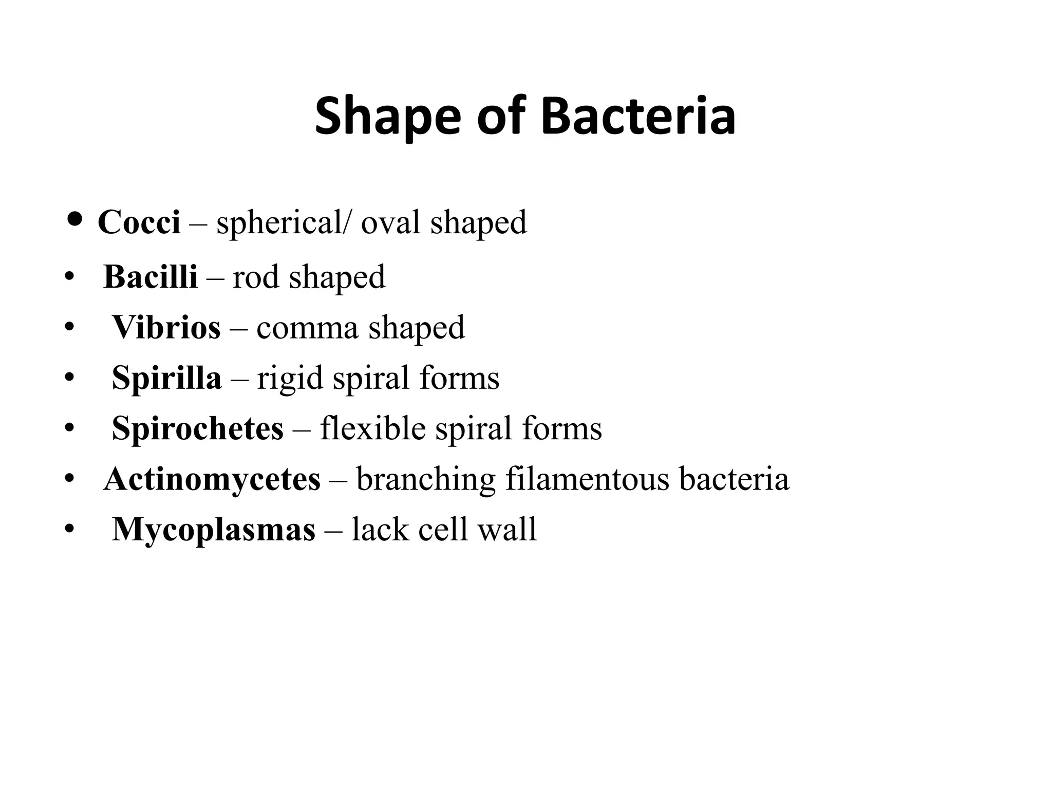
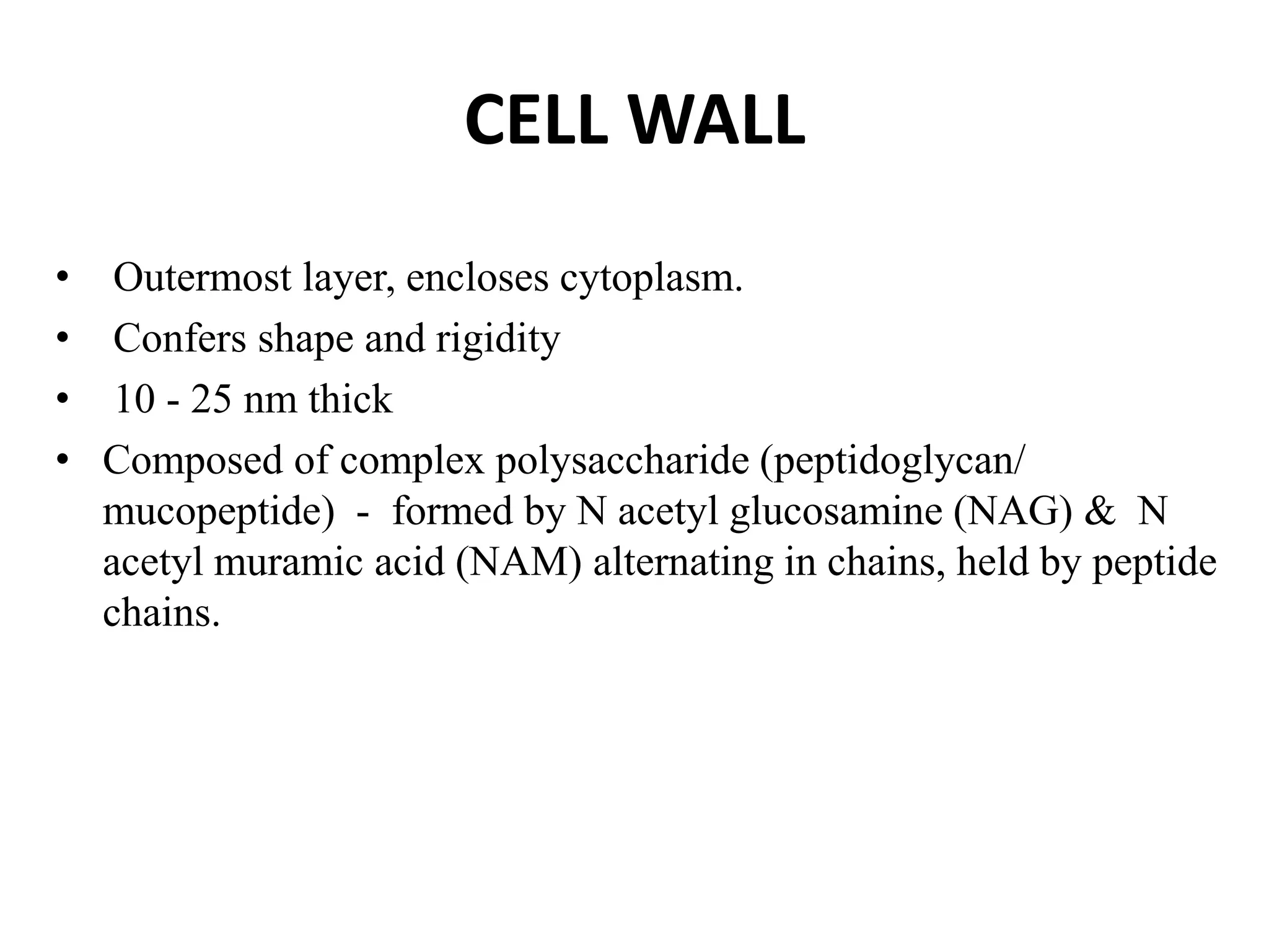
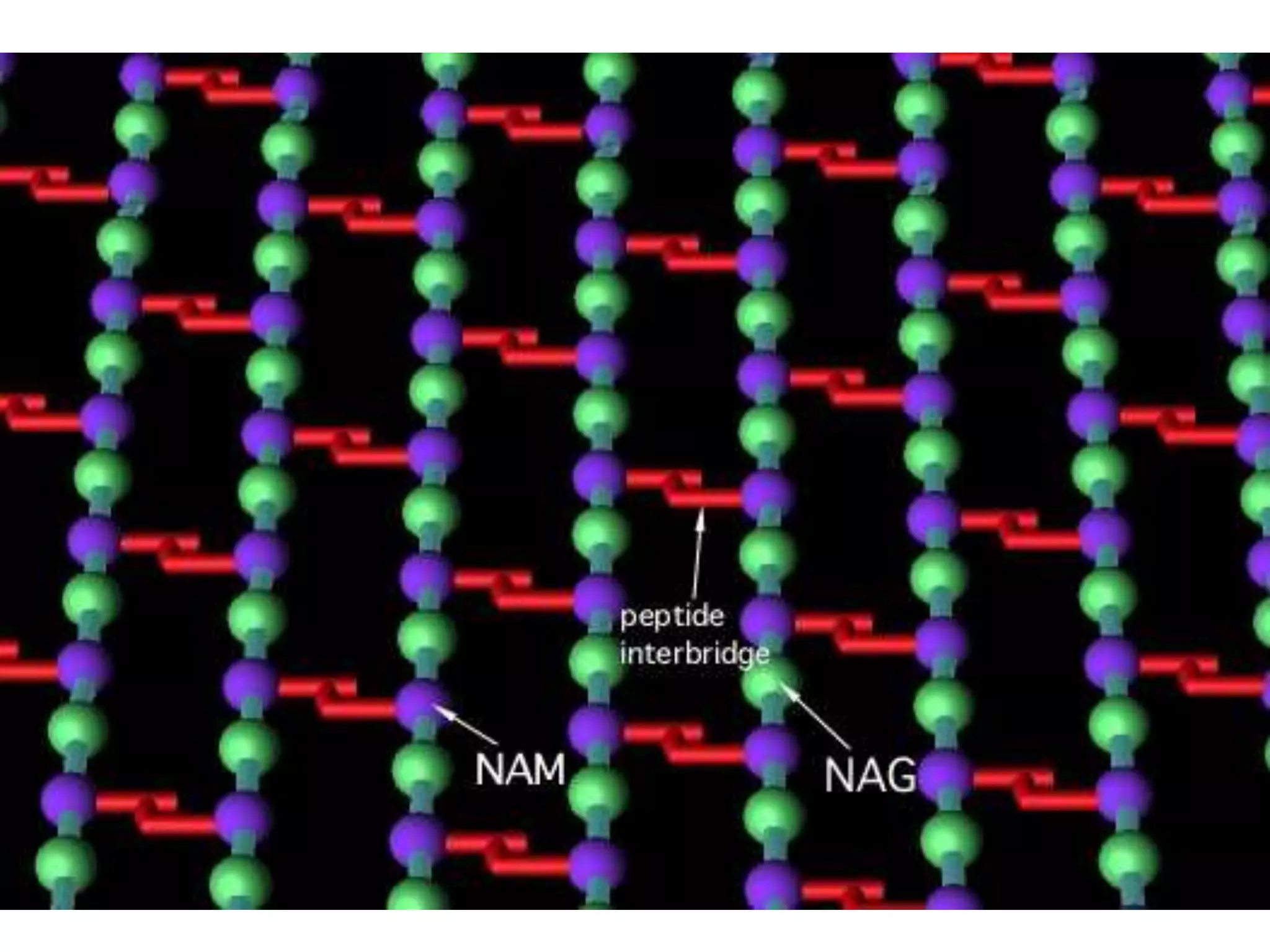
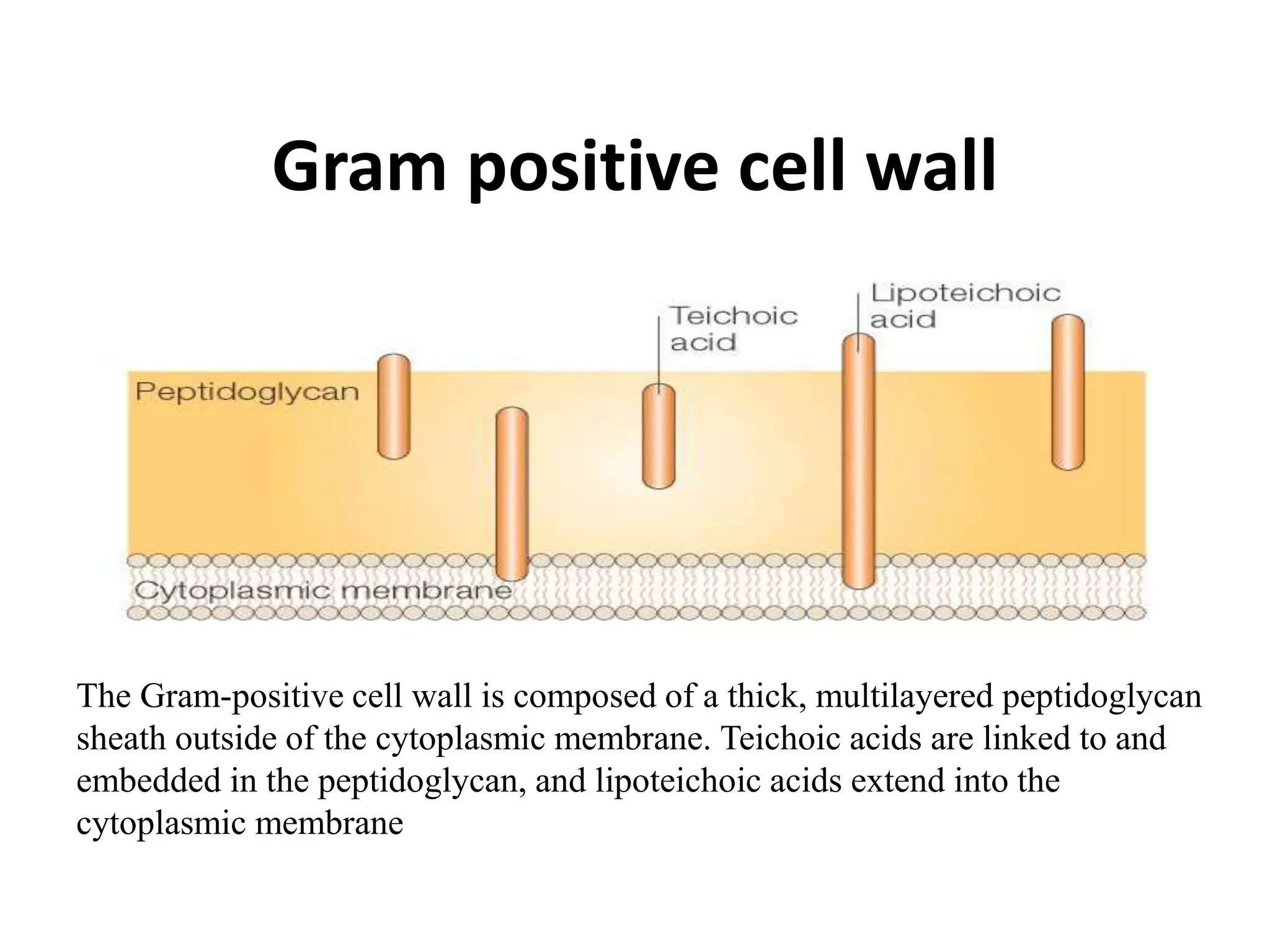
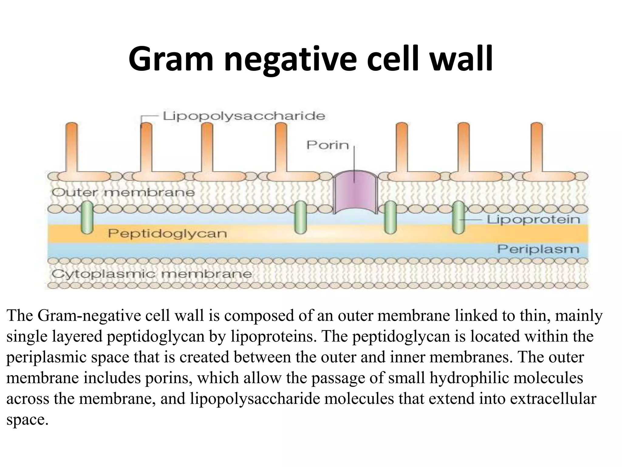
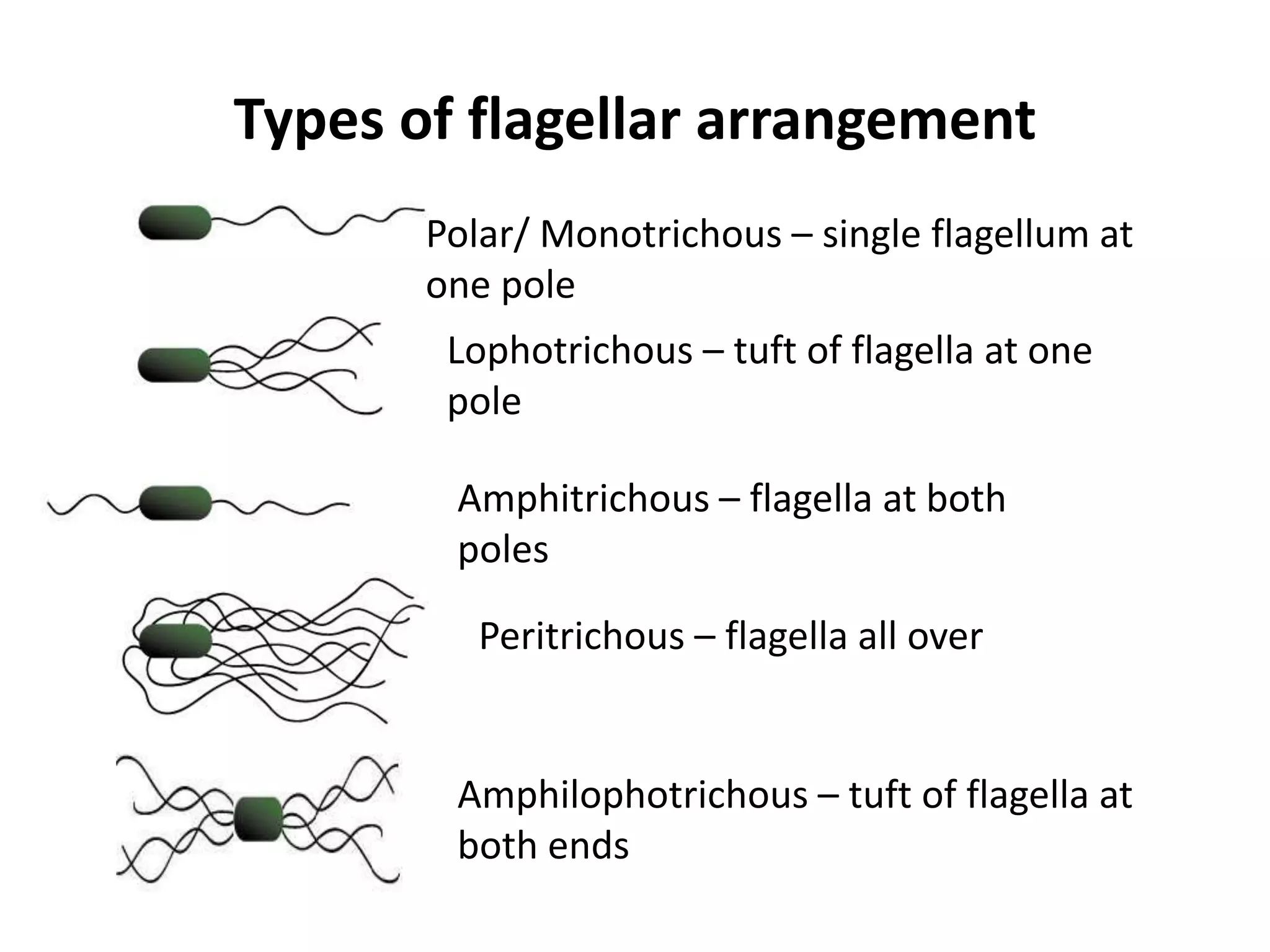
What is Physiology?
• Study of the mechanical, physical and biochemical
functions of living organisms.
Bacteria
• Bacteria are microscopic living organisms, usually
one celled , that can be found everywhere.
• They can be dangerous, such as when they cause
infection.
• Beneficial in the process of fermentation and
decomposition
Prokaryotes
• Bacteria are single celled microorganism containing a single
circular chromosome
• Bacteria lack Nuclear membrane, nucleolus, deoxyribo-
nucleoprotein.
• Mitotic division is absent in bacteria .
• Mitochondria, Lysosomes, Golgi apparatus, Endoplasmic
reticulum are absent in the bacterial cytoplasm.
Prokaryotic Cells
• Much smaller (microns) and more simple than eukaryotes .
• prokaryotes are molecules surrounded by a membrane and cell
wall.
• they lack a true nucleus and don’t have membrane bound
organelles like mitochondria, etc.
• large surface-to-volume ratio : nutrients can easily and rapidly
reach any part of the cells interior
Shape of Bacteria
• Cocci – spherical/ oval shaped
• Bacilli – rod shaped
• Vibrios – comma shaped
• Spirilla – rigid spiral forms
• Spirochetes – flexible spiral forms
• Actinomycetes – branching filamentous bacteria
• Mycoplasmas – lack cell wall
Structure & Function of Cell
Components
CELL WALL
• Outermost layer, encloses cytoplasm.
• Confers shape and rigidity
• 10 - 25 nm thick
• Composed of complex polysaccharide (peptidoglycan/
mucopeptide) - formed by N acetyl glucosamine (NAG) & N
acetyl muramic acid (NAM) alternating in chains, held by peptide
chains.
Cell wall…
• Carries bacterial antigens – important in virulence &
immunity.
• Chemical nature of the cell wall helps to divide bacteria into
two broad groups – Gram positive & Gram negative.
• Gram +ve bacteria have simpler chemical nature than Gram –
ve bacteria.
Gram positive cell wall
The Gram-positive cell wall is composed of a thick, multilayered peptidoglycan
sheath outside of the cytoplasmic membrane. Teichoic acids are linked to and
embedded in the peptidoglycan, and lipoteichoic acids extend into the
cytoplasmic membrane
Gram negative cell wall
The Gram-negative cell wall is composed of an outer membrane linked to thin, mainly
single layered peptidoglycan by lipoproteins. The peptidoglycan is located within the
periplasmic space that is created between the outer and inner membranes. The outer
membrane includes porins, which allow the passage of small hydrophilic molecules
across the membrane, and lipopolysaccharide molecules that extend into extracellular
space.
Cytoplasmic (Plasma) membrane
• Thin layer 5-10 nm, separates cell wall from cytoplasm.
• Acts as a semi permeable membrane, controls the inflow and
outflow of metabolites.
• Composed of lipoproteins with small amounts of
carbohydrates.
Other Cytoplasmic Components
• Ribosome's – protein synthesis.
• Mesosomes – Multilaminated structures formed as
invaginations of plasma membrane.
• Principal sites of respiratory enzymes.
• Coordinate nuclear & cytoplasmic division during binary
fission.
Nucleus
• No nucleolus.
• No nuclear membrane.
• Genome –
• single, circular double stranded DNA.
• Haploid.
• Divides by binary fission.
Additional Organelles
• Plasmid –
• Extranuclear genetic elements consisting of DNA.
• Transmitted to daughter cells during binary fission.
• May be transferred from one bacterium to another
• Not essential for life of the cell.
• Confer certain properties e.g. drug resistance, toxicity.
• Flagella –
• Long (3 to 12 μm), filamentous surface appendages.
• Organs of locomotion.
• Chemically, composed of proteins called flagellins.
• The number and distribution of flagella on the bacterial surface
are characteristic for a given species - hence are useful in
identifying and classifying bacteria.
Types of flagellar arrangement
Polar/ Monotrichous – single flagellum at
one pole
Lophotrichous – tuft of flagella at one
pole
Amphitrichous – flagella at both
poles
Peritrichous – flagella all over
Amphilophotrichous – tuft of flagella at
both ends
Spores
• Highly resistant resting stages.
• Endospores – formed inside cell.
• Due to deletion of exogenous nutrients / unfavourable environment
New microsoft office power point presentation (2)

New microsoft office power point presentation (2)

  • 2.
    What is Physiology? •Study of the mechanical, physical and biochemical functions of living organisms.
  • 3.
    Bacteria • Bacteria aremicroscopic living organisms, usually one celled , that can be found everywhere. • They can be dangerous, such as when they cause infection. • Beneficial in the process of fermentation and decomposition
  • 4.
    Prokaryotes • Bacteria aresingle celled microorganism containing a single circular chromosome • Bacteria lack Nuclear membrane, nucleolus, deoxyribo- nucleoprotein. • Mitotic division is absent in bacteria . • Mitochondria, Lysosomes, Golgi apparatus, Endoplasmic reticulum are absent in the bacterial cytoplasm.
  • 5.
    Prokaryotic Cells • Muchsmaller (microns) and more simple than eukaryotes . • prokaryotes are molecules surrounded by a membrane and cell wall. • they lack a true nucleus and don’t have membrane bound organelles like mitochondria, etc. • large surface-to-volume ratio : nutrients can easily and rapidly reach any part of the cells interior
  • 6.
    Shape of Bacteria •Cocci – spherical/ oval shaped • Bacilli – rod shaped • Vibrios – comma shaped • Spirilla – rigid spiral forms • Spirochetes – flexible spiral forms • Actinomycetes – branching filamentous bacteria • Mycoplasmas – lack cell wall
  • 7.
    Structure & Functionof Cell Components
  • 8.
    CELL WALL • Outermostlayer, encloses cytoplasm. • Confers shape and rigidity • 10 - 25 nm thick • Composed of complex polysaccharide (peptidoglycan/ mucopeptide) - formed by N acetyl glucosamine (NAG) & N acetyl muramic acid (NAM) alternating in chains, held by peptide chains.
  • 10.
    Cell wall… • Carriesbacterial antigens – important in virulence & immunity. • Chemical nature of the cell wall helps to divide bacteria into two broad groups – Gram positive & Gram negative. • Gram +ve bacteria have simpler chemical nature than Gram – ve bacteria.
  • 11.
    Gram positive cellwall The Gram-positive cell wall is composed of a thick, multilayered peptidoglycan sheath outside of the cytoplasmic membrane. Teichoic acids are linked to and embedded in the peptidoglycan, and lipoteichoic acids extend into the cytoplasmic membrane
  • 12.
    Gram negative cellwall The Gram-negative cell wall is composed of an outer membrane linked to thin, mainly single layered peptidoglycan by lipoproteins. The peptidoglycan is located within the periplasmic space that is created between the outer and inner membranes. The outer membrane includes porins, which allow the passage of small hydrophilic molecules across the membrane, and lipopolysaccharide molecules that extend into extracellular space.
  • 13.
    Cytoplasmic (Plasma) membrane •Thin layer 5-10 nm, separates cell wall from cytoplasm. • Acts as a semi permeable membrane, controls the inflow and outflow of metabolites. • Composed of lipoproteins with small amounts of carbohydrates.
  • 14.
    Other Cytoplasmic Components •Ribosome's – protein synthesis. • Mesosomes – Multilaminated structures formed as invaginations of plasma membrane. • Principal sites of respiratory enzymes. • Coordinate nuclear & cytoplasmic division during binary fission.
  • 15.
    Nucleus • No nucleolus. •No nuclear membrane. • Genome – • single, circular double stranded DNA. • Haploid. • Divides by binary fission.
  • 16.
    Additional Organelles • Plasmid– • Extranuclear genetic elements consisting of DNA. • Transmitted to daughter cells during binary fission. • May be transferred from one bacterium to another • Not essential for life of the cell. • Confer certain properties e.g. drug resistance, toxicity.
  • 17.
    • Flagella – •Long (3 to 12 μm), filamentous surface appendages. • Organs of locomotion. • Chemically, composed of proteins called flagellins. • The number and distribution of flagella on the bacterial surface are characteristic for a given species - hence are useful in identifying and classifying bacteria.
  • 18.
    Types of flagellararrangement Polar/ Monotrichous – single flagellum at one pole Lophotrichous – tuft of flagella at one pole Amphitrichous – flagella at both poles Peritrichous – flagella all over Amphilophotrichous – tuft of flagella at both ends
  • 19.
    Spores • Highly resistantresting stages. • Endospores – formed inside cell. • Due to deletion of exogenous nutrients / unfavourable environment