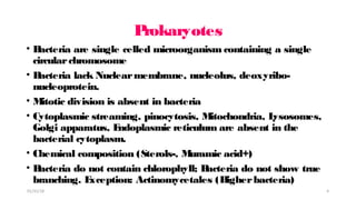
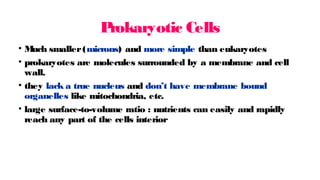
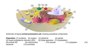
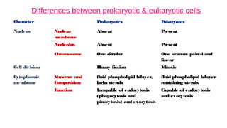
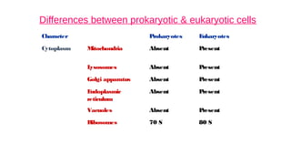
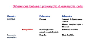
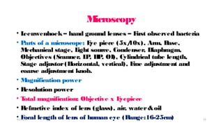
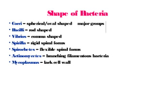
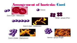
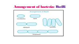
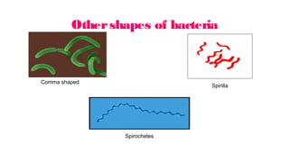
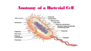
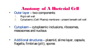
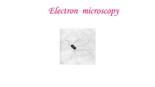
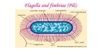
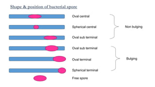
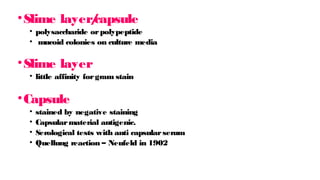
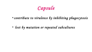
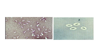
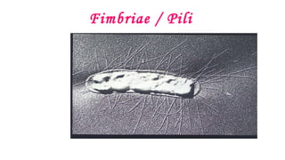
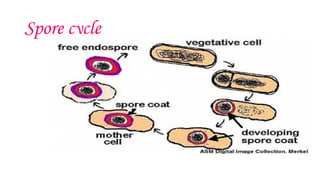
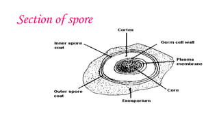
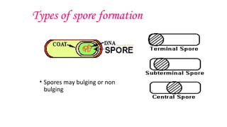
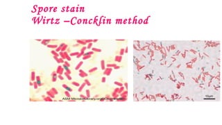
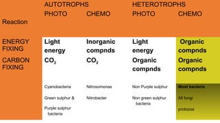
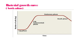
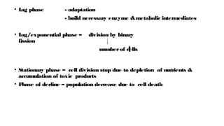
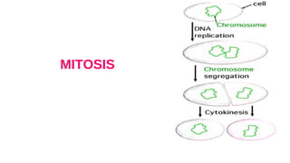
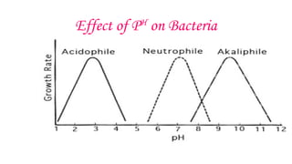
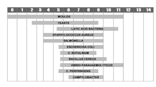
SlideShare a Scribd company logo
M
ORP
H
OL
OGY &
P
H
YSIOL
OGY OF
B
ACT
E
RIA
Dr. R. Someshwaran, M
D., Assistant professor,
Department of M
icrobiology, K
F
M
S&
R
Objectives of today’s class
T
o define the term prokaryote &eukaryote
T
o understand different shapes of bacteria
T
o describe bacterial anatomy in detail
T
o explain about bacterial growth curve
T
o know the factors affecting bacterial growth
01/31/18 2
Introduction
• L
iving organisms classified under3 kingdoms:
1.Plant kingdom
2.Animal kingdom
3.P
rotista
• Under Protista- Microorganismsareclassified as
i.P
ROK
ARYOT
E
S (Bacteria& Blue-green algae)
ii.E
UK
ARYOT
E
S (Fungi, Protozoa, slimemolds& other algae)
01/31/18 3
P
rokaryotes
• B
acteria are single celled microorganism containing a single
circularchromosome
• B
acteria lack Nuclearmembrane, nucleolus, deoxyribo-
nucleoprotein.
• M
itotic division is absent in bacteria
• Cytoplasmic streaming, pinocytosis, M
itochondria, L
ysosomes,
Golgi apparatus, E
ndoplasmic reticulum are absent in the
bacterial cytoplasm.
• Chemical composition (Sterols-, M
uramic acid+)
• B
acteria do not contain chlorophyll; B
acteria do not show true
branching. E
xception: Actinomycetales (H
igherbacteria)
01/31/18 4
P
rokaryotic Cells
• M
uch smaller(microns) and more simple than eukaryotes
• prokaryotes are molecules surrounded by a membrane and cell
wall.
• they lack a true nucleus and don’t have membrane bound
organelles like mitochondria, etc.
• large surface-to-volume ratio : nutrients can easily and rapidly
reach any part of the cells interior
Schematic of typical animal (eukaryotic) cell, showing subcellular components.
Organelles: (1) nucleolus (2) nucleus (3) ribosome (4) vesicle
(5) rough endoplasmic reticulum (ER) (6) Golgi apparatus (7) Cytoskeleton
(8) smooth ER (9) mitochondria (10) vacuole (11) cytoplasm (12)
lysosome (13) centrioles
Differences between prokaryotic & eukaryotic cells
Character Prokaryotes E
ukaryotes
Nucleus Nuclear
membrane
Absent Present
Nucleolus Absent Present
Chromosome One circular One ormore paired and
linear
Cell division B
inary fission M
itosis
Cytoplasmic
membrane
Structure and
Composition
fluid phospholipid bilayer,
lacks sterols
fluid phospholipid bilayer
containing sterols
F
unction Incapable of endocytosis
(phagocytosis and
pinocytosis) and exocytosis
Capable of endocytosis
and exocytosis
Differences between prokaryotic & eukaryotic cells
Character Prokaryotes E
ukaryotes
Cytoplasm M
itochondria Absent P
resent
L
ysosomes Absent P
resent
Golgi apparatus Absent P
resent
E
ndoplasmic
reticulum
Absent P
resent
Vacuoles Absent P
resent
Ribosomes 70 S 80 S
Differences between prokaryotic & eukaryotic cells
Character Prokaryotes E
ukaryotes
Cell W
all Present Animals &P
rotozoans –
Absent
P
lants, F
ungi &Algae -
P
resent
Composition Peptidoglycan –
complex carbohydrate
Cellulose orchitin
L
ocomotor
organelles
F
lagella F
lagella/Cilia
Size of bacteria
• Unit of measurement used in bacteriology is micron or
micrometre (µm)
• One micron is equal to one thousandth of millimetre
• One nanometre (nm) ormillimicron (mµ) is equal to
one thousandth of a micron orone millionth of a
millimetre
• One Angstrom unit (Å) is equal to one tenth of a
nanometre
01/31/18 10
L
imit of resolution
• L
imit of resolution with the unaided human eye is 200
microns (0.2mm)
• B
acteria are even much smallerand can be visualized
only undermagnification.
• B
acteria of medical importance generally measure 0.2-
1.5µm in diameterand about 3-5µm in length
01/31/18 11
M
icroscopy
• L
eeuwenhoek – hand ground lenses – F
irst observed bacteria
• P
arts of a microscope: E
ye piece (5x/
10x), Arm, B
ase,
M
echanical stage, L
ight source, Condenser, Diaphragm,
Objectives (Scanner, L
P
, H
P
, OI), Cylindrical tube length,
Stage adjustor(H
orizontal, vertical), F
ine adjustment and
coarse adjustment knob.
• M
agnification power
• Resolution power
• T
otal magnification: Objective x E
yepiece
• Refractive index of lens (glass), air, water&oil
• F
ocal length of lens of human eye (Range:16-25cm)
01/31/18 12
T
ypes of M
icroscopes
a. Optical orlight microscope
b. P
hase contrast microscope
c. F
luorescent microscope
d. E
lectron microscope
e. Dark field /Dark Ground microscope: 2 types namely
polarisation &interference microscopes
01/31/18 13
Staining techniques
• Simple stains providecolour contrast but impart samecolour
to all bacteria. Ex: Methyleneblue, Basic fuchsin.
• Negative staining provideuniformly coloured background
against which theunstained bacteriastand out in contrast. Ex:
Indiaink or Nigrosin.
• Impregnation methods for Cellsand structurestoo thin to be
seen under ordinary microscopethat may berendered visibleif
they arethickened by silver impregnation on thesurface. Used
for demonstration of Spirochetesand Bacterial flagella.
01/31/18 14
Staining techniques – Contd…
• Differential stains: Stainsimpart different colour to
different bacteriaor itsstructures. Ex: Gram stain & Acid fast
stain.
01/31/18 15
Shape of B
acteria
• Cocci – spherical/oval shaped majorgroups
• B
acilli – rod shaped
• Vibrios – comma shaped
• Spirilla – rigid spiral forms
• Spirochetes – flexible spiral forms
• Actinomycetes – branching filamentous bacteria
• M
ycoplasmas – lack cell wall
Arrangement of bacteria: Cocci
Cocci in pair – Diplococcus
Sarcina – groups of eight
Tetrad – groups of four
Cocci in chain - Streptococci
Cocci in cluster - Staphylococci
Coccus
Arrangement of bacteria: B
acilli
Othershapes of bacteria
Comma shaped
Spirochetes
Spirilla
Anatomy of a B
acterial Cell
Anatomy of A B
acterial Cell
• Outer layer – two components:
1. Rigid cell wall
2. Cytoplasmic (Cell/ Plasma) membrane– present beneath cell wall
• Cytoplasm – cytoplasmic inclusions, ribosomes,
mesosomesand nucleus
• Additional structures – plasmid, slimelayer, capsule,
flagella, fimbriae(pili), spores
Structure &F
unction of
Cell Components
CE
L
LW
AL
L
• Outermost layer, encloses cytoplasm
1. Confers shape and rigidity
2. 10 - 25 nm thick
3. Composed of complex polysaccharides (peptidoglycan/mucopeptide) -
formed by N acetyl glucosamine (NAG) & N acetyl muramic acid
(NAM
) alternating in chains, held by peptide chains.
• Cell wall –
4. Carries bacterial antigens – important in virulence &immunity
5. Chemical nature of the cell wall helps to divide bacteria into two broad
groups – Gram positive &Gram negative
6. Gram +ve bacteria have simplerchemical nature than Gram –ve
bacteria.
7. Several antibiotics may interfere with cell wall synthesis e.g. P
enicillin,
Cephalosporins
Gram positive cell wall
The Gram-positive cell wall is composed of a thick, multilayered peptidoglycan sheath outside of the
cytoplasmic membrane. Teichoic acids are linked to and embedded in the peptidoglycan, and
lipoteichoic acids extend into the cytoplasmic membrane
Gram negative cell wall
The Gram-negative cell wall is composed of an outer membrane linked to thin, mainly single-layered
peptidoglycan by lipoproteins. The peptidoglycan is located within the periplasmic space that is created
between the outer and inner membranes. The outer membrane includes porins, which allow the passage of
small hydrophilic molecules across the membrane, and lipopolysaccharide molecules that extend into
extracellular space.
Cell Wall
Differences between Gram positive &Gram
negative
Property of bacteria Gram Positive
Gram Negative
Thickness of wall 20-80 nm 10 nm
Number of layers in wall 1 2
Peptidoglycan content >50% 10-20%
Teichoic acid in wall + -
Lipid & lipoprotein content 0-3% 58%
Protein content 0% 9%
Lipopolysaccharide 0 13%
Sensitive to penicillin Yes Less sensitive
Digested by lysozyme Yes Weakly
Cytoplasmic (P
lasma) membrane
• T
hin layer5-10 nm, separates cell wall from cytoplasm
• Acts as a semipermeable membrane: controls the inflow and outflow
of metabolites
• Composed of lipoproteins with small amounts of carbohydrates
OtherCytoplasmic Components
• Ribosomes – protein synthesis
• M
esosomes –
1. M
ultilaminated structures formed as invaginations of plasma membrane
2. P
rincipal sites of respiratory enzymes
3. Coordinate nuclear&cytoplasmic division during binary fission
4. M
ore prominent in Gram +ve bacteria
• Intracytoplasmic inclusions – reserve of energy &phosphate forcell
metabolism e.g. M
etachromatic granules in diphtheria bacilli
Nucleus
• No nucleolus
• No nuclearmembrane
• Genome –
• single, circulardouble stranded DNA.
• H
aploid
• Divides by binary fission
Additional Organelles
1. P
lasmid –
• E
xtranucleargenetic elements consisting of DNA
• T
ransmitted to daughtercells during binary fission
• M
ay be transferred from one bacterium to another
• Not essential forlife of the cell
• Confercertain properties e.g. drug resistance, toxicity
Additional Organelles
2. Capsule &Slime layer–
• Viscous layersecreted around the cell wall.
• P
olysaccharide /polypeptide in nature
a) Capsule – sharply defined structure, antigenic in
nature
• P
rotects bacteria from lytic enzymes
• Inhibits phagocytosis
• Stained by negative staining using India Ink
• Can be demonstrated by Quellung reaction (capsule
swelling reaction)
a) Slime layer– loose undemarcated secretion
Additional Organelles
3. F
lagella –
L
ong (3 to 12 µm), filamentous surface appendages
Organs of locomotion
Chemically, composed of proteins called flagellins
T
he numberand distribution of flagella on the bacterial surface are characteristic fora
given species - hence are useful in identifying and classifying bacteria
F
lagella may serve as antigenic determinants (e.g. the Hantigens of Gram-negative
enteric bacteria)
P
resence shown by motility e.g. hanging drop preparation
T
ypes of flagellararrangement
Polar/ Monotrichous – single flagellum at one
pole
Lophotrichous – tuft of flagella at one pole
Peritrichous – flagella all over
Amphitrichous – flagella at both poles
Amphilophotrichous – tuft of flagella at both ends
Flagella
• Long unbranched sinuous filaments present on motile bacteria
• Made of 3 parts
• Filament, hook, and basal body
• Filament is external and hook basal body complex embedded in the cell
envelope
• Hook and basal body are antigenically different
• Detachment of flagella does not affect the viability of cell
• 3-20 m long, uniform diameter
μ
• M
ade up of a protein flagellin
• F
lagella of different genera - similarchemical composition they
are antigenically different
• F
lagellarantibodies help in serodiagnosis
Flagellar stains – Leifson stain
Electron microscopy
Flagella and fimbriae (Pili)
Shape & position of bacterial spore
Oval central
Spherical central
Oval sub terminal
Oval sub terminal
Oval terminal
Spherical terminal
Free spore
Non bulging
Bulging
Capsule/ slime layer
• B
acteria secrete viscid material
• organised into capsule - pneumococcus
• L
oose undemarcated secretion (slime layer) -
leuconostac
• M
icrocapsules – too thin to be seen underlight
microscope
• B
oth capsule and slime layermay be present
•Slime layer/
capsule
• polysaccharide orpolypeptide
• mucoid colonies on culture media
•Slime layer
• little affinity forgram stain
•Capsule
• stained by negative staining
• Capsularmaterial antigenic.
• Serological tests with anti capsularserum
• Quellung reaction – Neufeld in 1902
Capsule
•contribute to virulence by inhibiting phagocytosis
• lost by mutation or repeated subcultures
Fimbriae / Pili
• F
ine hairlike appendages
• Shorterand thinnerthan flagella projecting from cell membrane
• P
resent on both motile and non motile bacteria
• M
ade up of pilin
• 8 morphological types known
• Classified as common orsex pili
• Conferadhesive property to the cell
• Disappearfollowing subcultures
Properties
• Agglutinate red blood cells of guinea pigs, horses and pigs
strongly
• H
uman and sheep cells weakly
• Detected by hemagglutination
• Antigenically similarand may cause cross reaction in
serological tests
Sex pili
• L
ongerand fewer
• F
ound on male bacteria
• Attach to female bacteria
forming hollow conjugation
tubes through which genetic
material is transferred
Spores
• H
ighly resistant resting stages
• E
ndospores – formed inside cell
• Due to deletion of exogenous nutrients /unfavourable environment
• Young spores are seen attached to parent cell
• Appearas unstained area in gram stain
• Special spore stain can be used
Gram stain – Never comment
on spore
Spore cycle
Section of spore
Types of spore formation
• Spores may bulging or non
bulging
Clostridium tetani –
terminal spore
B
acillus anthracis –
central spore
Spore stain
Wirtz –Concklin method
Pleomorphism & involution forms
• Variation in shape and size of bacteria – pleomorphism
• Aberrant forms in ageing cultures – involution forms
• Due to defective cell wall synthesis
L forms
• Swollen cells and aberrant morphological forms observed by
Kleinberger Nobel at Lister institute, London
• Develop spontaneously or in the presence of agents that interfere
with cell wall synthesis
• May be stable or unstable
• Resemble mycobacteria
Gram variable bacteria
Pleomorphism & Involution forms
• P
leomorphism – great variation in shape &size of individual cells
e.g. Proteus species
• Involution forms – swollen &aberrant forms in ageing cultures,
especially in the presence of high salt concentration e.g. plague
bacillus
• Cause – defective cell wall synthesis
Bacterial Taxonomy
• Includes three components:
1. Classification : orderly arrangement
2. Identification of an unknown unit
3. Nomenclature: naming theunits
Bacterial Taxonomy: Classification
• Orderly arrangement :
• Kingdom – Division – Class– Order – Family – Tribe– Genus– Species
Phylogenetic classification – representsabranching treelikearrangement. One
characteristic being used for division at each branch or level
Molecular or Genetic classification – based on thedegreeof genetic relatednessof
different organisms
Intraspeciesclassification – based on biochemical properties(biotypes), antigenic
features(serotypes), bacteriophagesusceptibility (phagetypes)
Bacterial Taxonomy: Nomenclature
• T
wo kinds of name are given to bacteria
• Casual /common name – forlocal use, varies from country to country
e.g. “typhoid bacillus”
• Scientific /International Name – same all overworld, consists of two
words (in Italics) e.g. Salm
onellaty
phi,
Staphy
lococcus aureus
What is Physiology?
• Study of the mechanical, physical and biochemical functions
of living organisms.
Oxygen requirements
• Aerobic respiration is the release of energy from glucose or
anotherorganic substrate in the presence of Oxygen.
• Obligateaerobes-Pseudomonas
• Microaerophilic –Streptococcus
• Anaerobic respirationis oxidationof m
olecules intheabsenceof
oxy
gentoproduceenergy
• Obligateanaerobes - clostridia
• Facultativeanaerobes - vibrio
• Aerotolerantanaerobes - la
ctobacilli
• Ferm
entation
• conversionof carbohy
drates intoalcohols oracids underanaerobic
conditions.
• Eg: lactobacillus
Carbondioxide – factor affecting growth
• Small amount of CO2 is produced by most bacteria and used
endogenously formetabolism
• Capnophiles – bacteria which require a higherCO2 tension (5 –
10%) to grow.
• eg: B
rucellae, meningococcus , gonococcus
Bacterial vitamins
• Growth factors to be provided exogenously forgrowth.
• E
ssential
• Accessary
• H
.influenzae requires accessary factors X (haemin) and V
(NAD) present in blood to grow.
Effect of light
• Bacteria usually grow well in the dark.
• Phototrophs – need light to grow
• Pigmented bacteria like cyanobacteria
Reaction
AUTOTROPHS HETEROTROPHS
PHOTO CHEMO PHOTO CHEMO
ENERGY
FIXING
Light
energy
Inorganic
compnds
Light
energy
Organic
compnds
CARBON
FIXING
CO2 CO2 Organic
compnds
Organic
compnds
Cyanobacteria
Green sulphur &
Purple sulphur
bacteria
Nitrosomonas
Nitrobacter
Non Purple sulphur
Non green sulphur
bacteria
Most bacteria
All fungi
protozoa
B
acterial growth curve
( batch culture)
• L
ag phase - adaptation
- build necessary enzyme &metabolic intermediates
• L
og/exponential phase – division by binary
fission
numberof cells
• Stationary phase – cell division stop due to depletion of nutrients &
accumulation of toxic products
• P
hase of decline – population decrease due to cell death
MITOSIS
E
.coli M
.leprae
20 minutes 20 days
• At extremes we eitherhave a mass progeny (4000 tones) in
24 hours in a continuous culture (or) no replication at all.
• In laboratories it is always a batch culture where the nutrients
are limited and toxic products accumulate.
• GE
NE
RAT
ION T
IM
E
Bacterial count
• T
otal count – total numberof bacteria in the sample
• Direct counting
• Coultercounter
• Viable count – measures the numberof living organisms
• Dilution methods
• P
lating methods
Other factors affecting bacteria
• T
emperature
• P
H
• M
echanical stress
• Osmosis
Effect of T0
on
bacteria
Effect of PH
on Bacteria
Osmotic effect and mechanical stress
• Tolerant to osmotic variations esp Gram positive bacteria
• Sudden transfer from concentrated solution to distilled water –
plasmoptysis
• Mechanical stress like grinding, vigorous shaking with glass beads or
exposure ultrasonic vibration – rupture of cell
Summary
• P
rokaryote
• GPVs GN Difference
• P
leomorphic bacteria
• Lforms
• Cell appendages
01/31/18 83
T
hank you
01/31/18 84

More Related Content

Similar to morphphysioofbacteriadr-180131090251.pdf

Baterial morphology
Baterial morphologyBaterial morphology
Baterial morphology
ANUPAM KUMAR
 
Bacterial Morphology.ppt
Bacterial Morphology.pptBacterial Morphology.ppt
Bacterial Morphology.ppt
Dr. Rakesh Prasad Sah
 
Bacteria
BacteriaBacteria
Bacteria
S ghazal
 
Introduction to Bacteriology, Morphology & staining
Introduction to Bacteriology, Morphology & stainingIntroduction to Bacteriology, Morphology & staining
Introduction to Bacteriology, Morphology & staining
JNMC AMU, Aligarh, India
 
Bacterial amatomy.ppt
Bacterial amatomy.pptBacterial amatomy.ppt
Bacterial amatomy.ppt
BasraJaswinder
 
New microsoft office power point presentation (2)
New microsoft office power point presentation (2)New microsoft office power point presentation (2)
New microsoft office power point presentation (2)
salman ahmad
 
Morphology of bacteria
Morphology of bacteriaMorphology of bacteria
Morphology of bacteria
Snehal Patel
 
Ultra structure of Bacteria.......
Ultra   structure   of   Bacteria.......Ultra   structure   of   Bacteria.......
Ultra structure of Bacteria.......
Cherry
 
Ultra structure of bateria
Ultra structure of bateriaUltra structure of bateria
Ultra structure of bateria
HemantKurmi3
 
Bacteria 1
Bacteria 1  Bacteria 1
Bacteria 1
Pharmacy Universe
 
Morphologyof Bacteria.pptx
Morphologyof Bacteria.pptxMorphologyof Bacteria.pptx
Morphologyof Bacteria.pptx
OsmanHassan35
 
Lec 01. introduction to microbiology
Lec 01. introduction to microbiologyLec 01. introduction to microbiology
Lec 01. introduction to microbiology
Sebghatullah Mansoor
 
Lecture 2:- Bacterial morphology , bacterial cell structure
Lecture 2:- Bacterial morphology , bacterial cell structureLecture 2:- Bacterial morphology , bacterial cell structure
Lecture 2:- Bacterial morphology , bacterial cell structure
Raghda alomari
 
Structure of a prokaryotic cell
Structure of a prokaryotic cellStructure of a prokaryotic cell
Structure of a prokaryotic cell
Bahauddin Zakariya University lahore
 
Morophology of bacteria
Morophology of bacteriaMorophology of bacteria
structure of mo.pptx
structure of mo.pptxstructure of mo.pptx
structure of mo.pptx
DiptiPriya6
 
Presentation1.pptx
Presentation1.pptxPresentation1.pptx
Presentation1.pptx
SURYAPRAKASHDrKNModi
 
Chapter 4 functional anatomy of prok and euk partial
Chapter 4 functional anatomy of prok and euk partialChapter 4 functional anatomy of prok and euk partial
Chapter 4 functional anatomy of prok and euk partial
BilalHoushaymi
 
Morphology of Bacteriology
Morphology of BacteriologyMorphology of Bacteriology
Morphology of Bacteriology
hasini18052017
 
Bacterial morphology and classification
Bacterial morphology and classificationBacterial morphology and classification
Bacterial morphology and classification
Hariharan R
 

Similar to morphphysioofbacteriadr-180131090251.pdf (20)

Baterial morphology
Baterial morphologyBaterial morphology
Baterial morphology
 
Bacterial Morphology.ppt
Bacterial Morphology.pptBacterial Morphology.ppt
Bacterial Morphology.ppt
 
Bacteria
BacteriaBacteria
Bacteria
 
Introduction to Bacteriology, Morphology & staining
Introduction to Bacteriology, Morphology & stainingIntroduction to Bacteriology, Morphology & staining
Introduction to Bacteriology, Morphology & staining
 
Bacterial amatomy.ppt
Bacterial amatomy.pptBacterial amatomy.ppt
Bacterial amatomy.ppt
 
New microsoft office power point presentation (2)
New microsoft office power point presentation (2)New microsoft office power point presentation (2)
New microsoft office power point presentation (2)
 
Morphology of bacteria
Morphology of bacteriaMorphology of bacteria
Morphology of bacteria
 
Ultra structure of Bacteria.......
Ultra   structure   of   Bacteria.......Ultra   structure   of   Bacteria.......
Ultra structure of Bacteria.......
 
Ultra structure of bateria
Ultra structure of bateriaUltra structure of bateria
Ultra structure of bateria
 
Bacteria 1
Bacteria 1  Bacteria 1
Bacteria 1
 
Morphologyof Bacteria.pptx
Morphologyof Bacteria.pptxMorphologyof Bacteria.pptx
Morphologyof Bacteria.pptx
 
Lec 01. introduction to microbiology
Lec 01. introduction to microbiologyLec 01. introduction to microbiology
Lec 01. introduction to microbiology
 
Lecture 2:- Bacterial morphology , bacterial cell structure
Lecture 2:- Bacterial morphology , bacterial cell structureLecture 2:- Bacterial morphology , bacterial cell structure
Lecture 2:- Bacterial morphology , bacterial cell structure
 
Structure of a prokaryotic cell
Structure of a prokaryotic cellStructure of a prokaryotic cell
Structure of a prokaryotic cell
 
Morophology of bacteria
Morophology of bacteriaMorophology of bacteria
Morophology of bacteria
 
structure of mo.pptx
structure of mo.pptxstructure of mo.pptx
structure of mo.pptx
 
Presentation1.pptx
Presentation1.pptxPresentation1.pptx
Presentation1.pptx
 
Chapter 4 functional anatomy of prok and euk partial
Chapter 4 functional anatomy of prok and euk partialChapter 4 functional anatomy of prok and euk partial
Chapter 4 functional anatomy of prok and euk partial
 
Morphology of Bacteriology
Morphology of BacteriologyMorphology of Bacteriology
Morphology of Bacteriology
 
Bacterial morphology and classification
Bacterial morphology and classificationBacterial morphology and classification
Bacterial morphology and classification
 

More from dawitg2

bacterial plant pathogen jeyarajesh-190413122916.pptx
bacterial plant pathogen jeyarajesh-190413122916.pptxbacterial plant pathogen jeyarajesh-190413122916.pptx
bacterial plant pathogen jeyarajesh-190413122916.pptx
dawitg2
 
unit1_practical-applications-of-biotechnology.ppt
unit1_practical-applications-of-biotechnology.pptunit1_practical-applications-of-biotechnology.ppt
unit1_practical-applications-of-biotechnology.ppt
dawitg2
 
Introduction-to-Plant-Cell-Culture-lec1.ppt
Introduction-to-Plant-Cell-Culture-lec1.pptIntroduction-to-Plant-Cell-Culture-lec1.ppt
Introduction-to-Plant-Cell-Culture-lec1.ppt
dawitg2
 
fertilizers-150111082613-conversion-gate01.pdf
fertilizers-150111082613-conversion-gate01.pdffertilizers-150111082613-conversion-gate01.pdf
fertilizers-150111082613-conversion-gate01.pdf
dawitg2
 
sac-301-manuresfertilizersandsoilfertilitymanagement-210105122952.pdf
sac-301-manuresfertilizersandsoilfertilitymanagement-210105122952.pdfsac-301-manuresfertilizersandsoilfertilitymanagement-210105122952.pdf
sac-301-manuresfertilizersandsoilfertilitymanagement-210105122952.pdf
dawitg2
 
davindergill 135021014 -170426133338.pdf
davindergill 135021014 -170426133338.pdfdavindergill 135021014 -170426133338.pdf
davindergill 135021014 -170426133338.pdf
dawitg2
 
preparationofdifferentagro-chemicaldosesforfield-140203005533-phpapp02.pdf
preparationofdifferentagro-chemicaldosesforfield-140203005533-phpapp02.pdfpreparationofdifferentagro-chemicaldosesforfield-140203005533-phpapp02.pdf
preparationofdifferentagro-chemicaldosesforfield-140203005533-phpapp02.pdf
dawitg2
 
pesticide stoeage storage ppwscript.ppt
pesticide stoeage storage  ppwscript.pptpesticide stoeage storage  ppwscript.ppt
pesticide stoeage storage ppwscript.ppt
dawitg2
 
-pre-cautions--on--seed-storage--1-.pptx
-pre-cautions--on--seed-storage--1-.pptx-pre-cautions--on--seed-storage--1-.pptx
-pre-cautions--on--seed-storage--1-.pptx
dawitg2
 
Intgrated pest management 02 _ 06_05.pdf
Intgrated pest management 02 _ 06_05.pdfIntgrated pest management 02 _ 06_05.pdf
Intgrated pest management 02 _ 06_05.pdf
dawitg2
 
major potato and tomato disease in ethiopia .pptx
major potato and tomato disease in ethiopia .pptxmajor potato and tomato disease in ethiopia .pptx
major potato and tomato disease in ethiopia .pptx
dawitg2
 
pesticide formulation pp602lec1to2-211207073233.pdf
pesticide formulation pp602lec1to2-211207073233.pdfpesticide formulation pp602lec1to2-211207073233.pdf
pesticide formulation pp602lec1to2-211207073233.pdf
dawitg2
 
diseasespests-2013-130708184617-phpapp02.pptx
diseasespests-2013-130708184617-phpapp02.pptxdiseasespests-2013-130708184617-phpapp02.pptx
diseasespests-2013-130708184617-phpapp02.pptx
dawitg2
 
pesticide pp602lec1to2-211207073233.pptx
pesticide pp602lec1to2-211207073233.pptxpesticide pp602lec1to2-211207073233.pptx
pesticide pp602lec1to2-211207073233.pptx
dawitg2
 
agricultaraly important agrochemicals.ppt
agricultaraly important agrochemicals.pptagricultaraly important agrochemicals.ppt
agricultaraly important agrochemicals.ppt
dawitg2
 
agri pesticide chemistry-180722174627.pdf
agri pesticide chemistry-180722174627.pdfagri pesticide chemistry-180722174627.pdf
agri pesticide chemistry-180722174627.pdf
dawitg2
 
372922285 -important Fungal-Nutrition.ppt
372922285 -important Fungal-Nutrition.ppt372922285 -important Fungal-Nutrition.ppt
372922285 -important Fungal-Nutrition.ppt
dawitg2
 
disease development and pathogenesis-201118142432.pptx
disease development and pathogenesis-201118142432.pptxdisease development and pathogenesis-201118142432.pptx
disease development and pathogenesis-201118142432.pptx
dawitg2
 
ppp211lecture8-221211055228-824cf9da.pptx
ppp211lecture8-221211055228-824cf9da.pptxppp211lecture8-221211055228-824cf9da.pptx
ppp211lecture8-221211055228-824cf9da.pptx
dawitg2
 
defensemechanismsinplants-180308104711.pptx
defensemechanismsinplants-180308104711.pptxdefensemechanismsinplants-180308104711.pptx
defensemechanismsinplants-180308104711.pptx
dawitg2
 

More from dawitg2 (20)

bacterial plant pathogen jeyarajesh-190413122916.pptx
bacterial plant pathogen jeyarajesh-190413122916.pptxbacterial plant pathogen jeyarajesh-190413122916.pptx
bacterial plant pathogen jeyarajesh-190413122916.pptx
 
unit1_practical-applications-of-biotechnology.ppt
unit1_practical-applications-of-biotechnology.pptunit1_practical-applications-of-biotechnology.ppt
unit1_practical-applications-of-biotechnology.ppt
 
Introduction-to-Plant-Cell-Culture-lec1.ppt
Introduction-to-Plant-Cell-Culture-lec1.pptIntroduction-to-Plant-Cell-Culture-lec1.ppt
Introduction-to-Plant-Cell-Culture-lec1.ppt
 
fertilizers-150111082613-conversion-gate01.pdf
fertilizers-150111082613-conversion-gate01.pdffertilizers-150111082613-conversion-gate01.pdf
fertilizers-150111082613-conversion-gate01.pdf
 
sac-301-manuresfertilizersandsoilfertilitymanagement-210105122952.pdf
sac-301-manuresfertilizersandsoilfertilitymanagement-210105122952.pdfsac-301-manuresfertilizersandsoilfertilitymanagement-210105122952.pdf
sac-301-manuresfertilizersandsoilfertilitymanagement-210105122952.pdf
 
davindergill 135021014 -170426133338.pdf
davindergill 135021014 -170426133338.pdfdavindergill 135021014 -170426133338.pdf
davindergill 135021014 -170426133338.pdf
 
preparationofdifferentagro-chemicaldosesforfield-140203005533-phpapp02.pdf
preparationofdifferentagro-chemicaldosesforfield-140203005533-phpapp02.pdfpreparationofdifferentagro-chemicaldosesforfield-140203005533-phpapp02.pdf
preparationofdifferentagro-chemicaldosesforfield-140203005533-phpapp02.pdf
 
pesticide stoeage storage ppwscript.ppt
pesticide stoeage storage  ppwscript.pptpesticide stoeage storage  ppwscript.ppt
pesticide stoeage storage ppwscript.ppt
 
-pre-cautions--on--seed-storage--1-.pptx
-pre-cautions--on--seed-storage--1-.pptx-pre-cautions--on--seed-storage--1-.pptx
-pre-cautions--on--seed-storage--1-.pptx
 
Intgrated pest management 02 _ 06_05.pdf
Intgrated pest management 02 _ 06_05.pdfIntgrated pest management 02 _ 06_05.pdf
Intgrated pest management 02 _ 06_05.pdf
 
major potato and tomato disease in ethiopia .pptx
major potato and tomato disease in ethiopia .pptxmajor potato and tomato disease in ethiopia .pptx
major potato and tomato disease in ethiopia .pptx
 
pesticide formulation pp602lec1to2-211207073233.pdf
pesticide formulation pp602lec1to2-211207073233.pdfpesticide formulation pp602lec1to2-211207073233.pdf
pesticide formulation pp602lec1to2-211207073233.pdf
 
diseasespests-2013-130708184617-phpapp02.pptx
diseasespests-2013-130708184617-phpapp02.pptxdiseasespests-2013-130708184617-phpapp02.pptx
diseasespests-2013-130708184617-phpapp02.pptx
 
pesticide pp602lec1to2-211207073233.pptx
pesticide pp602lec1to2-211207073233.pptxpesticide pp602lec1to2-211207073233.pptx
pesticide pp602lec1to2-211207073233.pptx
 
agricultaraly important agrochemicals.ppt
agricultaraly important agrochemicals.pptagricultaraly important agrochemicals.ppt
agricultaraly important agrochemicals.ppt
 
agri pesticide chemistry-180722174627.pdf
agri pesticide chemistry-180722174627.pdfagri pesticide chemistry-180722174627.pdf
agri pesticide chemistry-180722174627.pdf
 
372922285 -important Fungal-Nutrition.ppt
372922285 -important Fungal-Nutrition.ppt372922285 -important Fungal-Nutrition.ppt
372922285 -important Fungal-Nutrition.ppt
 
disease development and pathogenesis-201118142432.pptx
disease development and pathogenesis-201118142432.pptxdisease development and pathogenesis-201118142432.pptx
disease development and pathogenesis-201118142432.pptx
 
ppp211lecture8-221211055228-824cf9da.pptx
ppp211lecture8-221211055228-824cf9da.pptxppp211lecture8-221211055228-824cf9da.pptx
ppp211lecture8-221211055228-824cf9da.pptx
 
defensemechanismsinplants-180308104711.pptx
defensemechanismsinplants-180308104711.pptxdefensemechanismsinplants-180308104711.pptx
defensemechanismsinplants-180308104711.pptx
 

Recently uploaded

The Roman Empire A Historical Colossus.pdf
The Roman Empire A Historical Colossus.pdfThe Roman Empire A Historical Colossus.pdf
The Roman Empire A Historical Colossus.pdf
kaushalkr1407
 
Supporting (UKRI) OA monographs at Salford.pptx
Supporting (UKRI) OA monographs at Salford.pptxSupporting (UKRI) OA monographs at Salford.pptx
Supporting (UKRI) OA monographs at Salford.pptx
Jisc
 
Lapbook sobre os Regimes Totalitários.pdf
Lapbook sobre os Regimes Totalitários.pdfLapbook sobre os Regimes Totalitários.pdf
Lapbook sobre os Regimes Totalitários.pdf
Jean Carlos Nunes Paixão
 
The basics of sentences session 5pptx.pptx
The basics of sentences session 5pptx.pptxThe basics of sentences session 5pptx.pptx
The basics of sentences session 5pptx.pptx
heathfieldcps1
 
Phrasal Verbs.XXXXXXXXXXXXXXXXXXXXXXXXXX
Phrasal Verbs.XXXXXXXXXXXXXXXXXXXXXXXXXXPhrasal Verbs.XXXXXXXXXXXXXXXXXXXXXXXXXX
Phrasal Verbs.XXXXXXXXXXXXXXXXXXXXXXXXXX
MIRIAMSALINAS13
 
Instructions for Submissions thorugh G- Classroom.pptx
Instructions for Submissions thorugh G- Classroom.pptxInstructions for Submissions thorugh G- Classroom.pptx
Instructions for Submissions thorugh G- Classroom.pptx
Jheel Barad
 
The French Revolution Class 9 Study Material pdf free download
The French Revolution Class 9 Study Material pdf free downloadThe French Revolution Class 9 Study Material pdf free download
The French Revolution Class 9 Study Material pdf free download
Vivekanand Anglo Vedic Academy
 
Acetabularia Information For Class 9 .docx
Acetabularia Information For Class 9  .docxAcetabularia Information For Class 9  .docx
Acetabularia Information For Class 9 .docx
vaibhavrinwa19
 
Unit 8 - Information and Communication Technology (Paper I).pdf
Unit 8 - Information and Communication Technology (Paper I).pdfUnit 8 - Information and Communication Technology (Paper I).pdf
Unit 8 - Information and Communication Technology (Paper I).pdf
Thiyagu K
 
Francesca Gottschalk - How can education support child empowerment.pptx
Francesca Gottschalk - How can education support child empowerment.pptxFrancesca Gottschalk - How can education support child empowerment.pptx
Francesca Gottschalk - How can education support child empowerment.pptx
EduSkills OECD
 
Language Across the Curriculm LAC B.Ed.
Language Across the  Curriculm LAC B.Ed.Language Across the  Curriculm LAC B.Ed.
Language Across the Curriculm LAC B.Ed.
Atul Kumar Singh
 
CLASS 11 CBSE B.St Project AIDS TO TRADE - INSURANCE
CLASS 11 CBSE B.St Project AIDS TO TRADE - INSURANCECLASS 11 CBSE B.St Project AIDS TO TRADE - INSURANCE
CLASS 11 CBSE B.St Project AIDS TO TRADE - INSURANCE
BhavyaRajput3
 
Thesis Statement for students diagnonsed withADHD.ppt
Thesis Statement for students diagnonsed withADHD.pptThesis Statement for students diagnonsed withADHD.ppt
Thesis Statement for students diagnonsed withADHD.ppt
EverAndrsGuerraGuerr
 
CACJapan - GROUP Presentation 1- Wk 4.pdf
CACJapan - GROUP Presentation 1- Wk 4.pdfCACJapan - GROUP Presentation 1- Wk 4.pdf
CACJapan - GROUP Presentation 1- Wk 4.pdf
camakaiclarkmusic
 
special B.ed 2nd year old paper_20240531.pdf
special B.ed 2nd year old paper_20240531.pdfspecial B.ed 2nd year old paper_20240531.pdf
special B.ed 2nd year old paper_20240531.pdf
Special education needs
 
A Strategic Approach: GenAI in Education
A Strategic Approach: GenAI in EducationA Strategic Approach: GenAI in Education
A Strategic Approach: GenAI in Education
Peter Windle
 
The approach at University of Liverpool.pptx
The approach at University of Liverpool.pptxThe approach at University of Liverpool.pptx
The approach at University of Liverpool.pptx
Jisc
 
The Accursed House by Émile Gaboriau.pptx
The Accursed House by Émile Gaboriau.pptxThe Accursed House by Émile Gaboriau.pptx
The Accursed House by Émile Gaboriau.pptx
DhatriParmar
 
The Challenger.pdf DNHS Official Publication
The Challenger.pdf DNHS Official PublicationThe Challenger.pdf DNHS Official Publication
The Challenger.pdf DNHS Official Publication
Delapenabediema
 
Mule 4.6 & Java 17 Upgrade | MuleSoft Mysore Meetup #46
Mule 4.6 & Java 17 Upgrade | MuleSoft Mysore Meetup #46Mule 4.6 & Java 17 Upgrade | MuleSoft Mysore Meetup #46
Mule 4.6 & Java 17 Upgrade | MuleSoft Mysore Meetup #46
MysoreMuleSoftMeetup
 

Recently uploaded (20)

The Roman Empire A Historical Colossus.pdf
The Roman Empire A Historical Colossus.pdfThe Roman Empire A Historical Colossus.pdf
The Roman Empire A Historical Colossus.pdf
 
Supporting (UKRI) OA monographs at Salford.pptx
Supporting (UKRI) OA monographs at Salford.pptxSupporting (UKRI) OA monographs at Salford.pptx
Supporting (UKRI) OA monographs at Salford.pptx
 
Lapbook sobre os Regimes Totalitários.pdf
Lapbook sobre os Regimes Totalitários.pdfLapbook sobre os Regimes Totalitários.pdf
Lapbook sobre os Regimes Totalitários.pdf
 
The basics of sentences session 5pptx.pptx
The basics of sentences session 5pptx.pptxThe basics of sentences session 5pptx.pptx
The basics of sentences session 5pptx.pptx
 
Phrasal Verbs.XXXXXXXXXXXXXXXXXXXXXXXXXX
Phrasal Verbs.XXXXXXXXXXXXXXXXXXXXXXXXXXPhrasal Verbs.XXXXXXXXXXXXXXXXXXXXXXXXXX
Phrasal Verbs.XXXXXXXXXXXXXXXXXXXXXXXXXX
 
Instructions for Submissions thorugh G- Classroom.pptx
Instructions for Submissions thorugh G- Classroom.pptxInstructions for Submissions thorugh G- Classroom.pptx
Instructions for Submissions thorugh G- Classroom.pptx
 
The French Revolution Class 9 Study Material pdf free download
The French Revolution Class 9 Study Material pdf free downloadThe French Revolution Class 9 Study Material pdf free download
The French Revolution Class 9 Study Material pdf free download
 
Acetabularia Information For Class 9 .docx
Acetabularia Information For Class 9  .docxAcetabularia Information For Class 9  .docx
Acetabularia Information For Class 9 .docx
 
Unit 8 - Information and Communication Technology (Paper I).pdf
Unit 8 - Information and Communication Technology (Paper I).pdfUnit 8 - Information and Communication Technology (Paper I).pdf
Unit 8 - Information and Communication Technology (Paper I).pdf
 
Francesca Gottschalk - How can education support child empowerment.pptx
Francesca Gottschalk - How can education support child empowerment.pptxFrancesca Gottschalk - How can education support child empowerment.pptx
Francesca Gottschalk - How can education support child empowerment.pptx
 
Language Across the Curriculm LAC B.Ed.
Language Across the  Curriculm LAC B.Ed.Language Across the  Curriculm LAC B.Ed.
Language Across the Curriculm LAC B.Ed.
 
CLASS 11 CBSE B.St Project AIDS TO TRADE - INSURANCE
CLASS 11 CBSE B.St Project AIDS TO TRADE - INSURANCECLASS 11 CBSE B.St Project AIDS TO TRADE - INSURANCE
CLASS 11 CBSE B.St Project AIDS TO TRADE - INSURANCE
 
Thesis Statement for students diagnonsed withADHD.ppt
Thesis Statement for students diagnonsed withADHD.pptThesis Statement for students diagnonsed withADHD.ppt
Thesis Statement for students diagnonsed withADHD.ppt
 
CACJapan - GROUP Presentation 1- Wk 4.pdf
CACJapan - GROUP Presentation 1- Wk 4.pdfCACJapan - GROUP Presentation 1- Wk 4.pdf
CACJapan - GROUP Presentation 1- Wk 4.pdf
 
special B.ed 2nd year old paper_20240531.pdf
special B.ed 2nd year old paper_20240531.pdfspecial B.ed 2nd year old paper_20240531.pdf
special B.ed 2nd year old paper_20240531.pdf
 
A Strategic Approach: GenAI in Education
A Strategic Approach: GenAI in EducationA Strategic Approach: GenAI in Education
A Strategic Approach: GenAI in Education
 
The approach at University of Liverpool.pptx
The approach at University of Liverpool.pptxThe approach at University of Liverpool.pptx
The approach at University of Liverpool.pptx
 
The Accursed House by Émile Gaboriau.pptx
The Accursed House by Émile Gaboriau.pptxThe Accursed House by Émile Gaboriau.pptx
The Accursed House by Émile Gaboriau.pptx
 
The Challenger.pdf DNHS Official Publication
The Challenger.pdf DNHS Official PublicationThe Challenger.pdf DNHS Official Publication
The Challenger.pdf DNHS Official Publication
 
Mule 4.6 & Java 17 Upgrade | MuleSoft Mysore Meetup #46
Mule 4.6 & Java 17 Upgrade | MuleSoft Mysore Meetup #46Mule 4.6 & Java 17 Upgrade | MuleSoft Mysore Meetup #46
Mule 4.6 & Java 17 Upgrade | MuleSoft Mysore Meetup #46
 

morphphysioofbacteriadr-180131090251.pdf

  • 1. M ORP H OL OGY & P H YSIOL OGY OF B ACT E RIA Dr. R. Someshwaran, M D., Assistant professor, Department of M icrobiology, K F M S& R
  • 2. Objectives of today’s class T o define the term prokaryote &eukaryote T o understand different shapes of bacteria T o describe bacterial anatomy in detail T o explain about bacterial growth curve T o know the factors affecting bacterial growth 01/31/18 2
  • 3. Introduction • L iving organisms classified under3 kingdoms: 1.Plant kingdom 2.Animal kingdom 3.P rotista • Under Protista- Microorganismsareclassified as i.P ROK ARYOT E S (Bacteria& Blue-green algae) ii.E UK ARYOT E S (Fungi, Protozoa, slimemolds& other algae) 01/31/18 3
  • 4. P rokaryotes • B acteria are single celled microorganism containing a single circularchromosome • B acteria lack Nuclearmembrane, nucleolus, deoxyribo- nucleoprotein. • M itotic division is absent in bacteria • Cytoplasmic streaming, pinocytosis, M itochondria, L ysosomes, Golgi apparatus, E ndoplasmic reticulum are absent in the bacterial cytoplasm. • Chemical composition (Sterols-, M uramic acid+) • B acteria do not contain chlorophyll; B acteria do not show true branching. E xception: Actinomycetales (H igherbacteria) 01/31/18 4
  • 5. P rokaryotic Cells • M uch smaller(microns) and more simple than eukaryotes • prokaryotes are molecules surrounded by a membrane and cell wall. • they lack a true nucleus and don’t have membrane bound organelles like mitochondria, etc. • large surface-to-volume ratio : nutrients can easily and rapidly reach any part of the cells interior
  • 6. Schematic of typical animal (eukaryotic) cell, showing subcellular components. Organelles: (1) nucleolus (2) nucleus (3) ribosome (4) vesicle (5) rough endoplasmic reticulum (ER) (6) Golgi apparatus (7) Cytoskeleton (8) smooth ER (9) mitochondria (10) vacuole (11) cytoplasm (12) lysosome (13) centrioles
  • 7. Differences between prokaryotic & eukaryotic cells Character Prokaryotes E ukaryotes Nucleus Nuclear membrane Absent Present Nucleolus Absent Present Chromosome One circular One ormore paired and linear Cell division B inary fission M itosis Cytoplasmic membrane Structure and Composition fluid phospholipid bilayer, lacks sterols fluid phospholipid bilayer containing sterols F unction Incapable of endocytosis (phagocytosis and pinocytosis) and exocytosis Capable of endocytosis and exocytosis
  • 8. Differences between prokaryotic & eukaryotic cells Character Prokaryotes E ukaryotes Cytoplasm M itochondria Absent P resent L ysosomes Absent P resent Golgi apparatus Absent P resent E ndoplasmic reticulum Absent P resent Vacuoles Absent P resent Ribosomes 70 S 80 S
  • 9. Differences between prokaryotic & eukaryotic cells Character Prokaryotes E ukaryotes Cell W all Present Animals &P rotozoans – Absent P lants, F ungi &Algae - P resent Composition Peptidoglycan – complex carbohydrate Cellulose orchitin L ocomotor organelles F lagella F lagella/Cilia
  • 10. Size of bacteria • Unit of measurement used in bacteriology is micron or micrometre (µm) • One micron is equal to one thousandth of millimetre • One nanometre (nm) ormillimicron (mµ) is equal to one thousandth of a micron orone millionth of a millimetre • One Angstrom unit (Å) is equal to one tenth of a nanometre 01/31/18 10
  • 11. L imit of resolution • L imit of resolution with the unaided human eye is 200 microns (0.2mm) • B acteria are even much smallerand can be visualized only undermagnification. • B acteria of medical importance generally measure 0.2- 1.5µm in diameterand about 3-5µm in length 01/31/18 11
  • 12. M icroscopy • L eeuwenhoek – hand ground lenses – F irst observed bacteria • P arts of a microscope: E ye piece (5x/ 10x), Arm, B ase, M echanical stage, L ight source, Condenser, Diaphragm, Objectives (Scanner, L P , H P , OI), Cylindrical tube length, Stage adjustor(H orizontal, vertical), F ine adjustment and coarse adjustment knob. • M agnification power • Resolution power • T otal magnification: Objective x E yepiece • Refractive index of lens (glass), air, water&oil • F ocal length of lens of human eye (Range:16-25cm) 01/31/18 12
  • 13. T ypes of M icroscopes a. Optical orlight microscope b. P hase contrast microscope c. F luorescent microscope d. E lectron microscope e. Dark field /Dark Ground microscope: 2 types namely polarisation &interference microscopes 01/31/18 13
  • 14. Staining techniques • Simple stains providecolour contrast but impart samecolour to all bacteria. Ex: Methyleneblue, Basic fuchsin. • Negative staining provideuniformly coloured background against which theunstained bacteriastand out in contrast. Ex: Indiaink or Nigrosin. • Impregnation methods for Cellsand structurestoo thin to be seen under ordinary microscopethat may berendered visibleif they arethickened by silver impregnation on thesurface. Used for demonstration of Spirochetesand Bacterial flagella. 01/31/18 14
  • 15. Staining techniques – Contd… • Differential stains: Stainsimpart different colour to different bacteriaor itsstructures. Ex: Gram stain & Acid fast stain. 01/31/18 15
  • 16. Shape of B acteria • Cocci – spherical/oval shaped majorgroups • B acilli – rod shaped • Vibrios – comma shaped • Spirilla – rigid spiral forms • Spirochetes – flexible spiral forms • Actinomycetes – branching filamentous bacteria • M ycoplasmas – lack cell wall
  • 17. Arrangement of bacteria: Cocci Cocci in pair – Diplococcus Sarcina – groups of eight Tetrad – groups of four Cocci in chain - Streptococci Cocci in cluster - Staphylococci Coccus
  • 19. Othershapes of bacteria Comma shaped Spirochetes Spirilla
  • 20. Anatomy of a B acterial Cell
  • 21. Anatomy of A B acterial Cell • Outer layer – two components: 1. Rigid cell wall 2. Cytoplasmic (Cell/ Plasma) membrane– present beneath cell wall • Cytoplasm – cytoplasmic inclusions, ribosomes, mesosomesand nucleus • Additional structures – plasmid, slimelayer, capsule, flagella, fimbriae(pili), spores
  • 23. CE L LW AL L • Outermost layer, encloses cytoplasm 1. Confers shape and rigidity 2. 10 - 25 nm thick 3. Composed of complex polysaccharides (peptidoglycan/mucopeptide) - formed by N acetyl glucosamine (NAG) & N acetyl muramic acid (NAM ) alternating in chains, held by peptide chains.
  • 24. • Cell wall – 4. Carries bacterial antigens – important in virulence &immunity 5. Chemical nature of the cell wall helps to divide bacteria into two broad groups – Gram positive &Gram negative 6. Gram +ve bacteria have simplerchemical nature than Gram –ve bacteria. 7. Several antibiotics may interfere with cell wall synthesis e.g. P enicillin, Cephalosporins
  • 25. Gram positive cell wall The Gram-positive cell wall is composed of a thick, multilayered peptidoglycan sheath outside of the cytoplasmic membrane. Teichoic acids are linked to and embedded in the peptidoglycan, and lipoteichoic acids extend into the cytoplasmic membrane
  • 26. Gram negative cell wall The Gram-negative cell wall is composed of an outer membrane linked to thin, mainly single-layered peptidoglycan by lipoproteins. The peptidoglycan is located within the periplasmic space that is created between the outer and inner membranes. The outer membrane includes porins, which allow the passage of small hydrophilic molecules across the membrane, and lipopolysaccharide molecules that extend into extracellular space.
  • 28. Differences between Gram positive &Gram negative Property of bacteria Gram Positive Gram Negative Thickness of wall 20-80 nm 10 nm Number of layers in wall 1 2 Peptidoglycan content >50% 10-20% Teichoic acid in wall + - Lipid & lipoprotein content 0-3% 58% Protein content 0% 9% Lipopolysaccharide 0 13% Sensitive to penicillin Yes Less sensitive Digested by lysozyme Yes Weakly
  • 29. Cytoplasmic (P lasma) membrane • T hin layer5-10 nm, separates cell wall from cytoplasm • Acts as a semipermeable membrane: controls the inflow and outflow of metabolites • Composed of lipoproteins with small amounts of carbohydrates
  • 30. OtherCytoplasmic Components • Ribosomes – protein synthesis • M esosomes – 1. M ultilaminated structures formed as invaginations of plasma membrane 2. P rincipal sites of respiratory enzymes 3. Coordinate nuclear&cytoplasmic division during binary fission 4. M ore prominent in Gram +ve bacteria • Intracytoplasmic inclusions – reserve of energy &phosphate forcell metabolism e.g. M etachromatic granules in diphtheria bacilli
  • 31. Nucleus • No nucleolus • No nuclearmembrane • Genome – • single, circulardouble stranded DNA. • H aploid • Divides by binary fission
  • 32. Additional Organelles 1. P lasmid – • E xtranucleargenetic elements consisting of DNA • T ransmitted to daughtercells during binary fission • M ay be transferred from one bacterium to another • Not essential forlife of the cell • Confercertain properties e.g. drug resistance, toxicity
  • 33. Additional Organelles 2. Capsule &Slime layer– • Viscous layersecreted around the cell wall. • P olysaccharide /polypeptide in nature a) Capsule – sharply defined structure, antigenic in nature • P rotects bacteria from lytic enzymes • Inhibits phagocytosis • Stained by negative staining using India Ink • Can be demonstrated by Quellung reaction (capsule swelling reaction) a) Slime layer– loose undemarcated secretion
  • 34. Additional Organelles 3. F lagella – L ong (3 to 12 µm), filamentous surface appendages Organs of locomotion Chemically, composed of proteins called flagellins T he numberand distribution of flagella on the bacterial surface are characteristic fora given species - hence are useful in identifying and classifying bacteria F lagella may serve as antigenic determinants (e.g. the Hantigens of Gram-negative enteric bacteria) P resence shown by motility e.g. hanging drop preparation
  • 35. T ypes of flagellararrangement Polar/ Monotrichous – single flagellum at one pole Lophotrichous – tuft of flagella at one pole Peritrichous – flagella all over Amphitrichous – flagella at both poles Amphilophotrichous – tuft of flagella at both ends
  • 36. Flagella • Long unbranched sinuous filaments present on motile bacteria • Made of 3 parts • Filament, hook, and basal body • Filament is external and hook basal body complex embedded in the cell envelope • Hook and basal body are antigenically different
  • 37.
  • 38. • Detachment of flagella does not affect the viability of cell • 3-20 m long, uniform diameter μ • M ade up of a protein flagellin • F lagella of different genera - similarchemical composition they are antigenically different • F lagellarantibodies help in serodiagnosis
  • 39. Flagellar stains – Leifson stain
  • 42. Shape & position of bacterial spore Oval central Spherical central Oval sub terminal Oval sub terminal Oval terminal Spherical terminal Free spore Non bulging Bulging
  • 43. Capsule/ slime layer • B acteria secrete viscid material • organised into capsule - pneumococcus • L oose undemarcated secretion (slime layer) - leuconostac • M icrocapsules – too thin to be seen underlight microscope • B oth capsule and slime layermay be present
  • 44. •Slime layer/ capsule • polysaccharide orpolypeptide • mucoid colonies on culture media •Slime layer • little affinity forgram stain •Capsule • stained by negative staining • Capsularmaterial antigenic. • Serological tests with anti capsularserum • Quellung reaction – Neufeld in 1902
  • 45. Capsule •contribute to virulence by inhibiting phagocytosis • lost by mutation or repeated subcultures
  • 46.
  • 48. • F ine hairlike appendages • Shorterand thinnerthan flagella projecting from cell membrane • P resent on both motile and non motile bacteria • M ade up of pilin • 8 morphological types known • Classified as common orsex pili • Conferadhesive property to the cell • Disappearfollowing subcultures
  • 49. Properties • Agglutinate red blood cells of guinea pigs, horses and pigs strongly • H uman and sheep cells weakly • Detected by hemagglutination • Antigenically similarand may cause cross reaction in serological tests
  • 50. Sex pili • L ongerand fewer • F ound on male bacteria • Attach to female bacteria forming hollow conjugation tubes through which genetic material is transferred
  • 51.
  • 52. Spores • H ighly resistant resting stages • E ndospores – formed inside cell • Due to deletion of exogenous nutrients /unfavourable environment • Young spores are seen attached to parent cell • Appearas unstained area in gram stain • Special spore stain can be used
  • 53. Gram stain – Never comment on spore
  • 56. Types of spore formation • Spores may bulging or non bulging
  • 57. Clostridium tetani – terminal spore B acillus anthracis – central spore
  • 59. Pleomorphism & involution forms • Variation in shape and size of bacteria – pleomorphism • Aberrant forms in ageing cultures – involution forms • Due to defective cell wall synthesis
  • 60. L forms • Swollen cells and aberrant morphological forms observed by Kleinberger Nobel at Lister institute, London • Develop spontaneously or in the presence of agents that interfere with cell wall synthesis • May be stable or unstable • Resemble mycobacteria
  • 62. Pleomorphism & Involution forms • P leomorphism – great variation in shape &size of individual cells e.g. Proteus species • Involution forms – swollen &aberrant forms in ageing cultures, especially in the presence of high salt concentration e.g. plague bacillus • Cause – defective cell wall synthesis
  • 63. Bacterial Taxonomy • Includes three components: 1. Classification : orderly arrangement 2. Identification of an unknown unit 3. Nomenclature: naming theunits
  • 64. Bacterial Taxonomy: Classification • Orderly arrangement : • Kingdom – Division – Class– Order – Family – Tribe– Genus– Species Phylogenetic classification – representsabranching treelikearrangement. One characteristic being used for division at each branch or level Molecular or Genetic classification – based on thedegreeof genetic relatednessof different organisms Intraspeciesclassification – based on biochemical properties(biotypes), antigenic features(serotypes), bacteriophagesusceptibility (phagetypes)
  • 65. Bacterial Taxonomy: Nomenclature • T wo kinds of name are given to bacteria • Casual /common name – forlocal use, varies from country to country e.g. “typhoid bacillus” • Scientific /International Name – same all overworld, consists of two words (in Italics) e.g. Salm onellaty phi, Staphy lococcus aureus
  • 66. What is Physiology? • Study of the mechanical, physical and biochemical functions of living organisms.
  • 67. Oxygen requirements • Aerobic respiration is the release of energy from glucose or anotherorganic substrate in the presence of Oxygen. • Obligateaerobes-Pseudomonas • Microaerophilic –Streptococcus
  • 68. • Anaerobic respirationis oxidationof m olecules intheabsenceof oxy gentoproduceenergy • Obligateanaerobes - clostridia • Facultativeanaerobes - vibrio • Aerotolerantanaerobes - la ctobacilli • Ferm entation • conversionof carbohy drates intoalcohols oracids underanaerobic conditions. • Eg: lactobacillus
  • 69. Carbondioxide – factor affecting growth • Small amount of CO2 is produced by most bacteria and used endogenously formetabolism • Capnophiles – bacteria which require a higherCO2 tension (5 – 10%) to grow. • eg: B rucellae, meningococcus , gonococcus
  • 70. Bacterial vitamins • Growth factors to be provided exogenously forgrowth. • E ssential • Accessary • H .influenzae requires accessary factors X (haemin) and V (NAD) present in blood to grow.
  • 71. Effect of light • Bacteria usually grow well in the dark. • Phototrophs – need light to grow • Pigmented bacteria like cyanobacteria
  • 72. Reaction AUTOTROPHS HETEROTROPHS PHOTO CHEMO PHOTO CHEMO ENERGY FIXING Light energy Inorganic compnds Light energy Organic compnds CARBON FIXING CO2 CO2 Organic compnds Organic compnds Cyanobacteria Green sulphur & Purple sulphur bacteria Nitrosomonas Nitrobacter Non Purple sulphur Non green sulphur bacteria Most bacteria All fungi protozoa
  • 73. B acterial growth curve ( batch culture)
  • 74. • L ag phase - adaptation - build necessary enzyme &metabolic intermediates • L og/exponential phase – division by binary fission numberof cells • Stationary phase – cell division stop due to depletion of nutrients & accumulation of toxic products • P hase of decline – population decrease due to cell death
  • 76. E .coli M .leprae 20 minutes 20 days • At extremes we eitherhave a mass progeny (4000 tones) in 24 hours in a continuous culture (or) no replication at all. • In laboratories it is always a batch culture where the nutrients are limited and toxic products accumulate. • GE NE RAT ION T IM E
  • 77. Bacterial count • T otal count – total numberof bacteria in the sample • Direct counting • Coultercounter • Viable count – measures the numberof living organisms • Dilution methods • P lating methods
  • 78. Other factors affecting bacteria • T emperature • P H • M echanical stress • Osmosis
  • 80. Effect of PH on Bacteria
  • 81.
  • 82. Osmotic effect and mechanical stress • Tolerant to osmotic variations esp Gram positive bacteria • Sudden transfer from concentrated solution to distilled water – plasmoptysis • Mechanical stress like grinding, vigorous shaking with glass beads or exposure ultrasonic vibration – rupture of cell
  • 83. Summary • P rokaryote • GPVs GN Difference • P leomorphic bacteria • Lforms • Cell appendages 01/31/18 83