A detail on CSF
INTRODUCTION
PROPERTIES
COMPOSITION
FORMATION OF (CSF)
CSF is formed by choroid plexuses, situated with in the ventricles.
Choroid plexuses are tuft of capillaries present inside the ventricles.
A large amount of CSF is formed in the lateral ventricles.
SUBSTANCES AFFECTING THE FORMATION OF (CSF)
PILOCARPINE, extract of pituitary gland stimulate the secretion of CSF.
Injection of isotonic saline also stimulates CSF formation.
Injection of hypotonic saline increases CSF formation.
Hypertonic saline decreases CSF formation and CSF pressure.
ABSORPTION OF (CSF)
CSF is mostly by the archnoid villi into dural sinuses and spinal veins.
Small amount is absorbed along the perineural spaces into cervical lymphatics and into perivascular spaces.
Normally , about 500 mL of CSF is formed everyday and an equal amount is absorbed.
FUNCTIONS OF (CSF)
COLLECTION OF CSF
APPLIED PHYSIOLOGY
CLINICAL CONSIDERATIONSNoncommunicating (obstructive) hydrocephalus occurs more frequentlyCSF of ventricles unable to reach subarachnoid spaceProduction of CSF continuesGyri are flattened against inside of skullIf skull is still pliable head may enlarge
CSF by Dr. Pandian M, Tutor, Dept of Physiology, DYPMCKOP, MH, This PPT for F...Pandian M
Introduction
The CSF contributes to homeostasis in three main ways
Composition of CSF
FORMATION OF CEREBROSPINAL FLUID
Cerebrospinal Fluid Pressure
Cerebrospinal fluid circulation
ABSORPTION OF CEREBROSPINAL FLUID
The Functions of the Cerebrospinal Fluid
Clinical applications
References
csf is the fluid which is present around the brain and spinal card as a shock absorber, provide nutrition and keep them wet. CSF analysis is an important tool in the diagnosis of many disease especially in meningitis and hemorrhages and for the diagnosis of many malignancy.
Cerebrospinal fluid (csf) D. ARAVINTH RAJ.ARAVINTH RAJ
The ppt is about CSF,
A. Introduction.
B. Physical properties and composition.
C. Functions.
D. Formation.
E. Circulation.
F. CSF analysis.
G. Blood-Brain Barrier.
H. Hydrocephalus.
TEST BANK for Operations Management, 14th Edition by William J. Stevenson, Ve...kevinkariuki227
TEST BANK for Operations Management, 14th Edition by William J. Stevenson, Verified Chapters 1 - 19, Complete Newest Version.pdf
Prix Galien International 2024 Forum ProgramLevi Shapiro
June 20, 2024, Prix Galien International and Jerusalem Ethics Forum in ROME. Detailed agenda including panels:
- ADVANCES IN CARDIOLOGY: A NEW PARADIGM IS COMING
- WOMEN’S HEALTH: FERTILITY PRESERVATION
- WHAT’S NEW IN THE TREATMENT OF INFECTIOUS,
ONCOLOGICAL AND INFLAMMATORY SKIN DISEASES?
- ARTIFICIAL INTELLIGENCE AND ETHICS
- GENE THERAPY
- BEYOND BORDERS: GLOBAL INITIATIVES FOR DEMOCRATIZING LIFE SCIENCE TECHNOLOGIES AND PROMOTING ACCESS TO HEALTHCARE
- ETHICAL CHALLENGES IN LIFE SCIENCES
- Prix Galien International Awards Ceremony
These lecture slides, by Dr Sidra Arshad, offer a quick overview of physiological basis of a normal electrocardiogram.
Learning objectives:
1. Define an electrocardiogram (ECG) and electrocardiography
2. Describe how dipoles generated by the heart produce the waveforms of the ECG
3. Describe the components of a normal electrocardiogram of a typical bipolar leads (limb II)
4. Differentiate between intervals and segments
5. Enlist some common indications for obtaining an ECG
Study Resources:
1. Chapter 11, Guyton and Hall Textbook of Medical Physiology, 14th edition
2. Chapter 9, Human Physiology - From Cells to Systems, Lauralee Sherwood, 9th edition
3. Chapter 29, Ganong’s Review of Medical Physiology, 26th edition
4. Electrocardiogram, StatPearls - https://www.ncbi.nlm.nih.gov/books/NBK549803/
5. ECG in Medical Practice by ABM Abdullah, 4th edition
6. ECG Basics, http://www.nataliescasebook.com/tag/e-c-g-basics
Anti ulcer drugs and their Advance pharmacology ||
Anti-ulcer drugs are medications used to prevent and treat ulcers in the stomach and upper part of the small intestine (duodenal ulcers). These ulcers are often caused by an imbalance between stomach acid and the mucosal lining, which protects the stomach lining.
||Scope: Overview of various classes of anti-ulcer drugs, their mechanisms of action, indications, side effects, and clinical considerations.
Explore natural remedies for syphilis treatment in Singapore. Discover alternative therapies, herbal remedies, and lifestyle changes that may complement conventional treatments. Learn about holistic approaches to managing syphilis symptoms and supporting overall health.
Title: Sense of Smell
Presenter: Dr. Faiza, Assistant Professor of Physiology
Qualifications:
MBBS (Best Graduate, AIMC Lahore)
FCPS Physiology
ICMT, CHPE, DHPE (STMU)
MPH (GC University, Faisalabad)
MBA (Virtual University of Pakistan)
Learning Objectives:
Describe the primary categories of smells and the concept of odor blindness.
Explain the structure and location of the olfactory membrane and mucosa, including the types and roles of cells involved in olfaction.
Describe the pathway and mechanisms of olfactory signal transmission from the olfactory receptors to the brain.
Illustrate the biochemical cascade triggered by odorant binding to olfactory receptors, including the role of G-proteins and second messengers in generating an action potential.
Identify different types of olfactory disorders such as anosmia, hyposmia, hyperosmia, and dysosmia, including their potential causes.
Key Topics:
Olfactory Genes:
3% of the human genome accounts for olfactory genes.
400 genes for odorant receptors.
Olfactory Membrane:
Located in the superior part of the nasal cavity.
Medially: Folds downward along the superior septum.
Laterally: Folds over the superior turbinate and upper surface of the middle turbinate.
Total surface area: 5-10 square centimeters.
Olfactory Mucosa:
Olfactory Cells: Bipolar nerve cells derived from the CNS (100 million), with 4-25 olfactory cilia per cell.
Sustentacular Cells: Produce mucus and maintain ionic and molecular environment.
Basal Cells: Replace worn-out olfactory cells with an average lifespan of 1-2 months.
Bowman’s Gland: Secretes mucus.
Stimulation of Olfactory Cells:
Odorant dissolves in mucus and attaches to receptors on olfactory cilia.
Involves a cascade effect through G-proteins and second messengers, leading to depolarization and action potential generation in the olfactory nerve.
Quality of a Good Odorant:
Small (3-20 Carbon atoms), volatile, water-soluble, and lipid-soluble.
Facilitated by odorant-binding proteins in mucus.
Membrane Potential and Action Potential:
Resting membrane potential: -55mV.
Action potential frequency in the olfactory nerve increases with odorant strength.
Adaptation Towards the Sense of Smell:
Rapid adaptation within the first second, with further slow adaptation.
Psychological adaptation greater than receptor adaptation, involving feedback inhibition from the central nervous system.
Primary Sensations of Smell:
Camphoraceous, Musky, Floral, Pepperminty, Ethereal, Pungent, Putrid.
Odor Detection Threshold:
Examples: Hydrogen sulfide (0.0005 ppm), Methyl-mercaptan (0.002 ppm).
Some toxic substances are odorless at lethal concentrations.
Characteristics of Smell:
Odor blindness for single substances due to lack of appropriate receptor protein.
Behavioral and emotional influences of smell.
Transmission of Olfactory Signals:
From olfactory cells to glomeruli in the olfactory bulb, involving lateral inhibition.
Primitive, less old, and new olfactory systems with different path
HOT NEW PRODUCT! BIG SALES FAST SHIPPING NOW FROM CHINA!! EU KU DB BK substit...GL Anaacs
Contact us if you are interested:
Email / Skype : kefaya1771@gmail.com
Threema: PXHY5PDH
New BATCH Ku !!! MUCH IN DEMAND FAST SALE EVERY BATCH HAPPY GOOD EFFECT BIG BATCH !
Contact me on Threema or skype to start big business!!
Hot-sale products:
NEW HOT EUTYLONE WHITE CRYSTAL!!
5cl-adba precursor (semi finished )
5cl-adba raw materials
ADBB precursor (semi finished )
ADBB raw materials
APVP powder
5fadb/4f-adb
Jwh018 / Jwh210
Eutylone crystal
Protonitazene (hydrochloride) CAS: 119276-01-6
Flubrotizolam CAS: 57801-95-3
Metonitazene CAS: 14680-51-4
Payment terms: Western Union,MoneyGram,Bitcoin or USDT.
Deliver Time: Usually 7-15days
Shipping method: FedEx, TNT, DHL,UPS etc.Our deliveries are 100% safe, fast, reliable and discreet.
Samples will be sent for your evaluation!If you are interested in, please contact me, let's talk details.
We specializes in exporting high quality Research chemical, medical intermediate, Pharmaceutical chemicals and so on. Products are exported to USA, Canada, France, Korea, Japan,Russia, Southeast Asia and other countries.
Report Back from SGO 2024: What’s the Latest in Cervical Cancer?bkling
Are you curious about what’s new in cervical cancer research or unsure what the findings mean? Join Dr. Emily Ko, a gynecologic oncologist at Penn Medicine, to learn about the latest updates from the Society of Gynecologic Oncology (SGO) 2024 Annual Meeting on Women’s Cancer. Dr. Ko will discuss what the research presented at the conference means for you and answer your questions about the new developments.
These simplified slides by Dr. Sidra Arshad present an overview of the non-respiratory functions of the respiratory tract.
Learning objectives:
1. Enlist the non-respiratory functions of the respiratory tract
2. Briefly explain how these functions are carried out
3. Discuss the significance of dead space
4. Differentiate between minute ventilation and alveolar ventilation
5. Describe the cough and sneeze reflexes
Study Resources:
1. Chapter 39, Guyton and Hall Textbook of Medical Physiology, 14th edition
2. Chapter 34, Ganong’s Review of Medical Physiology, 26th edition
3. Chapter 17, Human Physiology by Lauralee Sherwood, 9th edition
4. Non-respiratory functions of the lungs https://academic.oup.com/bjaed/article/13/3/98/278874
2. INTRODUCTION
The cerebrospinal fluid (CSF) is a dynamic,metabolically
active fluid surrounding the brain and spinal cord and has
many important functions.
It is very valuable as a diagnostic aid in the evaluation of
inflammatory conditions, infections involving the brain,
spinal cord, and subarachnoid haemorrhage.
3. FORMATION OF CSF/ANATOMY
Both the brain and spinal cord are covered by three
protective membranes referred to as the meninges. The
outermost layer is called the dura mater and is composed
of tough connective tissue.
• The middle layer is the arachnoid named for it spider web
like appearance.
• The delicate inner most layer which is in direct contact
with the brain and spinal cord is called the pia mater. An
inflammation of the meninges is referred to as meningitis.
7/
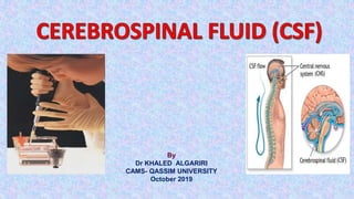
4. FORMATION OF CSF/ANATOMY
Between the arachnoid layer and the pia mater is a
space called the subarachnoid space. It contains a
clear, colorless fluid referred to as Cerebrospinal Fluid
(CSF).
• CSF is produced in the ventricles of the brain by a
collection of rich vascular protrusions called the
choroid plexus.
5.
6. Generally, the total volume of CSF
circulating throughout the adult Central
Nervous System (Brain and Spinal Cord) is
approximately 90 - 150 ml.
• In newborns this volume is 10 -60 ml.
• Excess CSF is continuously reabsorbed by
arachnoid villi and returned to the venous system
thus maintaining a consistent amount of fluid.
7. FUNCTIONS OF CSF
1-Protection : CSF protects the brain from damage by
buffering the brain. It acts as a cushion
2-Buoyancy: The actual mass of the human brain is about
1400 grams; however, the net weight of the brain suspended in
the CSF is equivalent to a mass of 25 grams. which allows
the brain to maintain its density without being impaired
by its own weight
8. FUNCTIONS OF CSF
3-Chemical stability/Homeostasis: CSF maintain the
distribution of necessary substance and waste product
between CNS and Blood stream
4-Prevention of brain ischemia: made by decreasing the amount of
CSF in the limited space inside the skull. This decreases total pressure .
transport of biomolecules to the brain
• Clearance of catabolites (CO2, lactate)
• Maintenance of constant intracranial pressure
5-Clearin of waste: Removes waste from the brain through the bllod
for elimination via kidneys.
10. Examination of CSF
(Physical examination)
Normal CSF is:
Colorless
Clear
Free of clots
Free of blood
If CSF is cloudy (turbid) perform microscopic
examination:
is usually due to leucocytes
may be due to micro-organisms
11. Blood & Hemoglobin pigments in CSF
Traumatic tap
bright red color
RBCS in decreasing
number as the fluid is sampled
Subarachnoid hemorrhage (SAH)
Xanthochromia (hemoglobin
breakdown pigments) = RBCs lysis
& metabolism previously occurred
(at least 2 hr earlier)
12. CSF glucose test
A CSF glucose test measures the amount of
sugar (glucose) in the cerebrospinal fluid
(CSF). CSF is a clear fluid that flows in the
space surrounding the spinal cord and brain.
13. Normal Values
The glucose level in the CSF should be 50 - 80 mg/100
mL (or greater than 2/3 of the blood sugar level).
Note: Normal value ranges may vary slightly among
different laboratories. Talk to your doctor about the
meaning of your specific test results
14. Abnormal CSF [Glucose]
↑ CSF [glucose]:
Not clinically informative
Provides only confirmation of hyperglycemia
15. ↓CSF [glucose] (hypoglycorrhachia):
1)Disorder in carrier-mediated transport
•e.g. TB meningitis
2)Active metabolism of glucose by cells or organisms:
•e.g. acute purulent, amebic, & fungal meningitis
3)Increased metabolism by the CNS
•e.g. by CNS neoplasm
In viral meningitis CSF [glucose] is usually normal
16. CSF/blood glucose ratio
Normal CSF glucose/ plasma glucose ratio is
approximately 0.6-0.7
(N.B. Ratio is decreased if plasma glucose is more than 500
mg/dl due to saturation of the glucose carrier system to CSF
CSF/blood glucose ratio may be a better single indicator for
bacterial meningitis. Since the CSF glucose and blood glucose
values are promptly and easily obtained from a lumbar puncture
17. Protein in CSF
Proteins, mostly albumin are found in the CSF
(0.15-0.45 g/L)
Source of CSF proteins:
• 80% from plasma by ultrafiltration
• 20% from intrathecal synthesis
18. ↑ CSF [total protein]:
Must be compared to the serum [protein]
Useful nonspecific indicator of pathological states:
Lysis of contaminant blood (traumatic tap)
↑ premeability of the epithelial membrane due to:
Bacterial or fungal infection
Cerebral hemorrhage
↑ production by CNS tissue in:
Multiple sclerosis (MS)
Subacute Sclerosing Panencephalitis (SSPE)
Obstruction e.g. in:
Tumors
Abscess
19. Albumin in CSF
Albumin is produced solely in the liver
Its presence in CSF must occur through BBB
Measured by the protein electrophoresis method
20. CSF Immunoglobulin
CSF IgG can arise:
from plasma cells within CSF
from the blood through BBB
↑ [IgG] and normal [Alb] of CSF suggests local
production of IgG, e.g.,
Multiple sclerosis (MS)
Subacute sclerosing panencephalitis (SSPE)
21. CSF LACTATE
In neonates (10-40mg/dl) .
In adult or older children (10-22 mg/dl)
Measurement of lactate concentrations in cerebrospinal fluid
(CSF) may be useful as part of the investigation of inborn errors
of metabolism in which lactic acidosis occurs.
This includes disorders of gluconeogenesis, pyruvate
dehydrogenase complex, the Krebs cycle and the mitochondrial
electron transport chain.
Levels greater than 35 mg/dl are frequently seen with bacterial
meningitis (due to increased glycolysis by bacteria &
inflammatory cells), whereas in viral meningitis, lactate levels
remain lower than 25 mg/dl.
22. CSF LACTATE cont
CSF lactate levels remain elevated during initial
treatment but fall rapidly when treatment is successful,
thus offering a sensitive method for evaluating the
effectiveness of antibiotic therapy.
Measurement of lactate in CSF has also been advocated
for investigating children with unexplained neurological
disease
24. CSF GLUTAMINE
Normal range 8-18 mg/dL
Glutamine is produced in the CNS by the brain cells from ammonia and
alpha-ketoglutarate. This process serves to remove the toxic metabolic waste
product ammonia from the CNS.
Glutamine synthesis helps to protect the CNS from the toxic effects of
increased ammonia.
Ammonia production is increase dramatically in patients with liver
failure.
Accordingly, CSF glutamine production is increased in cases of hepatic
encephalopathy
25. CSF ENZYMES
Lactate dehydrogenase
LDH – LD1, LD2, LD3, LD4, LD5
-Increase LD5 in metastatic brain tumor.
-Increase all fractions in primary brain tumor such as
Meningioma ( extra- exial)
Glioblastoma (intra – exial)
-Increase LD4,LD5 in bacterial meningitis.
28. Other Chemical Components of CSF
CSF [Calcium], [Potassium] & [Phosphates] are lower than their
levels in the blood
CSF [Chloride] & [Magnesium] are higher than their levels in the
blood
Abnormal CSF [Chloride]
marked in acute bacterial meningitis
slight in viral meningitis & brain tumors
29. How the Test is Performed
LUMBAR PUNCTURE
A lumbar puncture is a medical procedure where a needle is
inserted into the lower part of the spine to test for conditions
affecting the brain, spinal cord or other parts of the nervous
system
30. CSF is collected by lumbar puncture
between third, fourth, fifth lumbar vertebrae.
It requires certain precautions and careful
technique to prevent the introduction of
infection or the damaging of neural tissue.
Lumbar puncture
38. CSF is collected in three sterile tubes
•Tube 1 – used for chemical and serologic test:
centrifuge and use supernatant for chemistry analysis (glucose,
protein, enzymes and chloride) kept in freezer till performed.
• Tube 2 – used for microbiology lab kept in room
temperature
•Tube 3 – used for hematology (cell count) Cell
counts done as soon as possible after the fluid is collected as
cellular degradation occurs rapidly. If postponed, refrigerate for
up to one hour.