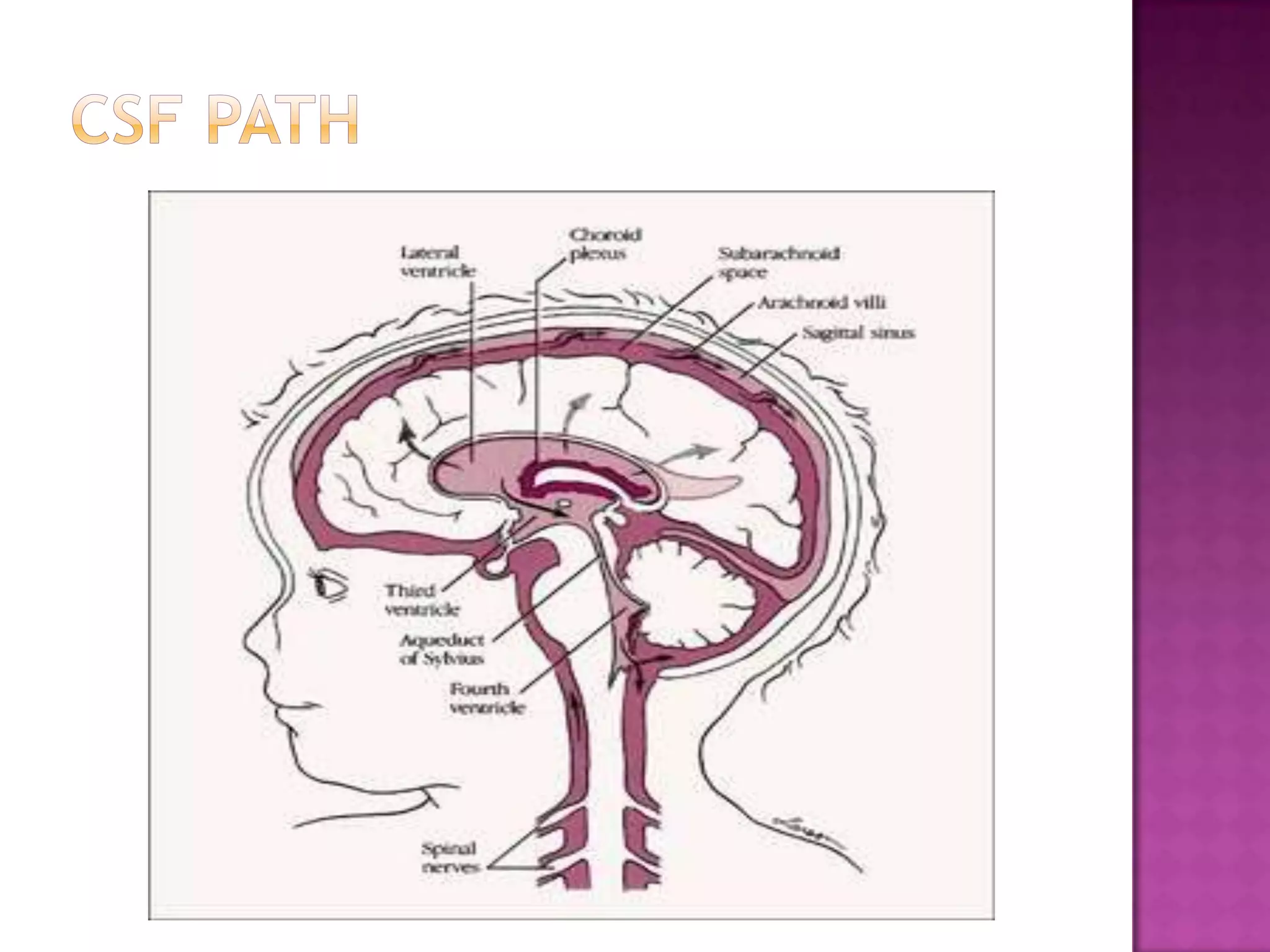
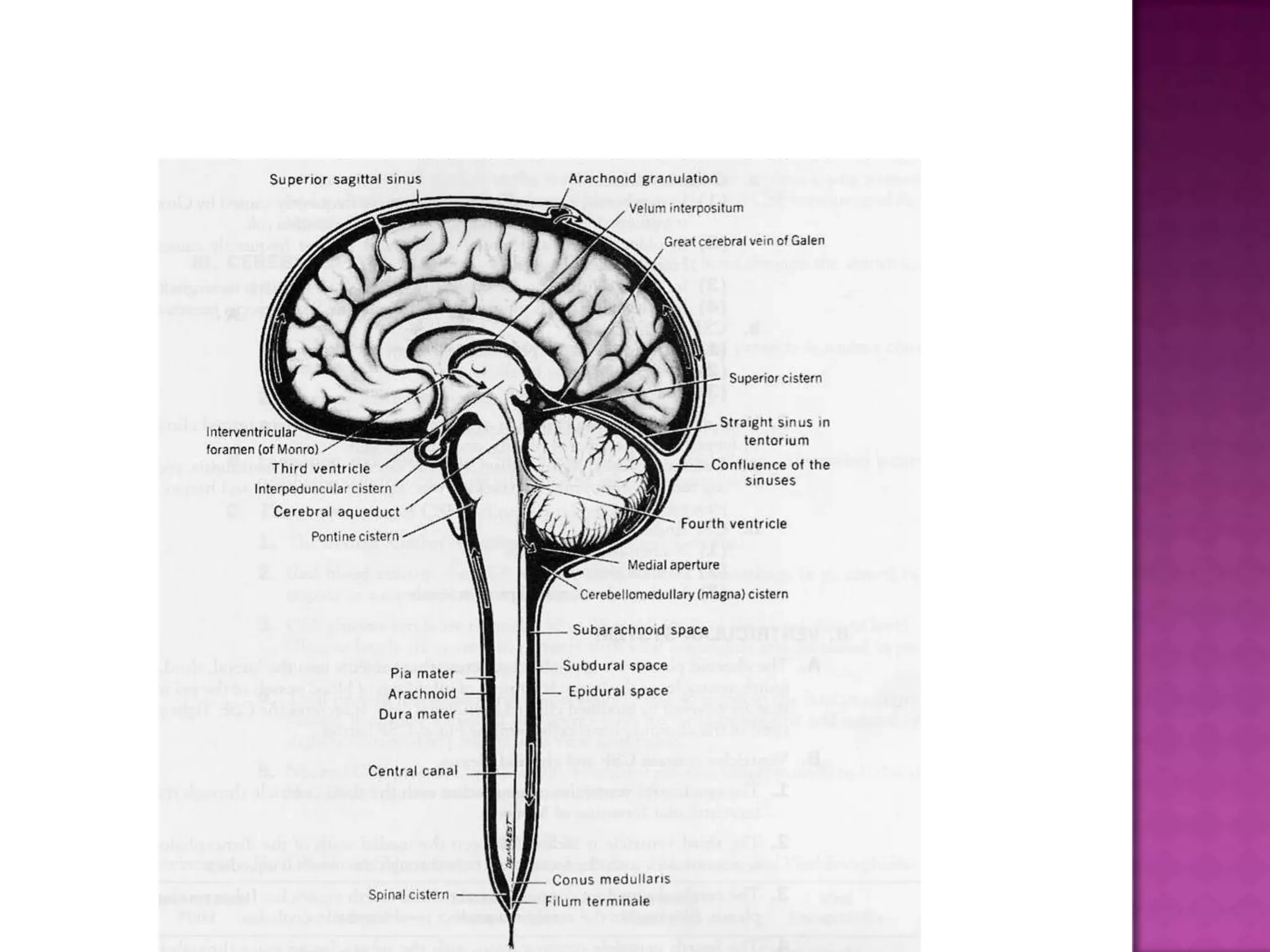
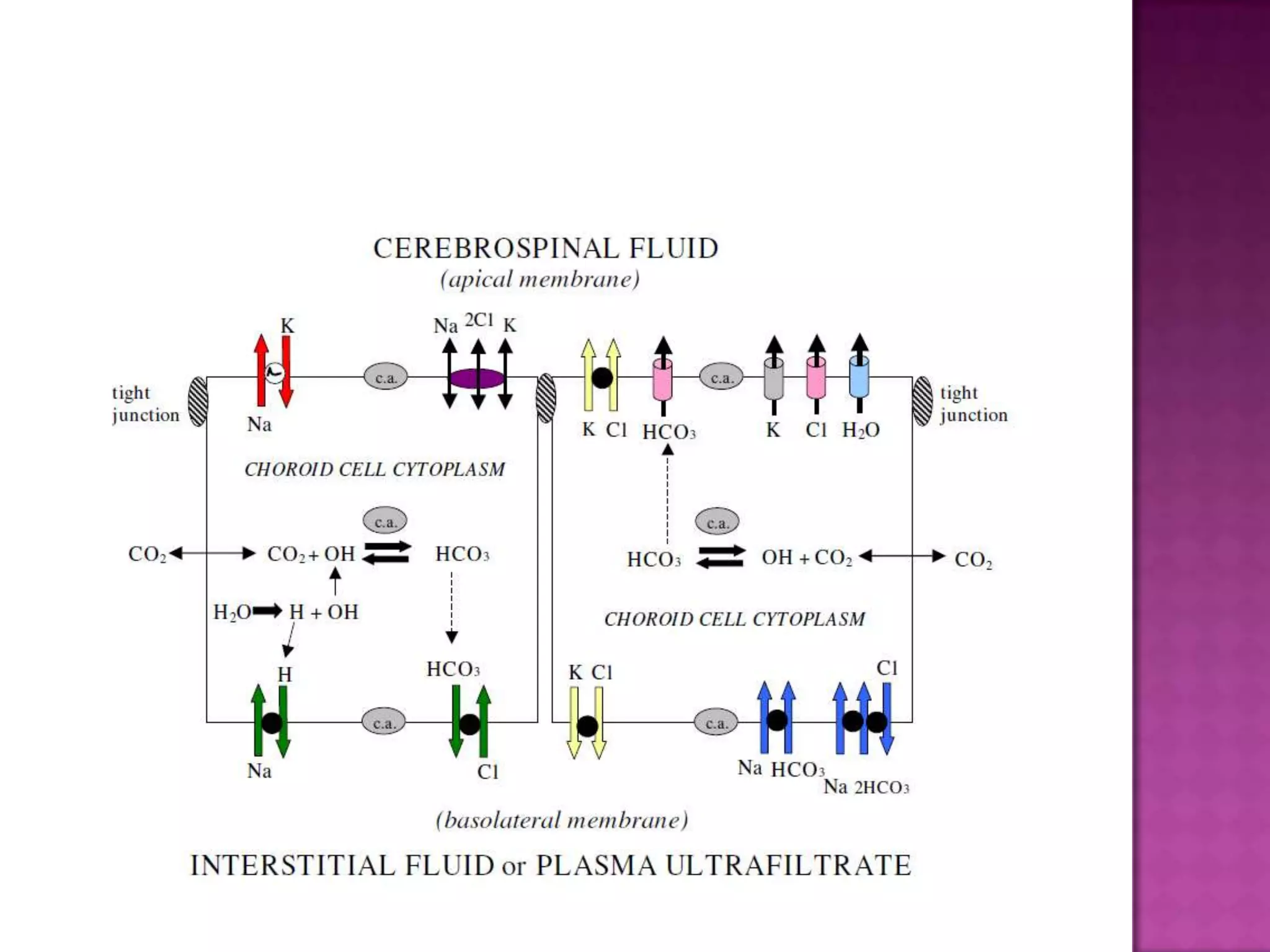
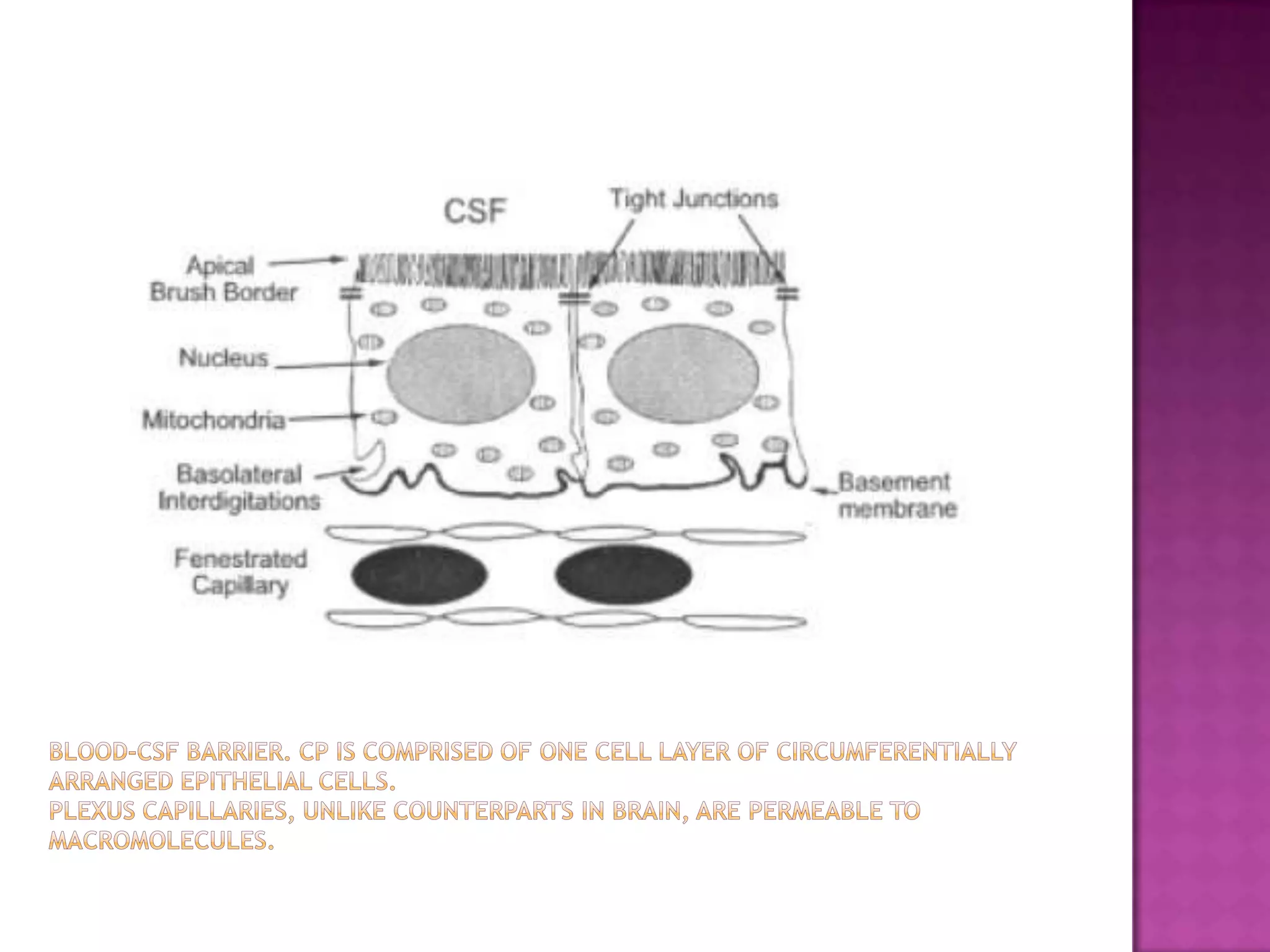
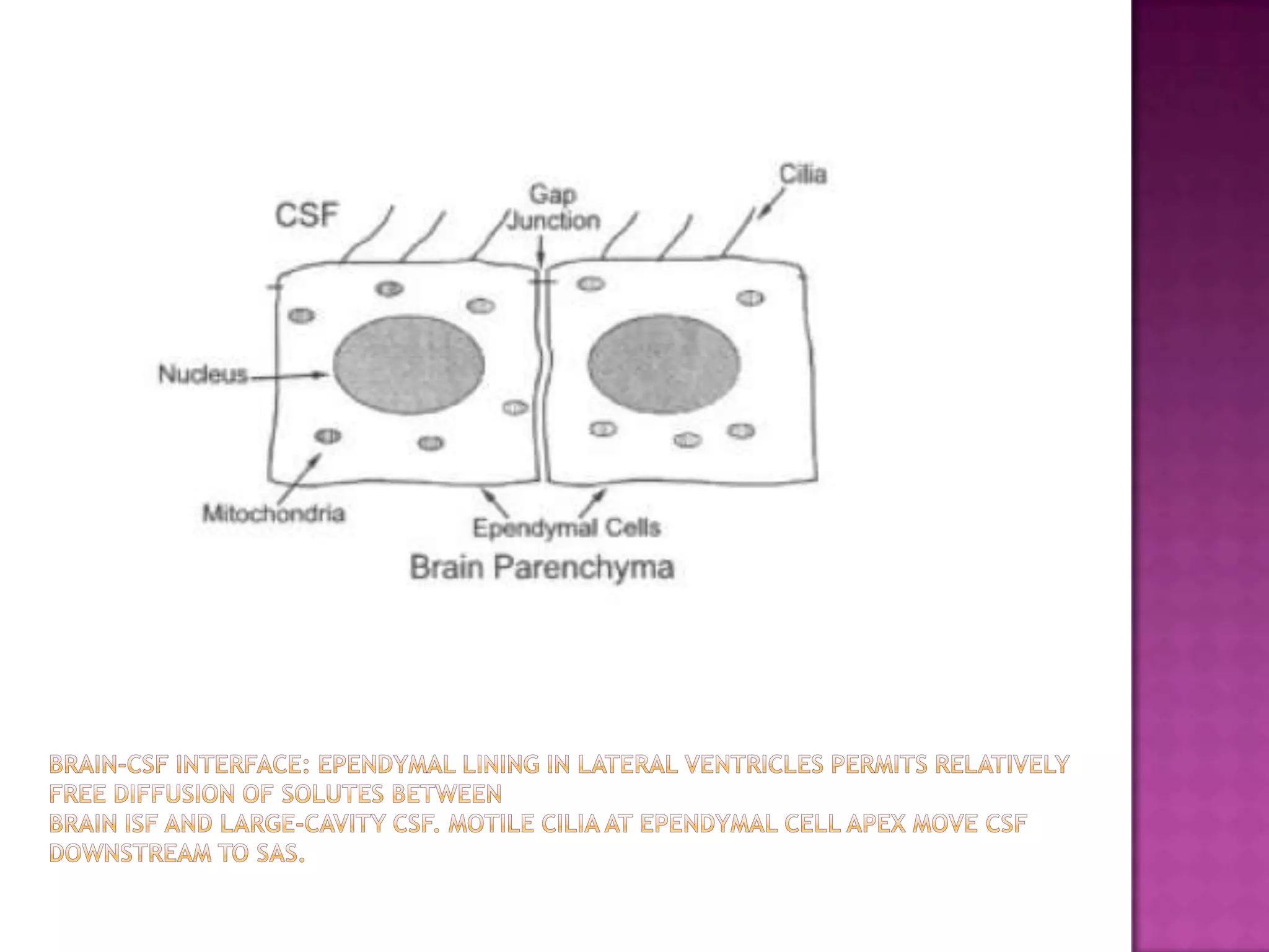
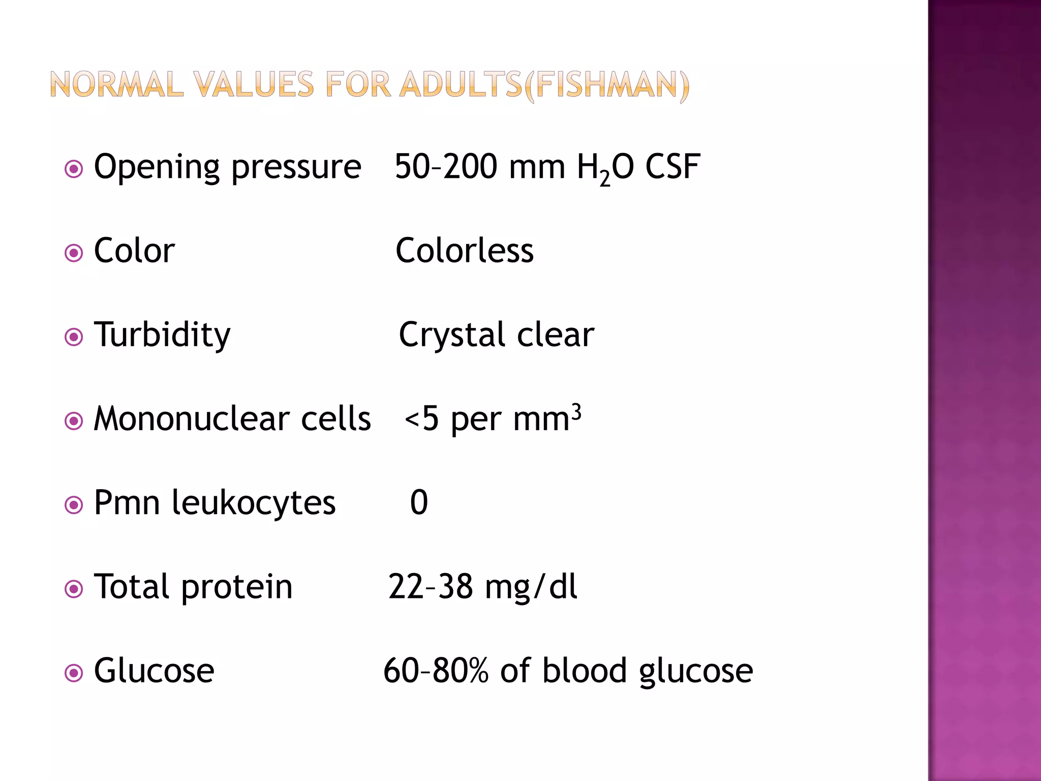
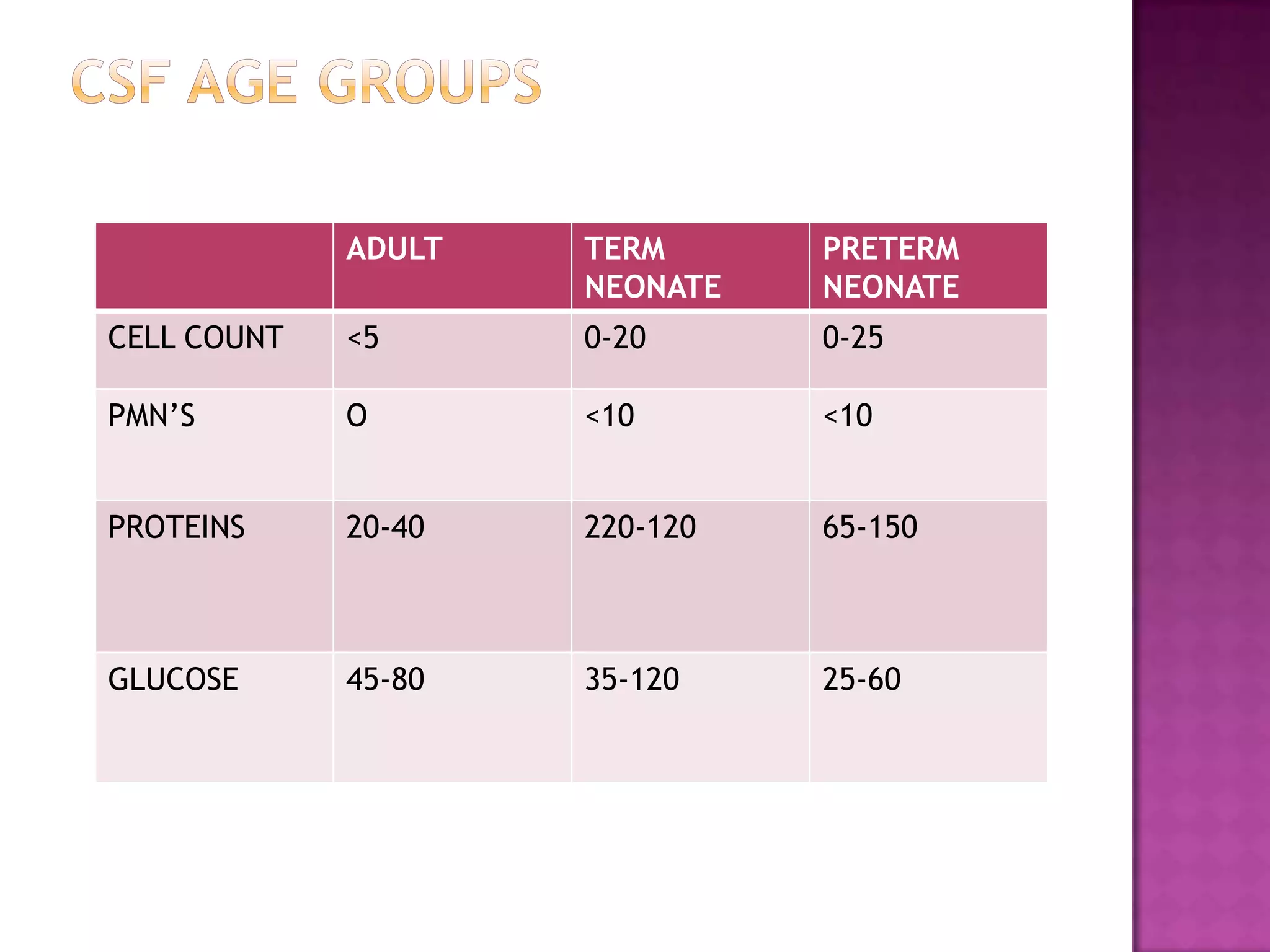
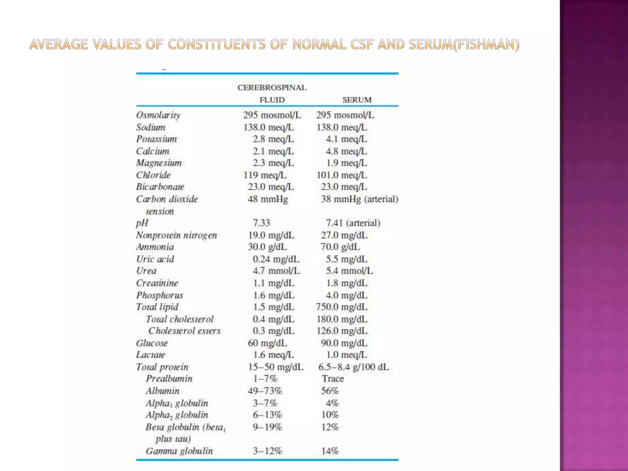
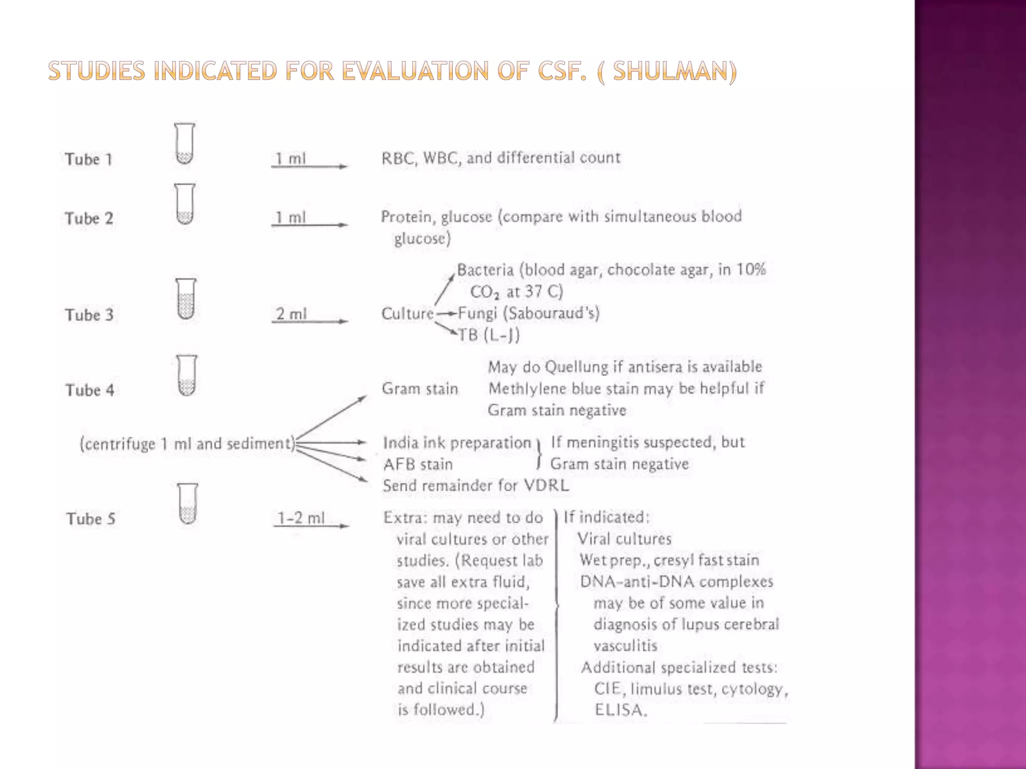
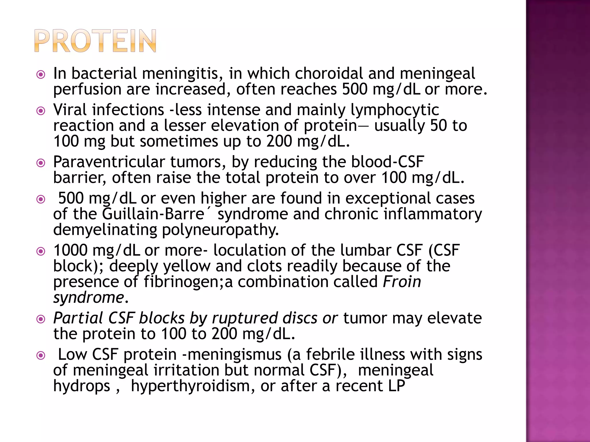
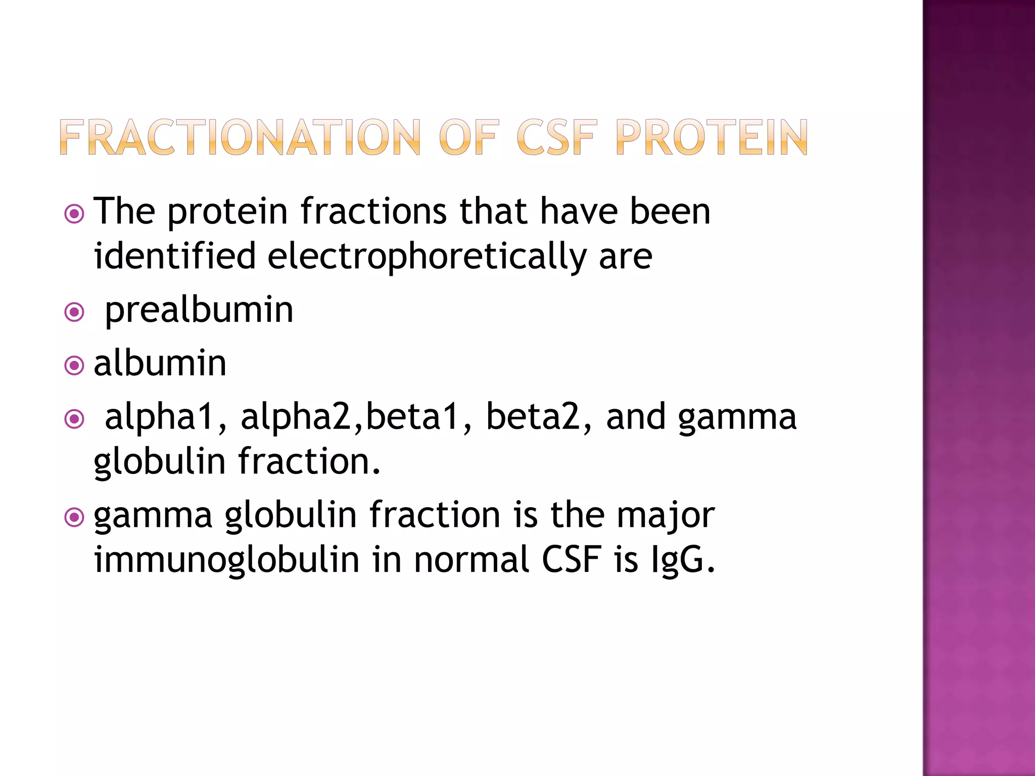
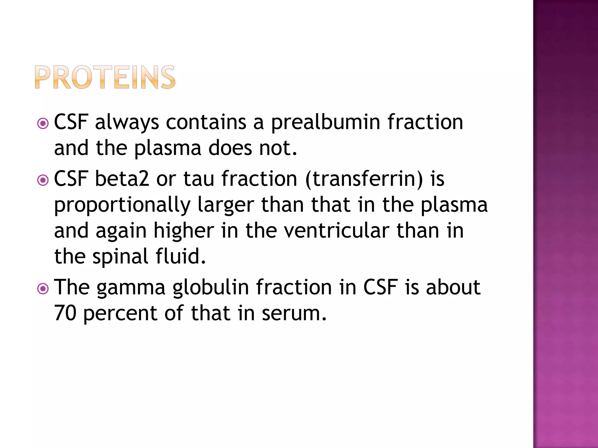
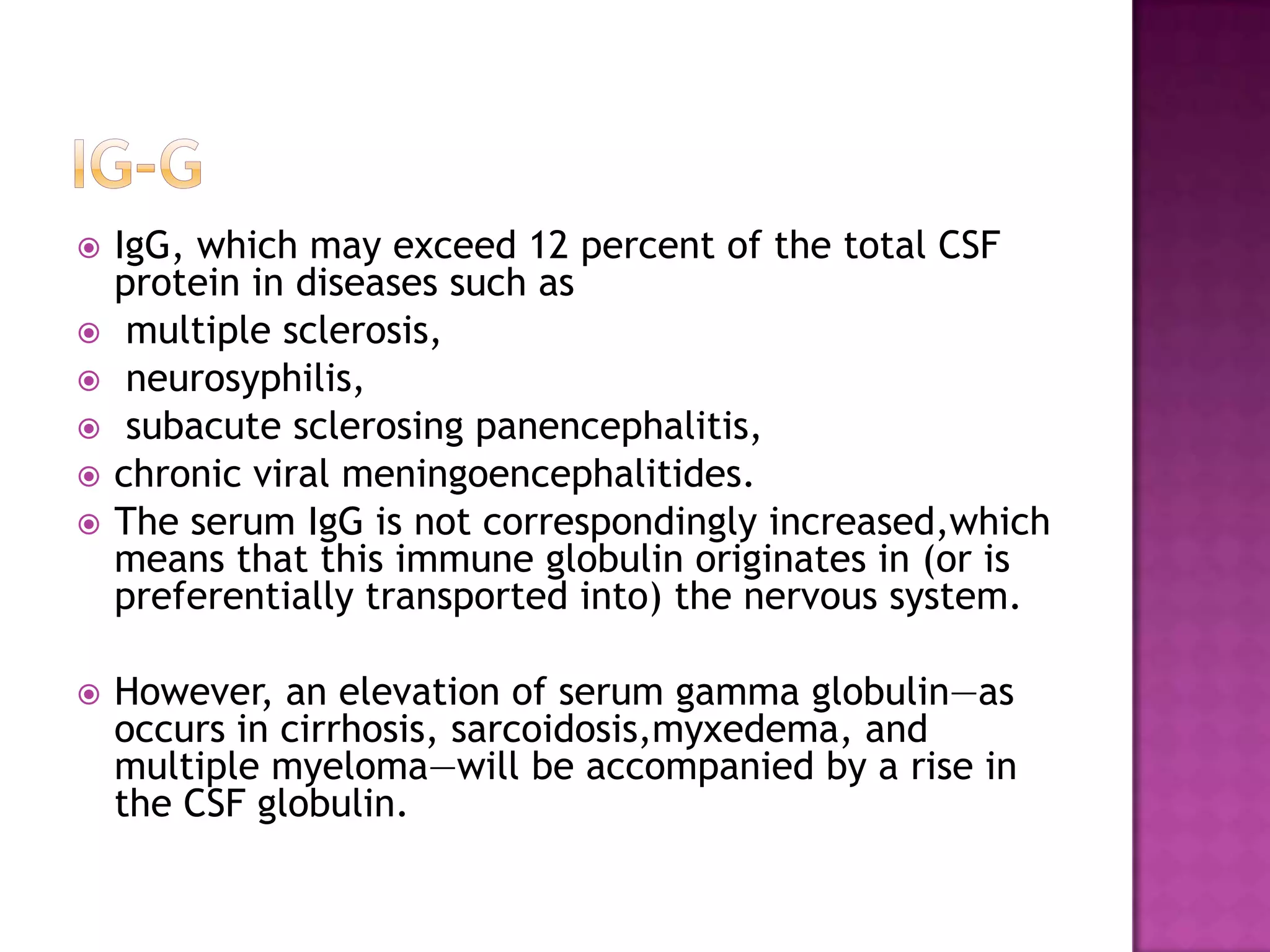
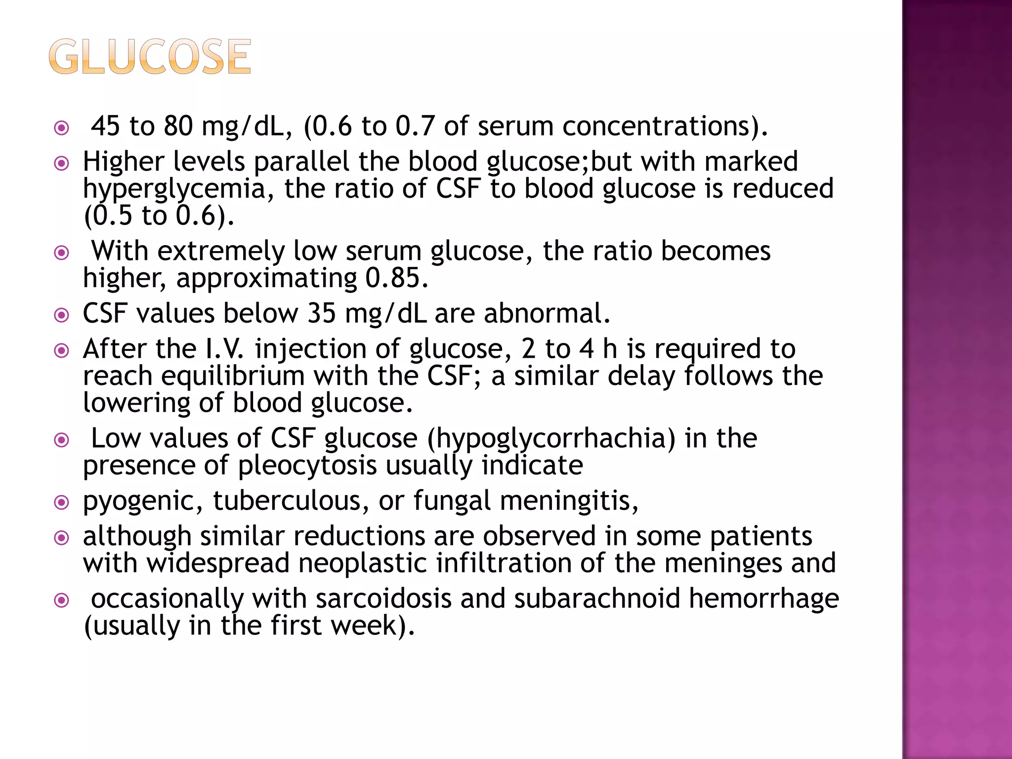
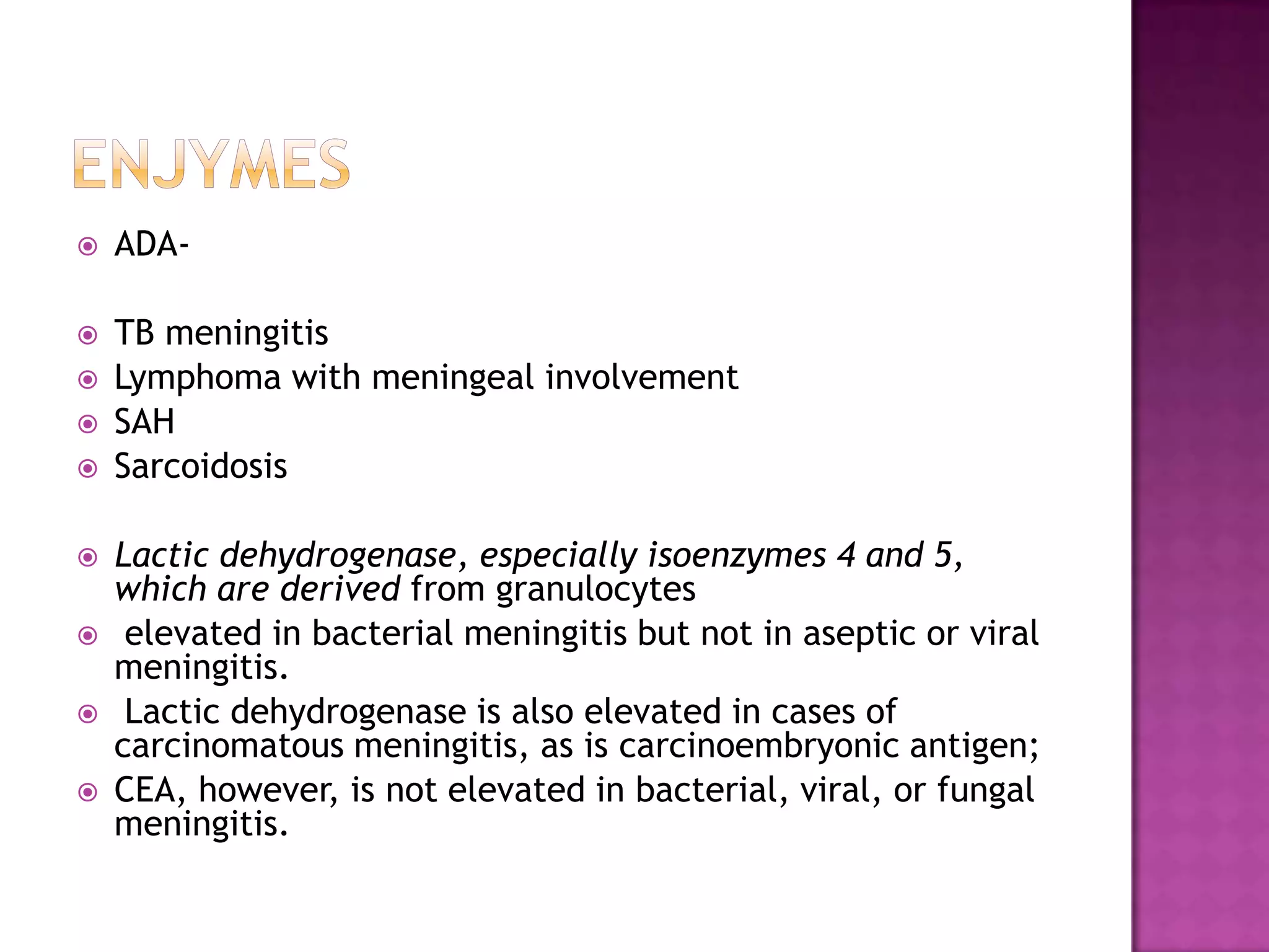
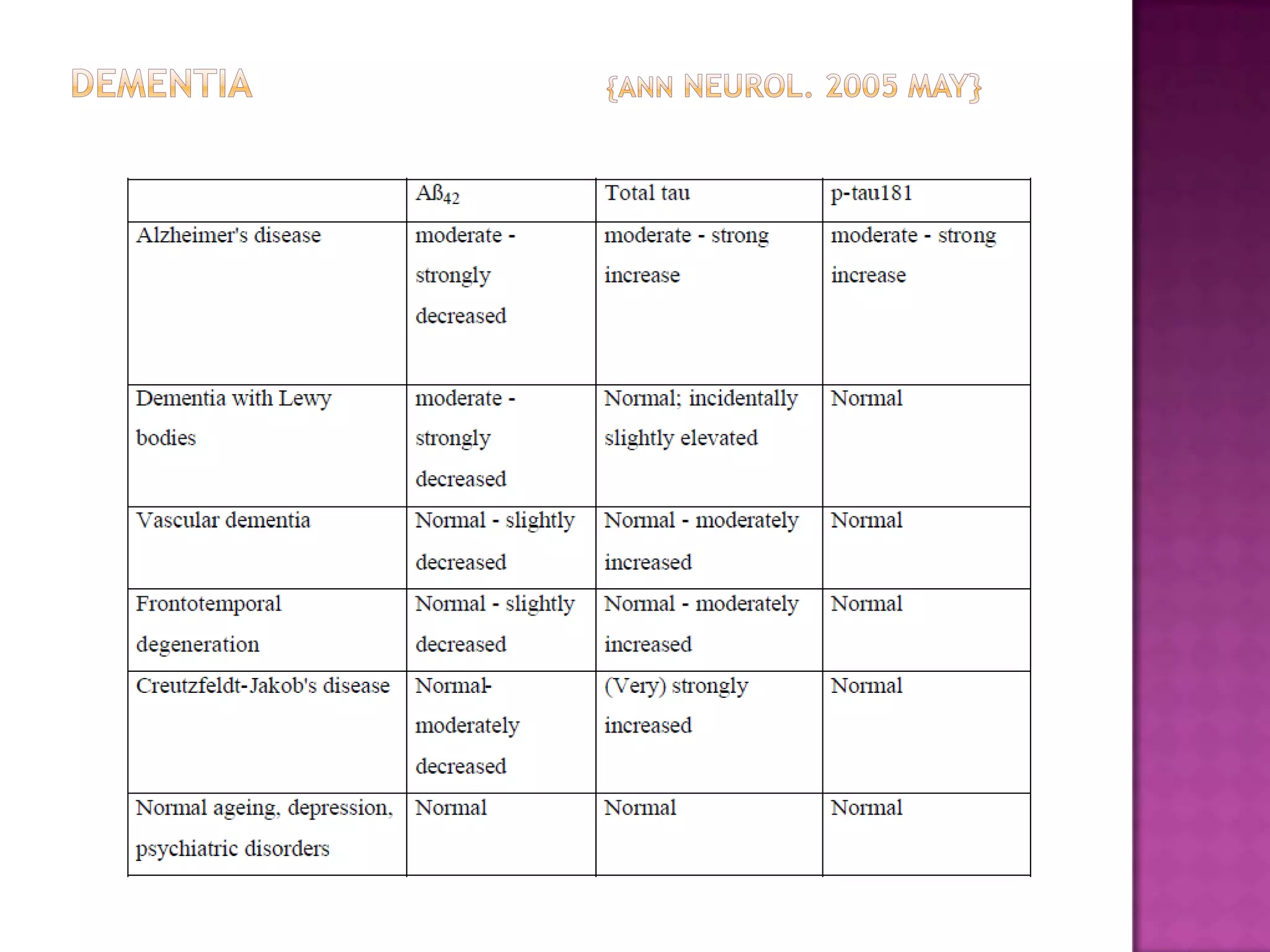
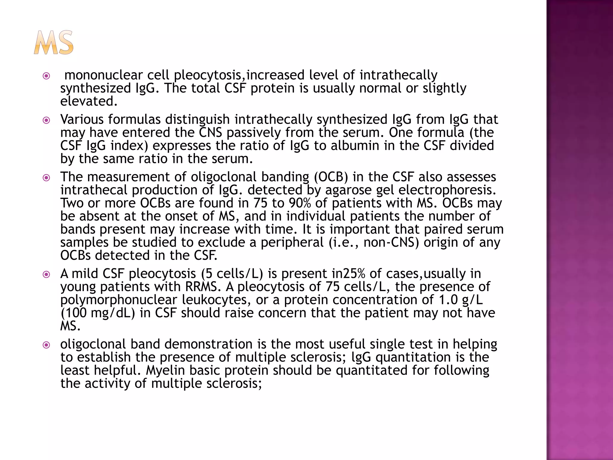
The document discusses cerebrospinal fluid (CSF), including its production and pathways in the central nervous system. CSF acts as a cushion or buffer for the brain, is produced by the choroid plexus at a rate of around 500 ml per day, and circulates through the ventricular system and subarachnoid space. Analysis of CSF provides valuable information for diagnosing neurological conditions through examination of cells, proteins, glucose, and other analytes. Abnormal levels can indicate infections, inflammatory conditions, and other disorders.
A detail on CSF
INTRODUCTION
PROPERTIES
COMPOSITION
FORMATION OF (CSF)
CSF is formed by choroid plexuses, situated with in the ventricles.
Choroid plexuses are tuft of capillaries present inside the ventricles.
A large amount of CSF is formed in the lateral ventricles.
SUBSTANCES AFFECTING THE FORMATION OF (CSF)
PILOCARPINE, extract of pituitary gland stimulate the secretion of CSF.
Injection of isotonic saline also stimulates CSF formation.
Injection of hypotonic saline increases CSF formation.
Hypertonic saline decreases CSF formation and CSF pressure.
ABSORPTION OF (CSF)
CSF is mostly by the archnoid villi into dural sinuses and spinal veins.
Small amount is absorbed along the perineural spaces into cervical lymphatics and into perivascular spaces.
Normally , about 500 mL of CSF is formed everyday and an equal amount is absorbed.
FUNCTIONS OF (CSF)
COLLECTION OF CSF
APPLIED PHYSIOLOGY
Cerebrospinal fluid (CSF) is a clear, colorless body fluid found within the tissue that surrounds the brain and spinal cord of all vertebrates. It replaces the body fluid found outside the cells of all bilateral animals.
The CSF is produced by specialized ependymal cells in the choroid plexuses of the ventricles of the brain, and absorbed in the arachnoid granulations. There is about 125 mL of CSF at any one time, and about 500 mL is generated every day. CSF acts as a cushion or buffer, providing basic mechanical and immunological protection to the brain inside the skull. CSF also serves a vital function in the cerebral autoregulation of cerebral blood flow.
The CSF occupies the subarachnoid space (between the arachnoid mater and the pia mater) and the ventricular system around and inside the brain and spinal cord. It fills the ventricles of the brain, cisterns, and sulci, as well as the central canal of the spinal cord. There is also a connection from the subarachnoid space to the bony labyrinth of the inner ear via the peri lymphatic duct where the perilymph is continuous with the cerebrospinal fluid. The ependymal cells of the choroid plexuses have multiple motile cilia on their apical surfaces that beat to move the CSF through the ventricles.
A sample of CSF can be taken via lumbar puncture. This can reveal the intracranial pressure, as well as indicate diseases including infections of the brain or its surrounding meninges.
Although noted by Hippocrates it was forgotten for centuries. It was discovered in the 18th century by Emanuel Swedenborg. In 1914 Harvey Cushing demonstrated that the CSF was secreted by the choroid plexus.
Cerebrospinal fluid is the fluid of Brian .in this presentation we will study about all over csf it's formation it's composition it's function there examination
New Directions in Targeted Therapeutic Approaches for Older Adults With Mantl...i3 Health
i3 Health is pleased to make the speaker slides from this activity available for use as a non-accredited self-study or teaching resource.
This slide deck presented by Dr. Kami Maddocks, Professor-Clinical in the Division of Hematology and
Associate Division Director for Ambulatory Operations
The Ohio State University Comprehensive Cancer Center, will provide insight into new directions in targeted therapeutic approaches for older adults with mantle cell lymphoma.
STATEMENT OF NEED
Mantle cell lymphoma (MCL) is a rare, aggressive B-cell non-Hodgkin lymphoma (NHL) accounting for 5% to 7% of all lymphomas. Its prognosis ranges from indolent disease that does not require treatment for years to very aggressive disease, which is associated with poor survival (Silkenstedt et al, 2021). Typically, MCL is diagnosed at advanced stage and in older patients who cannot tolerate intensive therapy (NCCN, 2022). Although recent advances have slightly increased remission rates, recurrence and relapse remain very common, leading to a median overall survival between 3 and 6 years (LLS, 2021). Though there are several effective options, progress is still needed towards establishing an accepted frontline approach for MCL (Castellino et al, 2022). Treatment selection and management of MCL are complicated by the heterogeneity of prognosis, advanced age and comorbidities of patients, and lack of an established standard approach for treatment, making it vital that clinicians be familiar with the latest research and advances in this area. In this activity chaired by Michael Wang, MD, Professor in the Department of Lymphoma & Myeloma at MD Anderson Cancer Center, expert faculty will discuss prognostic factors informing treatment, the promising results of recent trials in new therapeutic approaches, and the implications of treatment resistance in therapeutic selection for MCL.
Target Audience
Hematology/oncology fellows, attending faculty, and other health care professionals involved in the treatment of patients with mantle cell lymphoma (MCL).
Learning Objectives
1.) Identify clinical and biological prognostic factors that can guide treatment decision making for older adults with MCL
2.) Evaluate emerging data on targeted therapeutic approaches for treatment-naive and relapsed/refractory MCL and their applicability to older adults
3.) Assess mechanisms of resistance to targeted therapies for MCL and their implications for treatment selection
Lung Cancer: Artificial Intelligence, Synergetics, Complex System Analysis, S...Oleg Kshivets
RESULTS: Overall life span (LS) was 2252.1±1742.5 days and cumulative 5-year survival (5YS) reached 73.2%, 10 years – 64.8%, 20 years – 42.5%. 513 LCP lived more than 5 years (LS=3124.6±1525.6 days), 148 LCP – more than 10 years (LS=5054.4±1504.1 days).199 LCP died because of LC (LS=562.7±374.5 days). 5YS of LCP after bi/lobectomies was significantly superior in comparison with LCP after pneumonectomies (78.1% vs.63.7%, P=0.00001 by log-rank test). AT significantly improved 5YS (66.3% vs. 34.8%) (P=0.00000 by log-rank test) only for LCP with N1-2. Cox modeling displayed that 5YS of LCP significantly depended on: phase transition (PT) early-invasive LC in terms of synergetics, PT N0—N12, cell ratio factors (ratio between cancer cells- CC and blood cells subpopulations), G1-3, histology, glucose, AT, blood cell circuit, prothrombin index, heparin tolerance, recalcification time (P=0.000-0.038). Neural networks, genetic algorithm selection and bootstrap simulation revealed relationships between 5YS and PT early-invasive LC (rank=1), PT N0—N12 (rank=2), thrombocytes/CC (3), erythrocytes/CC (4), eosinophils/CC (5), healthy cells/CC (6), lymphocytes/CC (7), segmented neutrophils/CC (8), stick neutrophils/CC (9), monocytes/CC (10); leucocytes/CC (11). Correct prediction of 5YS was 100% by neural networks computing (area under ROC curve=1.0; error=0.0).
CONCLUSIONS: 5YS of LCP after radical procedures significantly depended on: 1) PT early-invasive cancer; 2) PT N0--N12; 3) cell ratio factors; 4) blood cell circuit; 5) biochemical factors; 6) hemostasis system; 7) AT; 8) LC characteristics; 9) LC cell dynamics; 10) surgery type: lobectomy/pneumonectomy; 11) anthropometric data. Optimal diagnosis and treatment strategies for LC are: 1) screening and early detection of LC; 2) availability of experienced thoracic surgeons because of complexity of radical procedures; 3) aggressive en block surgery and adequate lymph node dissection for completeness; 4) precise prediction; 5) adjuvant chemoimmunoradiotherapy for LCP with unfavorable prognosis.
Prix Galien International 2024 Forum ProgramLevi Shapiro
June 20, 2024, Prix Galien International and Jerusalem Ethics Forum in ROME. Detailed agenda including panels:
- ADVANCES IN CARDIOLOGY: A NEW PARADIGM IS COMING
- WOMEN’S HEALTH: FERTILITY PRESERVATION
- WHAT’S NEW IN THE TREATMENT OF INFECTIOUS,
ONCOLOGICAL AND INFLAMMATORY SKIN DISEASES?
- ARTIFICIAL INTELLIGENCE AND ETHICS
- GENE THERAPY
- BEYOND BORDERS: GLOBAL INITIATIVES FOR DEMOCRATIZING LIFE SCIENCE TECHNOLOGIES AND PROMOTING ACCESS TO HEALTHCARE
- ETHICAL CHALLENGES IN LIFE SCIENCES
- Prix Galien International Awards Ceremony
Acute scrotum is a general term referring to an emergency condition affecting the contents or the wall of the scrotum.
There are a number of conditions that present acutely, predominantly with pain and/or swelling
A careful and detailed history and examination, and in some cases, investigations allow differentiation between these diagnoses. A prompt diagnosis is essential as the patient may require urgent surgical intervention
Testicular torsion refers to twisting of the spermatic cord, causing ischaemia of the testicle.
Testicular torsion results from inadequate fixation of the testis to the tunica vaginalis producing ischemia from reduced arterial inflow and venous outflow obstruction.
The prevalence of testicular torsion in adult patients hospitalized with acute scrotal pain is approximately 25 to 50 percent
Flu Vaccine Alert in Bangalore Karnatakaaddon Scans
As flu season approaches, health officials in Bangalore, Karnataka, are urging residents to get their flu vaccinations. The seasonal flu, while common, can lead to severe health complications, particularly for vulnerable populations such as young children, the elderly, and those with underlying health conditions.
Dr. Vidisha Kumari, a leading epidemiologist in Bangalore, emphasizes the importance of getting vaccinated. "The flu vaccine is our best defense against the influenza virus. It not only protects individuals but also helps prevent the spread of the virus in our communities," he says.
This year, the flu season is expected to coincide with a potential increase in other respiratory illnesses. The Karnataka Health Department has launched an awareness campaign highlighting the significance of flu vaccinations. They have set up multiple vaccination centers across Bangalore, making it convenient for residents to receive their shots.
To encourage widespread vaccination, the government is also collaborating with local schools, workplaces, and community centers to facilitate vaccination drives. Special attention is being given to ensuring that the vaccine is accessible to all, including marginalized communities who may have limited access to healthcare.
Residents are reminded that the flu vaccine is safe and effective. Common side effects are mild and may include soreness at the injection site, mild fever, or muscle aches. These side effects are generally short-lived and far less severe than the flu itself.
Healthcare providers are also stressing the importance of continuing COVID-19 precautions. Wearing masks, practicing good hand hygiene, and maintaining social distancing are still crucial, especially in crowded places.
Protect yourself and your loved ones by getting vaccinated. Together, we can help keep Bangalore healthy and safe this flu season. For more information on vaccination centers and schedules, residents can visit the Karnataka Health Department’s official website or follow their social media pages.
Stay informed, stay safe, and get your flu shot today!
- Video recording of this lecture in English language: https://youtu.be/lK81BzxMqdo
- Video recording of this lecture in Arabic language: https://youtu.be/Ve4P0COk9OI
- Link to download the book free: https://nephrotube.blogspot.com/p/nephrotube-nephrology-books.html
- Link to NephroTube website: www.NephroTube.com
- Link to NephroTube social media accounts: https://nephrotube.blogspot.com/p/join-nephrotube-on-social-media.html
TEST BANK for Operations Management, 14th Edition by William J. Stevenson, Ve...kevinkariuki227
TEST BANK for Operations Management, 14th Edition by William J. Stevenson, Verified Chapters 1 - 19, Complete Newest Version.pdf
Knee anatomy and clinical tests 2024.pdfvimalpl1234
This includes all relevant anatomy and clinical tests compiled from standard textbooks, Campbell,netter etc..It is comprehensive and best suited for orthopaedicians and orthopaedic residents.
Report Back from SGO 2024: What’s the Latest in Cervical Cancer?bkling
Are you curious about what’s new in cervical cancer research or unsure what the findings mean? Join Dr. Emily Ko, a gynecologic oncologist at Penn Medicine, to learn about the latest updates from the Society of Gynecologic Oncology (SGO) 2024 Annual Meeting on Women’s Cancer. Dr. Ko will discuss what the research presented at the conference means for you and answer your questions about the new developments.
New Drug Discovery and Development .....NEHA GUPTA
The "New Drug Discovery and Development" process involves the identification, design, testing, and manufacturing of novel pharmaceutical compounds with the aim of introducing new and improved treatments for various medical conditions. This comprehensive endeavor encompasses various stages, including target identification, preclinical studies, clinical trials, regulatory approval, and post-market surveillance. It involves multidisciplinary collaboration among scientists, researchers, clinicians, regulatory experts, and pharmaceutical companies to bring innovative therapies to market and address unmet medical needs.
Couples presenting to the infertility clinic- Do they really have infertility...Sujoy Dasgupta
Dr Sujoy Dasgupta presented the study on "Couples presenting to the infertility clinic- Do they really have infertility? – The unexplored stories of non-consummation" in the 13th Congress of the Asia Pacific Initiative on Reproduction (ASPIRE 2024) at Manila on 24 May, 2024.
These simplified slides by Dr. Sidra Arshad present an overview of the non-respiratory functions of the respiratory tract.
Learning objectives:
1. Enlist the non-respiratory functions of the respiratory tract
2. Briefly explain how these functions are carried out
3. Discuss the significance of dead space
4. Differentiate between minute ventilation and alveolar ventilation
5. Describe the cough and sneeze reflexes
Study Resources:
1. Chapter 39, Guyton and Hall Textbook of Medical Physiology, 14th edition
2. Chapter 34, Ganong’s Review of Medical Physiology, 26th edition
3. Chapter 17, Human Physiology by Lauralee Sherwood, 9th edition
4. Non-respiratory functions of the lungs https://academic.oup.com/bjaed/article/13/3/98/278874
2. CSF Occupies the subarachnoid space and the ventricular system around and inside the brain. Occupies the space between the arachnoid mater and the pia mater. It constitutes the content of all ventricles, cisterns and sulci , as well as the central canal of the spinal cord. It acts as a "cushion" or buffer for the cortex, providing a basic mechanical and immunological protection to the brain inside the skull.
3. Definition Cerebrospinal fluid (CSF) is a biologic fluid, formed mainly in the ventricular choroid plexus, distributed within the ventricular system, basal cisterns, and subarachnoid space.
6. Csf production secreted by the choroid plexus - 500 ml per day, 0.35 ml per minute, and a turnover of 14% per hour. The usual volume is 150 ml. Formation occurs as a result of a two-step process. Fluid is first filtered through the core capillaries of the choroid plexus into the extracellular space surrounding the choroidal cells. This fluid is a plasma ultrafiltrate. Then sodium is actively transported by sodium–potassium activated adenosine triphosphatase (ATPase) across the choroidal cells into the CSF; water follows down an osmotic: gradient. Cholinergic stimulation increases production and adrenergic stimulation decreases production. Drugs that inhibit sodium–potassium ATPase or carbonic anhydrase decrease production. Furosemide also slows CSF production due to its effect on chloride flux. Some CSF is apparently produced in the ependyma of the brain, in addition to the choroid plexus.
9. Schema of main CNS compartments and interfaces. The blood-brain andblood-CSF barriers are true barriers with tight junctions between endothelial and epithelial cells, respectively. The brain-CSFinterface, because of gap junctions between ependymal (or pia-glial) cells, is more permeable than brain or spinal cord capillariesand choroid plexus.
10. Blood-CSF barrier. CP is comprised of one cell layer of circumferentially arranged epithelial cells.Plexus capillaries, unlike counterparts in brain, are permeable to macromolecules.
12. Brain-CSF interface: Ependymal lining in lateral ventricles permits relatively free diffusion of solutes betweenbrain ISF and large-cavity CSF. Motile cilia at ependymal cell apex move CSF downstream to SAS.
13. contraindications Absolute Infection in the skin overlying the access site. Relative Papilledema . A bleeding diathesis . A platelet count of 50,000 or lower . Severe pulmonary disease or respiratory difficulty in the patient.
14. Normal Values for Adults(fishman) Opening pressure 50–200 mm H2O CSF Color Colorless Turbidity Crystal clear Mononuclear cells <5 per mm3 Pmn leukocytes 0 Total protein 22–38 mg/dl Glucose 60–80% of blood glucose
17. Indications for lp To obtain pressure measurements and procure a sample of the CSF for cellular, cytologic, chemical, and bacteriologic examination. To aid in therapy by the administration of spinal anesthetics and occasionally antibiotics or antitumor agents, or by reduction of CSF pressure. To inject a radiopaque substance, as in myelography, or a radioactiveagent, as in radionuclide cisternography.
18. complications Brain herniation, 1 to 15% (Fishman, 1980). Headache,10% . Tourtellotte and associates (1964). Diplopia due to unilateral or bilateral sixth nerve palsy. Subarachnoid hemorrhage, or a "traumatic" tap. spinal epidural hematoma or spinal subdural hematoma.
20. Analysis of Xanthochromic CSF Technique-Compare CSF with a similar volume of water in an identical tube; look down the longitudinal axis of the tube, against a white background. Pigments seen in subarachnoid hemorrhage (SAH) Oxyhemoglobin-Pink or orange color; released into CSF in 2 hours after SAH, due to RBC lysis; may be released within 30 minutes if RBC greater than 150,000/mm3; maximum color in 36 hours, disappears in 7 to 10 days; cerebrospinal fluid must be examined immediately after the LP, since oxyhemoglobin can be produced by lysis of RBC in the test tube. Bilirubin-Produces the yellow pigment, or xanthochromia of CSF; produced in vivo by the conversion of free hemoglobin by macrophages and other leptomeningeal cells; not seen for 10 to 12 hours after the hemorrhage; reaches a maximum in 48 hours, and persists 2 to 4 hours. Methemoglobin- brown,appears when blood is loculated or encysted and isolated from the flow of CSF Other causes of xanthochromia Protein-Protein over 150 mg/dl produces xanthochroma, the intensity paralleling the amount of protein Red blood cells-RBC over 100,000/mm3 produce xanthochromia as a result of serum brought with them Jaundice-Serum bilirubin of 15 mg/dl produces xanthochromia. Carotene-Hypercarotenemia in food faddists produces xanthochromia Miscellaneous-Subdural hematomas, trauma, and clots in other locations will produce xanthochromia White blood cells-The WBC/RBC ratio is similar to that of the plasma in traumatic taps and fresh SAH; SAH that is a few days old will produce a chemical meningitis, which elevates the number of WBC Protein-Each 1000 RBC min raises CSF protein 1.5 mg/dl Traumatic tap-Tubes 1 to 3 show decreasing RBC.
21. Cellularity No cells or 1-5 lympho’s. Neonate-<30 pmn. EXAMINATION OF CELL Diagnosis of inf. Follow up in course of illness Response to treatment FALSE LOW COUNT >60 Mts Settlement of cells Lysis of cells Adsorbs to wall of bottle
26. Noninfectious Causes of CSF Pleocytosis Chemical meningitis Myelography Spinal anesthesia Intrathecal medication Ingestion of mercury or arsenic Vasculitis Subarachnoid hemorrhage Behcet's syndrome Lead encephalopathy Sarcoid Tumor (leukemia most common; glucose can drop to zero) Seizure activity (must diagnose only if other possibilities are ruled out and if pleocytosis is minimal and rapidly clears)
27. Proteins In contrast to the high protein content of blood (5500 to 8000 mg/dL), that of the lumbar spinal fluid is 45 mg/dL or less in the adult. Basal cisterns is 10 to 25 mg/dL , ventricles is 5 to 15 mg/dL, Reflecting a ventricular–lumbar gradient in the permeability of capillary endothelial cells to protein (blood-CSF barrier) and a lesser degree of fluid circulation in the lumbosacral region. Children,the protein concentration is less than 20 mg/dl. Higher Level -a pathologic process in or near the ependyma or meninges—in either the brain, spinal cord, or nerve roots.
28. SAH-PROTEIN Bleeding into the ventricles or subarachnoid space results in spillage not only of RBC but of serum proteins. If the serum protein concentrations are normal, the CSF protein should increase by about 1 mg per 1000 RBC provided that the same tube of CSF is used in determining the cell count and protein content. The same holds for a traumatic puncture that allows seepage of venous blood into the CSF at the puncture site. However, in the case of subarachnoid hemorrhage, due to the irritating effect of hemolyzed RBC upon the leptomeninges, the CSF protein may be increased by many times this ratio.
29. PROTEIN In bacterial meningitis, in which choroidal and meningeal perfusion are increased, often reaches 500 mg/dL or more. Viral infections -less intense and mainly lymphocytic reaction and a lesser elevation of protein— usually 50 to 100 mg but sometimes up to 200 mg/dL. Paraventricular tumors, by reducing the blood-CSF barrier, often raise the total protein to over 100 mg/dL. 500 mg/dL or even higher are found in exceptional cases of the Guillain-Barre´ syndrome and chronic inflammatory demyelinatingpolyneuropathy. 1000 mg/dL or more- loculation of the lumbar CSF (CSF block); deeply yellow and clots readily because of the presence of fibrinogen;a combination called Froin syndrome. Partial CSF blocks by ruptured discs or tumor may elevate the protein to 100 to 200 mg/dL. Low CSF protein -meningismus (a febrile illness with signs of meningeal irritation but normal CSF), meningealhydrops , hyperthyroidism, or after a recent LP
30. Fractionation of CSF Protein The protein fractions that have been identified electrophoretically are prealbumin albumin alpha1, alpha2,beta1, beta2, and gamma globulin fraction. gamma globulin fraction is the major immunoglobulin in normal CSF is IgG.
31. PROTEINS CSF always contains a prealbumin fraction and the plasma does not. CSF beta2 or tau fraction (transferrin) is proportionally larger than that in the plasma and again higher in the ventricular than in the spinal fluid. The gamma globulin fraction in CSF is about 70 percent of that in serum.
32. IG-g IgG, which may exceed 12 percent of the total CSF protein in diseases such as multiple sclerosis, neurosyphilis, subacutesclerosingpanencephalitis, chronic viral meningoencephalitides. The serum IgG is not correspondingly increased,which means that this immune globulin originates in (or is preferentially transported into) the nervous system. However, an elevation of serum gamma globulin—as occurs in cirrhosis, sarcoidosis,myxedema, and multiple myeloma—will be accompanied by a rise in the CSF globulin.
33. Glucose 45 to 80 mg/dL, (0.6 to 0.7 of serum concentrations). Higher levels parallel the blood glucose;but with marked hyperglycemia, the ratio of CSF to blood glucose is reduced (0.5 to 0.6). With extremely low serum glucose, the ratio becomes higher, approximating 0.85. CSF values below 35 mg/dL are abnormal. After the I.V. injection of glucose, 2 to 4 h is required to reach equilibrium with the CSF; a similar delay follows the lowering of blood glucose. Low values of CSF glucose (hypoglycorrhachia) in the presence of pleocytosis usually indicate pyogenic, tuberculous, or fungal meningitis, although similar reductions are observed in some patients with widespread neoplastic infiltration of the meninges and occasionally with sarcoidosis and subarachnoid hemorrhage (usually in the first week).
34. Serologic and Virologic Tests cryptococcal surface antigen Venereal Disease Research Laboratories (VDRL) slide flocculation test and rapid plasma reagin (RPR) agglutination test polymerase chain reaction (PCR) herpesviruses and cytomegalovirus
35. ammonia hepatic encephalopathy, inherited hyperammonemias, Reye syndrome. concentration corresponds roughly with the severity of the encephalopathy.
36. uric acid 5 percent of that in serum High gout. Uremia. meningitis . low Wilson disease
37. Urea& amino acids The urea concentration in the CSF is slightly lower than that in the serum;in uremia, it rises in parallel with that in the blood. An intravenous injection of urea raises the blood level immediately and the CSF level more slowly, exerting an osmotic dehydrating effect on the central nervous tissues and CSF. The concentration of amino acids in the CSF is about one-third that in plasma. Elevations of glutamine are found in hepatic coma and the Reye syndrome and of phenylalanine, histidine, valine, leucine, isoleucine,tyrosine, and homocystine in the corresponding aminoacidurias.
38. ENJYMES ADA- TB meningitis Lymphoma with meningeal involvement SAH Sarcoidosis Lactic dehydrogenase, especially isoenzymes 4 and 5, which are derived from granulocytes elevated in bacterial meningitis but not in aseptic or viral meningitis. Lactic dehydrogenase is also elevated in cases of carcinomatous meningitis, as is carcinoembryonic antigen; CEA, however, is not elevated in bacterial, viral, or fungal meningitis.
39. catecholamines Homovanillic acid (HVA), the major catabolite of dopamine, and 5-hydroxyindoleacetic acid (5-HIAA), the major catabolite of serotonin, are normally present in the spinal fluid; Both are five or six times higher in the ventricular than the lumbar CSF. The levels of both catabolites are reduced in patients with idiopathic and drug-induced parkinsonism
41. Dementia progression A study conducted by investigators at Washington University School of Medicine, in St. Louis, Missouri. Patients with very mild dementia of the Alzheimer's type (DAT), lower levels of the peptide amyloid beta 1-42 (Aβ42), high tau or phosphorylated tau at threonine 181 (ptau-181), or high tau-to-Aβ42 ratios quantitatively predict more rapid progression of cognitive deficits and dementia. {Arch Neurol. 2009;66:638-645}
42. ms mononuclear cell pleocytosis,increased level of intrathecally synthesized IgG. The total CSF protein is usually normal or slightly elevated. Various formulas distinguish intrathecally synthesized IgG from IgG that may have entered the CNS passively from the serum. One formula (the CSF IgG index) expresses the ratio of IgG to albumin in the CSF divided by the same ratio in the serum. The measurement of oligoclonal banding (OCB) in the CSF also assesses intrathecal production of IgG. detected by agarose gel electrophoresis. Two or more OCBs are found in 75 to 90% of patients with MS. OCBs may be absent at the onset of MS, and in individual patients the number of bands present may increase with time. It is important that paired serum samples be studied to exclude a peripheral (i.e., non-CNS) origin of any OCBs detected in the CSF. A mild CSF pleocytosis (5 cells/L) is present in25% of cases,usually in young patients with RRMS. A pleocytosis of 75 cells/L, the presence of polymorphonuclear leukocytes, or a protein concentration of 1.0 g/L (100 mg/dL) in CSF should raise concern that the patient may not have MS. oligoclonal band demonstration is the most useful single test in helping to establish the presence of multiple sclerosis; lgGquantitation is the least helpful. Myelin basic protein should be quantitated for following the activity of multiple sclerosis;