The cerebrospinal fluid is formed in the brain ventricles and surrounds the brain and spinal cord. CSF analysis provides important information for diagnosing central nervous system conditions like infections, tumors, and demyelinating diseases. A lumbar puncture is performed to collect CSF, which is then analyzed for properties, cells, chemicals, and microorganisms. Abnormalities in CSF parameters can indicate different conditions - for example, low glucose and high protein suggest bacterial meningitis while increased IgG with normal albumin indicates multiple sclerosis.


![CSF Immunoglobulin
• CSF IgG can arise:
– from plasma cells within CSF
– & from the blood through BBB
• ↑CSF [IgG] without concomitant ↑ in CSF [Alb] suggests local
production of IgG:
– multiple sclerosis (MS)
– subacute sclerosing panencephalitis (SPEE)
CSF IgG/Serum IgG
CSF serum /Albumin index
CSF IgG index:
Normally: < 0.7
=](https://image.slidesharecdn.com/7908180-230725160826-d0df5691/75/Csf-analysis-20-2048.jpg)


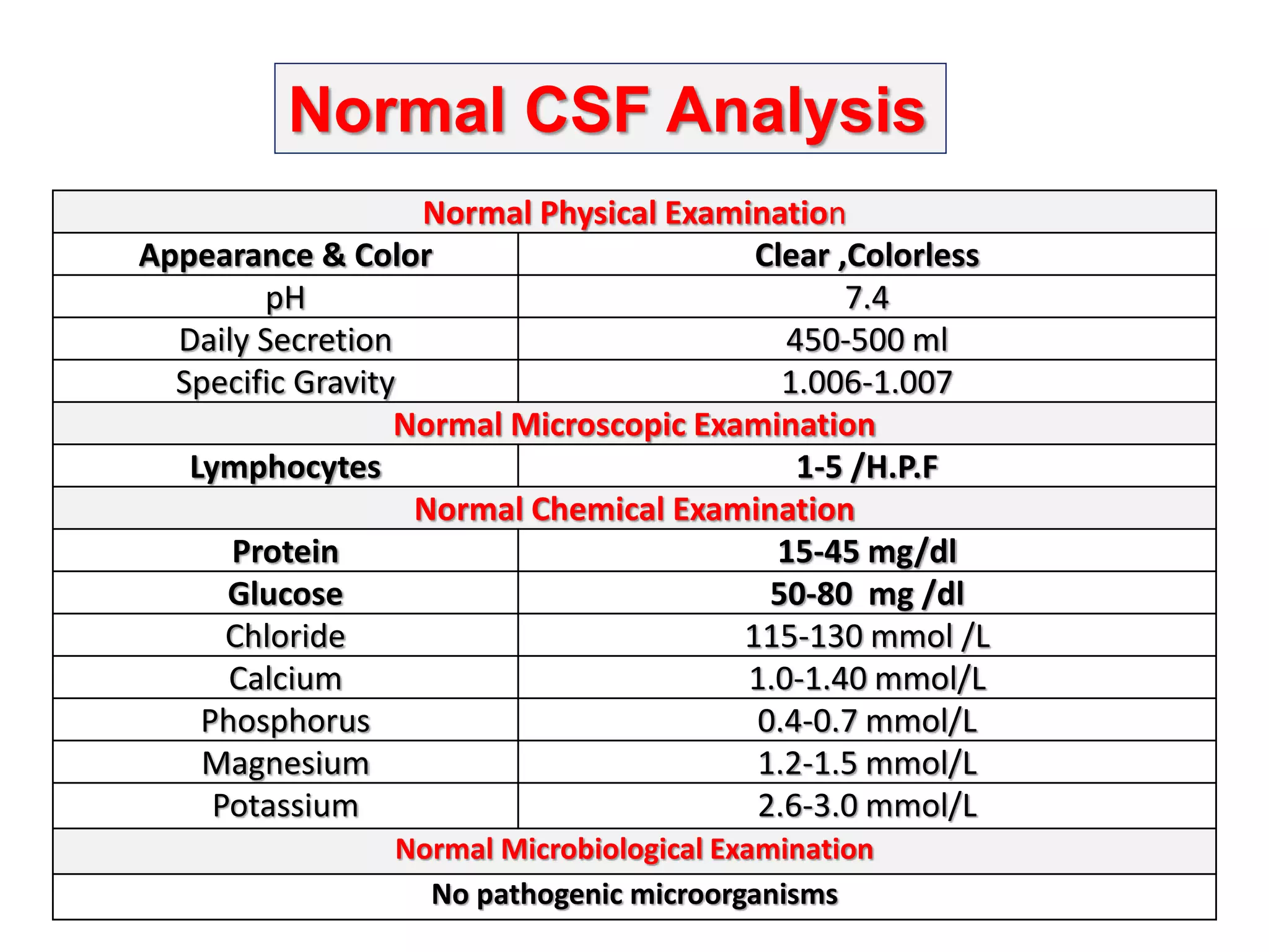
![Other Chemical Components of CSF
• CSF [Calcium], [Potassium] & [Phosphates] are lower
than their levels in the blood
• CSF [Chloride] & [Magnesium] are higher than their
levels in the blood
– Abnormal CSF [Chloride]
• marked in acute bacterial meningitis
• slight in viral meningitis & brain tumors](https://image.slidesharecdn.com/7908180-230725160826-d0df5691/75/Csf-analysis-25-2048.jpg)


