Downloaded 465 times

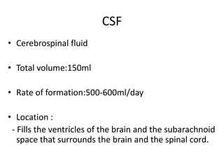
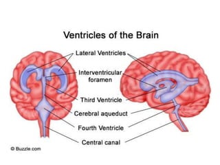
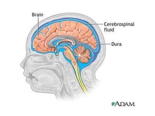
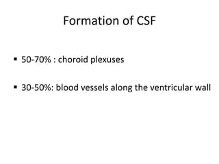
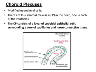

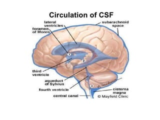

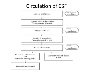
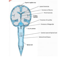


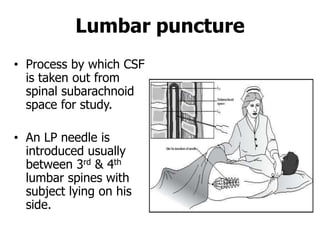
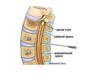
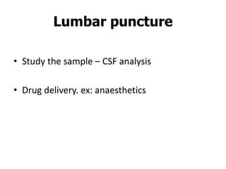
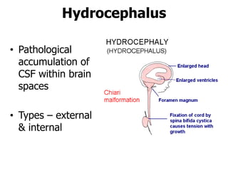


Cerebrospinal fluid (CSF) is formed in the brain ventricles and circulates around the brain and spinal cord. It is produced at a rate of around 500-600 ml per day, primarily by the choroid plexuses in the ventricles. CSF is absorbed into the venous blood through arachnoid villi and lymphatic vessels. It acts as a cushion and protects the brain from mechanical injury. CSF also helps remove waste from the brain and maintain homeostasis. Abnormal CSF accumulation can cause hydrocephalus, while lumbar puncture allows sampling of CSF for analysis.


![PERI-PROSTHETIC FRACTURE NAIL-PLATE CONSTRUCT [NPC].pptx](https://cdn.slidesharecdn.com/ss_thumbnails/drarunkumardrmohamedashrafperiprostheticfrasturenail-plateconstructnpc-260209164459-7e9d15a1-thumbnail.jpg?width=640&height=640&fit=bounds)


