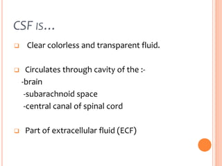
CEREBROSPINAL
FLUID
Prepared by:
Shaimaa sayed , Salma Yahya , Ghada Farah
CONTENTS …
Introduction
Physical properties
Functions
Formation
Circulation
CSF analysis
Hydrocephalus
CSF
CSF IS…
 Clear colorless and transparent fluid.
 Circulates through cavity of the :-
-brain
-subarachnoid space
-central canal of spinal cord
 Part of extracellular fluid (ECF)
CSF IS…
 Covered by : -
1. Pia meter (Inner membrane)
2. Arachnoid meter (center membrane)
3. Dura meter (Outer membrane)
THE MEMBRANS !!
CSF IS …
 Rate of formation :-
About 0.3 ml/min
And Normally 500ml of CSF is formed everyday
and equal amount is absorbed .
LOCATION OF CSF..
Two lateral ventricles
Third ventricle
Fourth ventricle
Spinal cord central canal
Subarachnoid space
Continuous with extracellular fluid
of brain parenchyma.
PHYSICAL PROPERTIES ….
1-Total volumes
 Adults :140 – 170 ml
Children : 10 – 60 ml
PHYSICAL PROPERTIES …
2-Spacific gravity : 1.005
3- ph : 7.28 - 7.32
4- appearance : clear
5-composition
PHYSICAL PROPERTIES…
For appearance : -
- clear & colorless is Normal
- bright red indicate presence of
blood
- turbid indicate presence of white
cells
FUNCTIONS OF CSF ..
Protects & lubricates the brain
Provides nutrients ,remove waste
Modulates pressure changes
 serves as a chemical buffer to maintain
constant ionic environment
Serves as a transport medium for
nutrients and metabolites ,endocrine
substansis and even neurotransmitters
FORMATION OF CSF ..
 The CSF is formed mainly in the brain
ventricles, and that the majority of the
remaining CSF is probably produced by
the ependymal surface of ventricles.
 It is generally accepted that CSF flows
unidirectional from the brain ventricles
to the subarachnoid space with the
exchange of various substances
between the CSF and interstitial
compartments.
FORMATION OF CSF ..
Choroid plexuses are the main site of
CSF production.
Then.. The passage of the plasma ultra
filtrate through the endothelium ,
facilitated by hydrostatic pressure.
Then.. Passes into the ventricles
An active metabolic process which
transforms the ultra filtrate into
secretion CSF.
CSF CIRCULATION ..
CSF CIRCULATION ..
The amount of CSF formation in the
day equal
to the amount of absorption.
Vf = Va
CSF CIRCULATION ..
CSF ANALYSIS
WHY?
CSF analysis can accurately distinguish
between a wide range of CNS diseases.
 Conditions found by CSF analysis can be
placed into four main categories:
1- infectious diseases.
2- hemorrhaging.
3- immune response disorders.
4- tumors.
CSF analysis may be ordered when a
patient has one or more of the following
symptoms :
 severe, unremitting headache
 stiff neck
 light sensitivity
 dizziness
 speaking difficulties
 trouble walking or poor coordination
 fatigue, lethargy, muscle weakness
How to colect the
sample to make
analysis ??
It’s easy by
Lumber Puncture
LUMBER PUNCTURE
A lumbar puncture (or LP, and
colloquially known as a spinal tap) is
a diagnostic procedure that is
performed in order to collect a
sample of cerebrospinal fluid (CSF)
for biochemical , microbiological,
and cytological analysis .
APPEARANCE OF CSF
In normal cases, CSF is clear and
colorless.
In cases of malady, the appearance
change according to the case.
We will discuss some of these in the
following:
1- Xanthochromic :
(Bright red or yellow)
This indicates the presence of blood.
The appearance of fresh blood in all
tubes supports the diagnosis of a
subarachnoid hemorrhage.
2- Turbid :
This indicates the presence of white cells
and is suggestive of a CNS infection.
CELL COUNTS
1- Increased Neutrophils in CSF
This indicates a bacterial meningitis,
cerebral abscess.
2- Increased Lymphocytes in CSF:
 This indicates a viral meningitis,
tuberculosis, syphilis, fungal and
parasitic infections.
 Degenerative diseases of the CNS,
such as multiple sclerosis, will also
generate elevated lymphocyte counts.
CSF DISORDERS
1- CSF Glucose:
 CSF glucose is derived from blood glucose
hence, ideally CSF glucose level should be
compared with fasting plasma glucose level
for adequate clinical interpretatio
 Decrease duo to hypoglycemia , tuberculosis.
Increase duo to high blood sugar.
2- CSF Pressure:
Increase duo to increases intra cranial
pressure
Decrease due to shock ,fainting or
diabetic coma.
3- CSF Protein :
Increase duo to blood in CSF ,tumor.
Decrease duo to rapid CSF production.
Hydrocephalus
(water on brain)
HYDROCEPHALUS
 The term hydrocephalus is derived from the
Greek words "hydro" meaning water and
"cephalus" meaning head.
 It is an abnormal collection of cerebrospinal
fluid (CSF) resulting in abnormal widening of
spaces in the brain.
 Causes of “Hydrocephalus” may be:
-Excessive production
-Decreased absorbtion
-Obstruction
TYPES OF HYDROCEPHALUS
Communicating and Non-
communicating
Acute and chronic
Congenital and acquired
(A)1- Non-communicating (obstructive):
 CSF circulation is blocked at or proximal to fourth
ventricular outlet foramina.(enlargement of
ventricles proximal to the block)
2- Communicating (non-obstructive):
 due to excessive formation of CSF or lack of
absorption
 the obstruction of CSF flow is in the
subarachnoid space from prior bleeding or
meningitis.
 This causes thickening of the arachnoid leading
to blockage of the return-flow channels.
(B) 1- Acute :
 hydrocephalus caused by tumor
 Develops within days or few weeks
 Manifests with rapid progression of symptoms
 Requires early attention and treatment .
2- Chronic :
 Over months (or even years)
 Subtle signs of memory impairment,
walking difficulty, urinary incontinence.
 Chronic hydrocephalus can present acutely
because of changes in the pathophysiology of
the CSF absorption or flow.
(C) 1- Congenital :
 Present at birth or few weeks/months after
birth.
2- Acquired
 Infection (post-meningitis)
 Post – hemorrhagic (SAH,IVH)
 Tumors
Treatment
Diversion of CSF:
1- Ventriculoperitoneal shunt
2- Lumboperitoneal shunt
Cerebrospinal fluid
Cerebrospinal fluid

Cerebrospinal fluid

  • 1.
  • 2.
  • 3.
  • 4.
    CSF IS…  Clearcolorless and transparent fluid.  Circulates through cavity of the :- -brain -subarachnoid space -central canal of spinal cord  Part of extracellular fluid (ECF)
  • 5.
    CSF IS…  Coveredby : - 1. Pia meter (Inner membrane) 2. Arachnoid meter (center membrane) 3. Dura meter (Outer membrane)
  • 6.
  • 7.
    CSF IS … Rate of formation :- About 0.3 ml/min And Normally 500ml of CSF is formed everyday and equal amount is absorbed .
  • 8.
    LOCATION OF CSF.. Twolateral ventricles Third ventricle Fourth ventricle Spinal cord central canal Subarachnoid space Continuous with extracellular fluid of brain parenchyma.
  • 9.
    PHYSICAL PROPERTIES …. 1-Totalvolumes  Adults :140 – 170 ml Children : 10 – 60 ml
  • 10.
    PHYSICAL PROPERTIES … 2-Spacificgravity : 1.005 3- ph : 7.28 - 7.32 4- appearance : clear 5-composition
  • 11.
    PHYSICAL PROPERTIES… For appearance: - - clear & colorless is Normal - bright red indicate presence of blood - turbid indicate presence of white cells
  • 13.
    FUNCTIONS OF CSF.. Protects & lubricates the brain Provides nutrients ,remove waste Modulates pressure changes  serves as a chemical buffer to maintain constant ionic environment Serves as a transport medium for nutrients and metabolites ,endocrine substansis and even neurotransmitters
  • 14.
    FORMATION OF CSF..  The CSF is formed mainly in the brain ventricles, and that the majority of the remaining CSF is probably produced by the ependymal surface of ventricles.  It is generally accepted that CSF flows unidirectional from the brain ventricles to the subarachnoid space with the exchange of various substances between the CSF and interstitial compartments.
  • 15.
    FORMATION OF CSF.. Choroid plexuses are the main site of CSF production. Then.. The passage of the plasma ultra filtrate through the endothelium , facilitated by hydrostatic pressure. Then.. Passes into the ventricles An active metabolic process which transforms the ultra filtrate into secretion CSF.
  • 17.
  • 18.
    CSF CIRCULATION .. Theamount of CSF formation in the day equal to the amount of absorption. Vf = Va
  • 19.
  • 20.
  • 21.
    WHY? CSF analysis canaccurately distinguish between a wide range of CNS diseases.  Conditions found by CSF analysis can be placed into four main categories: 1- infectious diseases. 2- hemorrhaging. 3- immune response disorders. 4- tumors.
  • 22.
    CSF analysis maybe ordered when a patient has one or more of the following symptoms :  severe, unremitting headache  stiff neck  light sensitivity  dizziness  speaking difficulties  trouble walking or poor coordination  fatigue, lethargy, muscle weakness
  • 23.
    How to colectthe sample to make analysis ?? It’s easy by Lumber Puncture
  • 24.
    LUMBER PUNCTURE A lumbarpuncture (or LP, and colloquially known as a spinal tap) is a diagnostic procedure that is performed in order to collect a sample of cerebrospinal fluid (CSF) for biochemical , microbiological, and cytological analysis .
  • 28.
    APPEARANCE OF CSF Innormal cases, CSF is clear and colorless. In cases of malady, the appearance change according to the case. We will discuss some of these in the following:
  • 29.
    1- Xanthochromic : (Brightred or yellow) This indicates the presence of blood. The appearance of fresh blood in all tubes supports the diagnosis of a subarachnoid hemorrhage. 2- Turbid : This indicates the presence of white cells and is suggestive of a CNS infection.
  • 30.
    CELL COUNTS 1- IncreasedNeutrophils in CSF This indicates a bacterial meningitis, cerebral abscess.
  • 31.
    2- Increased Lymphocytesin CSF:  This indicates a viral meningitis, tuberculosis, syphilis, fungal and parasitic infections.  Degenerative diseases of the CNS, such as multiple sclerosis, will also generate elevated lymphocyte counts.
  • 32.
    CSF DISORDERS 1- CSFGlucose:  CSF glucose is derived from blood glucose hence, ideally CSF glucose level should be compared with fasting plasma glucose level for adequate clinical interpretatio  Decrease duo to hypoglycemia , tuberculosis. Increase duo to high blood sugar.
  • 33.
    2- CSF Pressure: Increaseduo to increases intra cranial pressure Decrease due to shock ,fainting or diabetic coma. 3- CSF Protein : Increase duo to blood in CSF ,tumor. Decrease duo to rapid CSF production.
  • 34.
  • 36.
    HYDROCEPHALUS  The termhydrocephalus is derived from the Greek words "hydro" meaning water and "cephalus" meaning head.  It is an abnormal collection of cerebrospinal fluid (CSF) resulting in abnormal widening of spaces in the brain.  Causes of “Hydrocephalus” may be: -Excessive production -Decreased absorbtion -Obstruction
  • 39.
    TYPES OF HYDROCEPHALUS Communicatingand Non- communicating Acute and chronic Congenital and acquired
  • 40.
    (A)1- Non-communicating (obstructive): CSF circulation is blocked at or proximal to fourth ventricular outlet foramina.(enlargement of ventricles proximal to the block) 2- Communicating (non-obstructive):  due to excessive formation of CSF or lack of absorption  the obstruction of CSF flow is in the subarachnoid space from prior bleeding or meningitis.  This causes thickening of the arachnoid leading to blockage of the return-flow channels.
  • 41.
    (B) 1- Acute:  hydrocephalus caused by tumor  Develops within days or few weeks  Manifests with rapid progression of symptoms  Requires early attention and treatment . 2- Chronic :  Over months (or even years)  Subtle signs of memory impairment, walking difficulty, urinary incontinence.  Chronic hydrocephalus can present acutely because of changes in the pathophysiology of the CSF absorption or flow.
  • 42.
    (C) 1- Congenital:  Present at birth or few weeks/months after birth. 2- Acquired  Infection (post-meningitis)  Post – hemorrhagic (SAH,IVH)  Tumors
  • 43.
    Treatment Diversion of CSF: 1-Ventriculoperitoneal shunt 2- Lumboperitoneal shunt